Stability of Chlorophyll as Natural Colorant: A Review for Suji (Dracaena Angustifolia Roxb.) Leaves’ Case
 , , 2*, , 2, , 2
 1Department of Food Science and Technology, Faculty of Agricultural Engineering and Technology, Bogor Agricultural University, Bogor 16002, Indonesia2Southeast Asian Food and Agricultural Science and Technology (SEAFAST) Center,
Bogor Agricultural University, Bogor 16680, Indonesia
Corresponding Author Email: andarwulan@apps.ipb.ac.id
DOI : http://dx.doi.org/10.12944/CRNFSJ.6.3.04
Download this article as:
![]()
Suji (Dracaena angustifolia (Medik.) Roxb.) leaves are famous chlorophyll source used as food colorant in Indonesia and other south-east Asian countries. Its chlorophyll has unique characteristics which can degrade through enzymatic and non-enzymatic reactions. This article summarizes traditional application of Suji leaves, the characteristics of Suji leaf chlorophyll, postharvest stability, and several ways to retain its green color. Potential development of Suji leaf extract as food colorant or food ingredients are also discussed.
KEYWORDS:Degradation; Enzymes; Food; Green; Kinetic; Postharvest; Storage
Introduction
Chlorophyll, as a natural pigment, plays an important role for the green color appearance in plants. The green color of chlorophyll has been long time used as a natural colorant.1 Chlorophyll demand increases inline with increasing awareness for using natural colorants.2,3 From health aspect, chlorophyll and chlorophyllin are provide benefits to human body.4,5 Chlorophyll have antioxidant and antiinflamatory properties that prevent chronic diseases such as cancer.6–8 However, changes of chlorophyll structure into its derivatives made it loses its activities.9–12
Exploration for chlorophyll content in several plants has been done worldwide.13–17 One of the green plants that contain high chlorophyll content is Suji.18,19 Suji (Dracaena angustifolia (Medik.) Roxb.) plant is famous for its color and as medicinal plant. Suji own fresh green color and easily extracted using water.20 Chlorophyll water extract of Suji leaf is added to the food processing as natural colorant in various food and non food products.
Chlorophyll in Suji and other plants easily degraded due to enzymatic reactions and non-enzymatic reactions influenced by environmental conditions.10 Because of its role as a light capturing unit in photosynthesis process,21 the presence of light and heat radiation will excite the transfer of energy and increase the reactivity of chlorophyll thus its easily to undergo degradation process. Chlorophyll degradation progress rapidly as the chlorophyll structure changes into its derivative compounds that result in of its green color. Chlorophyll degradation during storage or post-harvest processing of horticultural produces causes significant losses.22,23 The discoloration of green vegetables to dark-green, yellow, or even black primarily due to the of chlorophyll.
Although potential as natural colorant, due to its chlorophyll content, Suji plants are only grown as ornamental in indoor or garden and has not been utilized optimally. In this review, authors will describe Suji leaf characteristics; postharvest chlorophyll stability and its possible degradation pathway and kinetics; and ways to maintain green color stability. Further, the potency of development natural green colorant from Suji leaf will be explained at the end of this review.
Botany of Suji
The genus Dracaena consists of about 150 species.24 The Suji (Dracaena angustifolia (Medik.) Roxb.) plants has been classified in the family of Agaveceae among the of flowers with six stamens, inflorescences, and plants with rosettes of fleshy fibrous leaves.25 Others recognized it as a distinct family, Dracaenaceae, along with the genera Sansevieria and Pleomele.26 Suji plant image is presented in Fig. 1.
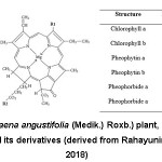 |
Figure 1: Suji (Dracaena angustifolia Roxb.) plant, chlorophyll structure and its derivatives |
Suji is an evergreen shrubby plant with a rhizomatous rootstock. The stems are grayish, smooth, and 1-3 meters tall with no or few branches.27 The plant has linear-lanceolate leaves, acuminate, length of 15-30 cm, 2-4 cm broad, sessile, margin entire. Its ribbon-shaped leaves elongated with size 17×2.5 cm and has dark green color. The leaf is smooth in both surface and sticking alternately in the stem with intervals of 0.5 cm28 followed by 2-5 yellowish-white flowers together in terminal wide-spreading panicles and globose compressed 3-lobed berry.29,30 Dracaenas propagation can be done with seeds, transplantation, and grafting28 but commercially they propagated using vegetative method by cutting relatively large stems.
Dracaena plants easily found in tropical zone from Africa to the Pacific islands and cultivated intensively in Southeast Asia, including Indonesia, Malaysia, and Vietnam. It has different names including Nam ginseng (Vietnam), Suji (Indonesia, Malaysia), chang hua long xue shue (Chinese), saiheva and si-ei (Papua New Guinea).27,29 Common names for this plant is Dragon’s blood palm according to the deep red liquid exudes from injured bark.25 Some literature also named Suji as Pleomele angustifolia Roxb. or Pleomele angustifolia N.E. Brown.8,20 It is planted near rivers and water stream, or as decorative plant in a garden. The preferred habitats are rainforests or semi-deserts.31 Various species of Dracaena have an importance in floriculture, medicinal, or social functions in many society. Despite this importance, the plant is still underutilized and not cultivated intensively yet.
Traditional Application of Dracaena Plants
Suji has been use as medicinal plant in Asia region since ancient times. Diseases are treated using various parts of this plant. Known locally as Nam ginseng, Suji roots and rhizomes are used as tonic and leukemia treatment in Vietnam.32 Its roots is effective in treating stomachache while its leaves are used for anti-inflammatory and anti-dysentry.28 The roots are applied to prevent insect bites by Philippines.27 Sundanese, who inhabit West Java region in Indonesia, treated cough diseases and lung disorders using Suji leaves.33 Boiled juice of squeezed leaves is given to asthma patients who have shortness of breath. Leaf decoction is drunk to increase appetite and body weight.29 In the field of cosmetics, Suji extract is believed to help fertilize hair growth, render the hair long and pliant.27
Green color from Suji leaves extract has been use for coloring food and non-food. In Indonesia and Malaysia, green color of Suji leaves water extract has been added in food preparations. The extract give fresh color for various drinks, sweets, puddings, and desserts. Suji leaves usually mixed with Pandanus leaves extract as the best combination to provide fresh green flavored desserts.34 Suji leaf water extract is used for coloring an Indian pastry made of glutinous rice. The leaf extract also use in preparation of Tumpeng ponco warno is rice featuring identical mountain cones and has five colors: red, blue, yellow, green, and white.35 Besides as coloring agent used in porridge and traditional cakes, Balinese people cooked Suji leaf shoots and eaten as a side dish with rice.36 For non-food applications, green color of Suji leaf extract is used as dye for paper, castor oil, and coconut oil. Suji extract has been applied for coloring fabrics in home-made Indonesian batik production.37
Phytochemicals in Suji Plants
Suji plants contain many phytochemical components. The presence of alkaloids, flavonoids, tannins, terpenoids, saponins, polyphenol, monoterpenoid, sesquiterpenoid, and glycosides are found in its root, rhizome, stem, and leaf based on qualitative screening.28,29,38,39 The ergosterol peroxide, linoleic acid, and E-phytol content in Suji plant showed its ability as antituberculosis.27 The methanol extract of underground parts of Suji plants own eight steroidal saponin and several recent compounds, including three spirostanol sapogenenins (namogenins A-C), four spirostanol saponins (namonins A-D), two furostanol saponin (namonins E-F), and a pregnan glycoside (namonin F). Some of them indicated effective ability as antiproliferative against HT-1080 fibrosarcoma cell cultured in vitro.32,40 Min et al., (2010) isolated six steroidal saponins from fresh stems of Suji plant, named angudracanosides A-F. Those compounds had antifungal activity against Cryptococcus neoformans. The methanol extract of whole Suji plant has two steroidal saponins, named drangustosides A-B. These compounds showed anti-inflammatory activity due to superoxide formation and elastase discharge by human neutrophils in reaction to formyl-L-methionyl-L-leucyl-L-phenylalanine, fMLP/CB.42 Saponins in the form of steroidal saponins in Dracaena plant suspected to have anti-inflammatory and analgesic characteristics.27 The ethanol extract of Suji leaves has antibacterial activity against bacteria S. dysenteriae ATCC 13313 and potential as a supplier of potassium in patients with hypokalemia dysentry.39
Dracaena angustifolia leaf extracts could activate cholinesterase activity and could be applied to build reactivators of cholinesterase prevented by organic phosphorous compounds.43 Suji leaves have an in vitro cholesterol-lowering activity. The compounds estimated to play a role in lowering cholesterol levels are phenolics, flavonoids, and vitamin C. Hydroxyl groups in cholesterol react with ketones in flavonoids to form hemiasetal. The research used a spectrophotometer to measure free cholesterol, not flavonoid-bound cholesterol. Carbonyl groups on flavonoids reacted with hydroxyl groups on cholesterol to form hydrogen bonds. The compound that bounded with a sample, or called free cholesterol, reacted with anhydrous acetic acid and concentrated sulfuric acid.44
Although comprehensive information on the chemical composition of the dragon’s blood resin is available, too few studies of the relationship to its medical function, specific compounds, and systematics have been completed.25 Further research needed to study the mechanism of specific compound from Suji plant responsible to health benefits.
Suji Leaf Chlorophyll Characteristics
Green color of Suji plants comes from the chlorophyll compound contained inside. Chlorophyll located in the intercellular lamella organel called a chloroplast.45,46 Its existence is protected by proteins that form a protein-chlorophyll complex.47–49 The complex is surrounded by protein-lipid bilayer thus making chlorophyll stable in it.50
Based on its structure, chlorophyll is a porphyrin containing a tetrapyrrole base ring that binds to each other through a methyne bridge (-C=) and binds Mg ion in the center.10 Close to the third pyrrole ring is located the fifth isocyclic ring while in the fourth ring is attached the propionic acid substituent esterified by a hydrophobic phytol group.3,51,52 Chlorophyll in plants mostly consists of two basic types namely chlorophyll a and chlorophyll b.53,55 Chlorophyll a has a methyl group (CH3) attached to R1 position (Fig. 1) thus its chemical formula is C55H72O5N4Mg and has blue-green color. Chemical formula of chlorophyll b is C55H70O6N4Mg which binds to the formyl group (CHO) on R1 position (Fig. 1), and has green-yellow color.52,56 Although Chlorophyll a is less polar than chlorophyll b, both are insoluble in water but very soluble in ethanol and methanol.20 Chemical structure of chlorophyll and its derivatives are given in Fig. 1.
Table 1: Chlorophyll Content of Several Plants
| Plant | Chlorophyll content (mg/kg fresh weight) | |||
| a | b | Total | Ratio a/b | |
| Chicory (cv. Anivip)17 | 2383.1 | 897.4 | 3280.5 | 2.7 |
| Chicory (cv. Monivip)17 | 1422.6 | 581.8 | 2004.4 | 2.4 |
| Dandelion leaf17 | 1805.4 | 677.1 | 2482.5 | 2.7 |
| Garden rocket17 | 2612.4 | 983.8 | 3596.2 | 2.6 |
| Wild rocket17 | 2160.1 | 872.2 | 3032.3 | 2.5 |
| Black locust leaf50 | 12864.6* | 3385.4* | 16250.0 | 3.8 |
| Scots pine50 | 2908.6* | 881.4* | 3790.0 | 3.3 |
| Sow thistle leaf50 | 10652.8* | 4097.2* | 14750.0 | 2.6 |
| Green peas109 | 140.1 | 90.6 | 230.7 | 1.5* |
| Pistachio15 | 3.6 | 1.8 | 5.4* | 2.0* |
| Spinach60 | 790.7 | 292.7 | 1083.4* | 2.7* |
| Green papric60 | 57.9 | 28.2 | 86.1* | 2.0* |
| Broccoli110 | 218.0 | 90.6 | 308.6 | 2.4* |
| Pink Lady apple (flesh)13 | 0.4 | 0.1 | 0.5* | 3.2* |
| Fuji (I) apple (flesh)13 | 0.9 | 0.2 | 1.1* | 3.6* |
| Reina de Reineta apple (flesh)13 | 1.2 | 0.3 | 1.5* | 3.8* |
| Green Golden Delicious apple (flesh)13 | 3.5 | 0.9 | 4.4* | 3.8* |
| Green Doncella apple (flesh)13 | 5.2 | 1.1 | 6.3* | 4.7* |
| Granny Smith apple (flesh)13 | 6.3 | 1.8 | 8.1* | 3.5* |
| Golden Rosett apple (peel)13 | 4.9 | 1.7 | 6.6* | 2.9* |
| Golden Montana apple (peel)13 | 4.5 | 1.2 | 5.7* | 3.9* |
| Golden Delicious apple (peel)13 | 18.3 | 5.3 | 23.6* | 3.5* |
| Royal Gala apple (peel)13 | 7.8 | 2.6 | 10.4* | 2.9* |
| Fuji (F) apple (peel)13 | 14.2 | 3.7 | 17.9* | 3.8* |
| Ariane apple (peel)13 | 9.0 | 2.2 | 11.2* | 4.0* |
| Starking Red Chief apple (peel)13 | 28.7 | 8.0 | 36.7* | 3.6* |
| Pink Lady apple (peel)13 | 31.8 | 8.5 | 40.3* | 3.7* |
| Fuji (I) apple (peel)13 | 51.5 | 15.1 | 66.6* | 3.4* |
| Reina de Reineta apple (peel)13 | 29.4 | 8.9 | 38.3* | 3.3* |
| Green Golden Delicious apple (peel)13 | 35.4 | 11.0 | 46.4* | 3.2* |
| Green Doncella apple (peel)13 | 52.8 | 11.3 | 64.1* | 4.7* |
| Granny Smith apple (peel)13 | 237.1 | 70.0 | 307.1* | 3.4* |
*calculated based on chlorophyll a, chlorophyll b, and total chlorophyll content or ratio of chlorophyll a/b.
In higher plants, chlorophyll generally composes 0.6 to 1.2%-wt of leaf wight on a dry matter basis.57 The chlorophyll content of several plants (vegetables, fruits, and nuts) are varied and the ratio of chlorophyll a/b in plants ranged from 1.5-4.7 with an average value of 3.1 (Table 1). The ratio can be varies due to growth conditions and external factors, especially high-light intensity and sun exposure.58
Clustered of chlorophyll content and its chlorophyll a/b ratio of several greeny plants was evaluated using Principal Component Analysis (PCA) to explore its relationship. The vertical axis indicates chlorophylls content (right: higher chlorophyll content, left: lower chlorophyll content) whereas the horizontal axis illustrated its chlorophyll a/b ratio (upper: higher ratio, lower: lower ratio). PCA plot projected by PC1 and PC2 showed variance occurred for different plants (Fig. 2). Suji leaf (Pr1 and X1) separated from others and located in negative quadrant. It revealed that Suji leaf has higher chlorophyll content than other plants with high chlorophyll b content that made its chlorophyll a/b ratio is low.
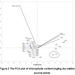 |
Figure 2:The PCA plot of chlorophylls content (mg/kg dry matter) of several plants |
(Note. Pr1: Suji leaf 19; X1: Suji leaf 111; Zn1: Chicory (cv. Anivip), Zn2: Chicory (cv. Monivip), Zn3: Dandelion leaf, Zn4: Garden rocket, Zn5: Wild rocket 17; dR1: Broccoli 110; Dp1: Golden Rosett apple (flesh), Dp2: Golden Montana apple (flesh), Dp3: Golden Delicious apple (flesh), Dp4: Royal Gala apple (flesh), Dp5: Fuji (F) apple (flesh), Dp6: Ariane apple (flesh), Dp7: Starking Red Chief apple (flesh), Dp8: Pink Lady apple (flesh), Dp9: Fuji (I) apple (flesh), Dp10: Reina de Reineta apple (flesh), Dp11: Green Golden Delicious apple (flesh), Dp12: Green Doncella apple (flesh), Dp13: Granny Smith apple (flesh), Dp14: Golden Rosett apple (peel), Dp15: Golden Montana apple (peel), Dp16: Golden Delicious apple (peel), Dp17: Royal Gala apple (peel), Dp18: Fuji (F) apple (peel), Dp19: Ariane apple (peel), Dp20: Starking Red Chief apple (peel), Dp21: Pink Lady apple (peel), Dp22: Fuji (I) apple (peel), Dp23: Reina de Reineta apple (peel), Dp24: Green Golden Delicious apple (peel), Dp25: Green Doncella apple (peel), Dp26: Granny Smith apple (peel) 13)
The difference of chlorophyll content in green leaf is influenced by growth location, leaf age in one tree, and leaf position.57 Leaf age is determined from the position of leaf out on the stem from the shoot. The older age leaves position will be further away from the shoots. Jokopriyambodo et al., (2014) examined that the chlorophyll content of mature Suji leaf extract was twice as of immature leaf extract. Ozgen and Sekerci (2011) research on spinach plant found that the end portion of a leaf far from the stem also has higher chlorophyll and phytochemical content than the base of the leaf attached to the stem.
Suji leaves have 73.25% moisture content which contains 2524.6 ppm chlorophyll a and 1250.3 chlorophyll b.18 Whilst spinach, the common source of chlorophyll, is contain 1083.4 ppm of total chlorophyll.60 The high amount or chlorophyll content, especially chlorophyll a, has implication on the green appearance of plant and a large ratio of chlorophyll a/b indicates the convenience of chlorophyll to dissolve in organic solvents (more hydrophobic). For example, black locust leaf and Fuji (F) apple (peel) which have the same chlorophyll a/b ratio of,3.813,50 however chlorophyll a and chlorophyll b of black locust leaf were higher than those in apple peel thus this leaf has greener color than apple skin.
The Suji leaves have high chlorophyll content with small difference between chlorophyll a and b (Table 1). The high content shown by Suji leaves dark green appearance while its low a/b ratio make Suji’s chlorophyll can be easily extracted using water. These two properties made Suji leaves has good potency to be utilize as food colorant or food ingredient.
The lower chlorophyll a/b ratio indicates the convenience of chlorophyll to dissolve in solvent. Chlorophyll a is less polar than chlorophyll b thus lower ratio of chlorophyll a/b resulted in higher solubility in water-based solvent. This property is important in food processing whereas in food processing because water is widely used as a medium to dissolve food products. Cassava leaf and Suji leaf contained high chlorophyll content with different chlorophyll a/b ratio. Cassava leaf has higher chlorophyll a/b ratio than it in Suji leaf.14 The lower ratio made chlorophyll in Suji leaves more convenient to be extracted using aqueous solvent. It is characterized by the juice of Suji leaves that are darker green color than the water of the cassava leaves or spinach leaves at the same amount.
Postharvest Stability of Suji Leaf Chlorophyll
Similar to other green leafy plants, degradation of chlorophyll in Suji leaf occurs during senescence or post harvest processing.61 Degradation was directly happened after leaves were harvested through enzymatic and non-enzymatic reactions.62–64 Enzymatic degradation of chlorophyll occurs due to the presence of chlorophyll degradation enzymes that are endogenously present in plant tissues. The enzymes degrade chlorophyll during senescence or ripening of green fruits/vegetables.65 While non-enzymatic degradation affected by environmental factors and usually develop during post-harvest processing.66
Enzymatic Degradation of Chlorophyll
Several types of identified chlorophyll-degradation enzymes present in plant tissues include peroxidase,67 Mg-dechelatase,68 pheophorbide a oxygenase, red chlorophyll-reductase catabolite,69 and chlorophyllase.70 Hörtensteiner and Kräutler (2011) and Ankita and Prasad (2015) mentioned that in the plant tissue, the initial phase of chlorophyll degradation begins with activity of chlorophyll b reductase enzyme and 7-hydroxy methyl chlorophyll-reductase which transforms chlorophyll b to chlorophyll a. The conversion encrypted by Non Yellow Coloring 1 (NYC1) gene62,72 While peroxidase enzyme starts chlorophyll degradation process by oxidizing chlorophyll a to 132-hydroxychlorophyll a.73
Chlorophyll a lost its phytol group due to the activity of the enzyme and forms chlorophyllide a.74,75 Enzyme is actually a glycoprotein compound in the thylakoid membrane. Schwartz et al., (2008) explains that is an esterase enzyme that catalyzes of phytol groups from chlorophyll and pheophytin structures. It activity is limited to porphyrins with carboxyloxy groups at C-10 and hydrogen at C-7 and C-8 positions. The enzyme is active in temperature range 60-82.2°C and it is damaged at higher temperatures (>100°C).10 The presence of chlorophyllide is desired to maintain the green color in heated vegetables.
Chlorophyllide which lost its Mg2+ called pheophorbide.75 The Mg-dechelatase enzyme or Mg-dechelatase substance will release Mg2+ in chlorophyll a and form pheophorbide a structure.73 Mg-dechelatase is active on artificial chlorophyll substrate (chlorophyllin) but not on natural compounds of chorophyllide.69 While Mg-dechelatase substance reacts to chlorophyllin and chorophyllide. It indicates that on in vivo the loss of Mg atoms from the chorophyllide structure is more due to the activity of low molecular weight compounds.62 Pheophorbide a decomposes into a chorophyll catabolite, which is generally colorless, through the formation of red chlorophyll catabolite by pheophorbide a oxygenase enzyme and red chlorophyll reductase.76
Schelbert et al., (2009) reported the role of pheophytinase enzymes (pheophytin pheophorbide hydrolase, PPH) in converting pheophytin to pheophorbide. The presence of lipoxygenase, peroxidase, and polyphenol oxidase (PPO) enzymes in plant tissues could degrade the quality of vegetables/fruits and responsible in chlorophyll degradation.62,67,78–80 The lipoxygenase plays an important role in the establishment of hydroperoxides and free radicals that induce yellow color or colorless appearance.10 Peroxidase enzyme is an oxidoreductase enzyme found in many organelles including chloroplasts.81 Although the peroxidase enzyme has a significant role in fruit ripening, it is also responsible for chlorophyll degradation through oxidation of phenolic compounds by hydrogen peroxide.67,82,83 Further, the phenoxy radical oxidize chlorophyll into colorless compounds.10 The chlorophyll-oxydase enzyme is located in the thylakoid membrane of chloroplasts.83 This enzyme oxydize chlorophyll into chlorophyll a-1 compounds that are suspected to be intermediates in the chlorophyll degradation process.73
Non-enzymatic Degradation of Chlorophyll
Environmental factors or processing of horticultural products (green fruits and vegetables) makes chlorophyll unstable. The presence of heat, acid or low pH, light, or microbial agents made chlorophyll degraded through several processes.66,84,85 The replacement of Mg2+ with two H+ ion in chlorophyll structure known as pheophytinization.86 The change of chlorophyll into pheophytin is influenced by pH or acid condition. Eckardt (2009) asserted that formation of pheophytin becomes the first stage of chlorophyll degradation during senescence, followed by the release of phytol groups. The original blue-green chlorophyll a turns into a gray-colored pheophytin a and green-yellow chlorophyll b turns into a brown-colored pheophytin b. The conformation of pheophytin from chlorophyll a occurs 2.5-10 times faster than that of chlorophyll b.57
The green color of chlorophyll in the vegetables/fruits was turning to olive-green during food processing. The organic acids from the food product were released during processing and pH while chlorophyll is stable at alkaline condition.84 During fermentation, chlorophylls change to become pheophytins and pyropheophytins. Decreasing pH and green color changing accelerated with the presence of acid-producing microbes.88 The presence of heat during fermentation is thought to affect pyropheophytin formation. Pheophytins and pheophorbides as derivatives products from chlorophyll degradation were found in the final product.57
Thermal process makes protein in the chlorophyll-protein complex denatured thus enzymes meet their substrate. Its reaction de-esterificate phytol groups in chlorophyll and produces chlorophyllid structure.74 The chromophore character of chlorophyll in de-esterification reaction is unchanged and the color remains green.3 The phytol group (which makes chlorophyll fat soluble) will be detached from the chlorophyll structure thus increasing the polarity of chlorophyll. Minguez-Mosquera et al., (2008) mentioned that pheophytin can also be a substrate to de-esterification reaction and form pheophorbide structure. Ramírez et al., (2015) found that all cholorophyll in processed olives contain no Mg ion (pheophytin form). Pumilia et al. (2014) mentioned that 85% decrease of pheophytin a and b levels during 60 min roasting of pistachio nuts as well as an increase in pyropheophytin a and b levels 10-12 times higher than in raw pistachio. The addition of time resulted in decreased levels of chlorophyll a and b in spinach and increased levels of chlorophyll a and b and pheophytin a and b due to pheophytinization reactions.90 In refrigerating and freezing process, which generally maintains post-harvest quality of horticultural products, chlorophyll damage still occurs. Spinach stored at freezing temperatures continues to undergo changes in its green color.91
High-pressure treatments do not cause chlorophyll degradation in some vegetables, while combination of high-pressure and high-temperature treatment rapidly degrade chlorophyll.60 Although chlorophyll b is more resilient than chlorophyll a at 70°C but both are degraded at higher temperatures. The alternative thermal processing to extend fruit shelf-life is microwave heating. Nevertheless, processing of kiwi fruit with microwave still eliminates most of its chlorophyll content and pheophytin a becoming the major component in the processed kiwi.11
Chlorophyll degradation reactions occur in the isocyclic ring (cyclopentanone/ring V) are differentiated into epimerization, decarbomethoxylation, and allomerization.89 Epimerization is an isomerization reaction between H and CO2CH3 molecules on an isocyclic ring of chlorophyll. This reaction does not cause discoloration. Decarbometoxylation is an oxidative reaction that replaces the carbomethoxy group in C-132 (COOCH3) with H ion without altering the basic porphyrin structure. This reaction produces pyro-derivative compounds that have same color and spectroscopic character as their precursors (chlorophyll, pheophytin, chlorophyllid, and pheophorbide). While allomerization reactions occur when the isocyclic ring is oxidized by an oxygen triplet molecule (3O2) or release of carbomethoxy group at C-132,57
Chemical degradation of chlorophyll in Suji leaf and other green plants can go through several processes, but in general the hypothetical pathway of chlorophyll degradation in Suji leaf suggested takes place through two paths. The first pathway is through the formation of pheophytin due to the activity of enzymes or acid conditions and the second pathway is through the formation of chlorophyllide as a result of specific reactions by enzymes (Fig. 3). Pheophytin and chlorophyllide then degraded into pheophorbide which loss it green color property. Pheophorbide can be further degraded to colorless products at a final step.
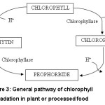 |
Figure 3: General pathway of chlorophyll degradation in plant or processed food |
Chlorophyll Degradation Kinetic
Degradation kinetics of chlorophyll and its derivative products during processing can be used as a model for predicting shelf life.92 Kinetic model are able to estimate the shelf life of a product based on different variables that can affect food degradation.93 Kinetics of chemical reaction described as the measurement of reaction velocity and analysis of experimental data to determine factors affecting the reaction, such as reaction rate, reaction order, equations that give information about the speed dependence on product concentration that will affect the reaction, and the effect of temperature on reaction rate.94
Chlorophyll a damages 2.5 times faster than chlorophyll b with activation energy ranged from 4.80±0.91 to 14.0±0.71 kcal/mol for chlorophyll a and 6.84±0.29 to 11.0±1.06 kcal/mol for chlorophyll b at various pH values.84 Chlorophyll’s green color loss, due to heat and oxidation, mostly follows the Arrhenius first order reaction. A slight change in temperature will leads to increasing chlorophyll kinetic.93
Various factors that affect the rate and kinetics of chlorophyll degradation were thermal and acidic condition, microbial growth, and light intensity.88,95–98 The green color of blanched green peas was rapidly disappearing with decreasing pH.84 Gunawan and Barringer (2000) found that acid compounds containing benzene rings induced color changes faster than acid compounds with simple carbon chains because of their hydrophobicity. Their study showed that microbial growth accelerates the color change by produce acid and cause of holes in the broccoli surface. Ghidouche et al., (2013) predicted the shelf life of multiple colorants by increasing the intensity of light. The result concluded that the light treatment has higher damage effect to the chlorophyll structure than temperature treatment. The increase of light intensity would increase the chlorophyll degradation rate. Photodegradation rate of chlorophyll was easily to changes of light intensity and can be described using the first order reaction model.94 The presence of lipid protected chlorophyll from photo-oxidation. Triolein acid gave better protection and retard the kinetic rate of photodegradation than oleic acid in a paraffin oil system, possibly due to rivalry between lipids and chlorophyll for singlet oxygen.99
A first-order kinetic mechanism was suitable for illustrating the processes of virgin olive oils under non-oxygen thermal auto-oxidation.93 The kinetic constants for chlorophylls degradation was 3.6 times lower than the respective constants for carotenoids. Chlorophyll has higher activation energy (16.03 ± 0.26 kcal.mol-1) than carotenoids (15.45 ± 0.17 kcal.mol-1). It indicates that small changes in temperature can increase the kinetic constant of chlorophyll. Chlorophyll structure is also more resistant to color changes due to heat.93 Decreased retention of Zn-chlorophyll pigments followed first-order kinetics with velocity constants (k) 1.5×10⁻³ day-1 and half-life predictive for more than 15 months.
Although specific kinetic study on chlorophyll in Suji leaf is not available yet, its predictive thermal degradation might followed the first-order kinetic of Arrhenius reaction. Process due to photodegradation of chlorophyll in Suji leaf will also follows the first-order reaction. The shelf-life model for based photosensitive food, chlorophyll from Suji leaves, could be predicted using formula by Manzocco et al., (2008) as follows:
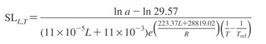
where SLL,T is the product shelf-life at certain light intensity (L) temperature value (T), a is the final color value, T is the absolute temperature (K), Tref is the reference temperature, and R is the molar gas constant (8.31 Jmol-1K-1).
The detail mechanism of chlorophyll degradation of Suji leaf has not been studied. Information on chlorophyll thermal- and photodegradation of Suji leaf will give better understanding on degradation mechanism thus leads to more effective ways to maintain chlorophyll stability.
Retaining Green Color Stability of Suji Leaf Chlorophyll
Utilization of liquid chlorophyll extract from Suji leaf as colorant has been done since long time ago in Indonesia. But it is less practical and the color has lower stability than synthetic colorants.1 Chlorophyll extraction on a household scale usually done by grinding green leaves then extracted using water. The liquid extract then filtered and added to the food and beverage processing process. Liquid extraction method has a disadvantage that liquid extract should be directly used. If the liquid extract is not immediately used then the green color will turn brown.
Previous researches on preserving green color of chlorophyll extract were mainly focused on maintaining chlorophyll structure, formation of chlorophyll-derived compounds that were still green and more resistant to heat (chlorophyllid), or making metallo-chlorophyll complexes.101 These efforts include: heat treatment to inactivate chlorophyllase enzyme and minimize chlorophyll conversion to pheophytin, formation of bright green metallo-chlorophyll complex by adding Cu2+ Zn2+ salt, control to the process conditions (temperature, pH, and ionic strength of food products), and addition of surface active agents.66 However previous studies on Suji leaves extract are mainly focused on producing metallo-chlorophyll complexes with addition of Zn to substitute Mg in the center of chlorophyll structure.18,20,102
Addition of CaCO3 and MgCO3 could retained green color of chlorophyll from Suji extract (Caesar et al., 2018). Those salts completely neutralized the acid in the plant tissue and avoid acidification thus inhibit the arrangement of pheophytins in chlorophyll extract.104,105 Jokopriyambodo et al., (2014) produced powdered colorant from water-insoluble Suji leaf extract using dried oven. Since powdered Suji extract has low solubility, the encapsulated powder by addition various coating materials was expected to increase its solubility and protect chlorophyll from environment damage. Therefore, development of natural colorant through chlorophyll drying process to become powder made it more convenient for storage and application.Combination of extraction using ZnCl2 and selected coating agent by spray drying method resulted best characteristics in green color, total chlorophyll, and antioxidant activity. A 30% n-octenyl succinic anhydride (OSA) modified starch as filler was able to retain green color of chlorophyll extract.106 Other extraction technique was maceration with aqueous solvent and NaHCO3 as and thus powdered using maltodextrin by freeze dryer.18 Despite green color of chlorophyll powder was faded due to addition of coating agents, encapsulation method made it more easily to dissolve in food.
Research on Suji leaf extraction method found that optimum extraction process of natural pigment from Suji leaf was done by stabilized aqueous extraction method with the addition of 700 ppm ZnCl2 combined with experiment parameters (pH 7 and temperature of 85°C). These method yields total chlorophyll content of 47.2975 mg/100 g fresh weight.20
Chlorophyll degradation into pheophytin can also be affected by the presence of surface active agents for example detergent. Detergent absorbed on surface of thylakoid cell membrane, where the chlorophyll is located, affects the H+ permeability through the membrane. The presence of cationic detergents will block the H+ ions on surface membrane diffusing into the cells to prevent chlorophyll degradation. In contrast, anionic detergents will attract H+ ions, increase their concentration on surface membrane, and increase their diffusing rate. Thus the presence of anionic detergents can accelerate chlorophyll degradation. While non-ionic detergents on surface membrane will reduce its negative charge so that H+ ions will be attracted to the surface and consequently chlorophyll degradation will decrease.57 Addition of Tween 80 increased the total chlorophyll content of extract and retained its green color. As non-ionic detergent, Tween 80 suppressed pheophytin formation, help the lipophilic chlorophyll emulsified in water, and facilitate contact with enzymes.19
Potential Development of Natural Green Colorant from Suji Leaf
Efforts to develop natural colorant from Suji leaves have not been able to maintain its stability. Further research needs to be done so that the green color of colorant is similar to fresh leaf. A comprehensive quantitative study also needs to estimate shelf-life and its application in food and non-food products. Industrial scale-up also needs to be intensively undertaken so that the production of natural colorant can be massively produced and of has high economic value.
Suji leaves have better characteristics than other chlorophyll sources. The Suji leaves have no gel-forming components which could prevent the extraction of chlorophyll from leaf tissue. In addition, Suji leaves also have non-toxic components such as cyanide on cassava leaves.107 Its leaf is not bitter but rather has specific scent.28 Compared to basil, Suji leaves have a distinctive lighter leaf scent that is favored and widely applied to various food products without disturbing the original aroma of the product. Utilization of Suji plants was limited recently, as an ornamental plant and food coloring in the household scale. Nonetheless Suji plants are promising to be cultivated intensively because its propagation is easy by stem cuttings or grafting.
Green pigments content in Suji plants is potential to be used as food coloring materials as well as functional food ingredients. Marquez and Sinnecker (2008) states that the requirement for an economical material to be used as a source of chlorophyll are its high pigment content, its availability, the ease of harvesting and drying, efficient extraction mechanisms, and the low activity of chlorophyllase enzymes. Suji leaf have most of the requirement as described above thus its support the potential of Suji leaves to be developed as a source of chlorophyll that can be used as food coloring as well as functional food raw materials.
Conclusion
Chlorophyll from Suji leaves extract has unique due to its content and ratio of a/b. This ratio makes Suji’s chlorophyll easier to dissolve in solvent than other sources. Chlorophyll might be turns into its derivatives through enzymatic and non-enzymatic degradation during processing followed first-order of Arrhenius kinetics reaction. The degradation was prevent by heat treatment modification, formation of metallo-chlorophyll complex, control of processing condition, and addition of antioxidant compounds. Chlorophyll extract was developed from liquid form to encapsulated powder using several methods. The chlorophyll pigments contain in Suji plants is potential to be used as colorant or food ingredients due to no gel-forming property, non-toxic components, and easy to cultivate. Due to limited research on chlorophyll from Suji leaf, further research needed to optimize the utilization of its potency and stability.
Acknowledgements
The authors thank the Ministry of Research, Technology and Higher Education of the Republic of Indonesia for research funding and Southeast Asian Food and Agricultural Science and Technology (SEAFAST) Center, Bogor Agricultural University for supporting the publication.
References
- Shahid M., Shahid-Ul-Islam., Mohammad F. Recent advancements in natural dye applications: A review. J Clean Prod. 2013;53:310-331. doi:10.1016/j.jclepro.2013.03.031
CrossRef - Hung S. M., Hsu B. D., Lee S. Modelling of isothermal chlorophyll extraction from herbaceous plants. J Food Eng. 2014;128:17-23. doi:10.1016/j.jfoodeng.2013.12.005
CrossRef - Sigurdson G. T., Tang P., Giusti M. M. Natural Colorants: Food Colorants from Natural Sources. Annu Rev Food Sci Technol. 2017;8(1):261-280. doi:10.1146/annurev-food-030216-025923
CrossRef - Ghidouche S., Rey B., Michel M., Galaffu N. A Rapid tool for the stability assessment of natural food colours. Food Chem. 2013;139(1-4):978-985. doi:10.1016/j.foodchem.2012.12.064
CrossRef - Tumolo T., Lanfer-Marquez U. M. Copper chlorophyllin: A food colorant with bioactive properties? Food Res Int. 2012;46(2):451-459. doi:10.1016/j.foodres.2011.10.031
CrossRef - Subramoniam A., Asha V. V., Nair S. A. et al. Chlorophyll revisited: Anti-inflammatory activities of chlorophyll a and inhibition of expression of TNF-α gene by the same. Inflammation. 2012;35(3):959-966. doi:10.1007/s10753-011-9399-0
CrossRef - McQuistan T. J., Simonich M. T., Pratt M. M. et al. Cancer chemoprevention by dietary chlorophylls: A 12,000-animal dose–dose matrix biomarker and tumor study. Food Chem Toxicol. 2012;50(2):341-352. doi:10.1016/j.fct.2011.10.065
CrossRef - Jokopriyambodo W., Sudarsono., Rohman A. The Antiradical Activity of Insoluble Water Suji (Pleomele angustifolia N . E . Brown) Leaf Extract and Its Application as Natural Colorant in Bread product. J Food Pharmaceitical Sci. 2014;2:52-56.
- Albanese D., Russo L., Cinquanta L., Brasiello A., Di Matteo M. Physical and chemical changes in minimally processed green asparagus during cold-storage. Food Chem. 2007;101(1):274-280. doi:10.1016/j.foodchem.2006.01.048
CrossRef - Yilmaz C., Gokmen V. Chlorophyll. In: Caballero B, Finglas PM, Toldra F, eds. Encyclopedia of Food and Health. Waltham, MA: Academic Press. 2016:37-41.
CrossRef - Benlloch-Tinoco M., Kaulmann A., Corte-Real J., Rodrigo D., Martínez-Navarrete N., Bohn T. Chlorophylls and carotenoids of kiwifruit puree are affected similarly or less by microwave than by conventional heat processing and storage. Food Chem. 2015;187:254-262. doi:10.1016/j.foodchem.2015.04.052
CrossRef - Comunian T. A., Monterrey-Quintero E. S., Thomazini M. et al., Assessment of production efficiency, physicochemical properties and storage stability of spray-dried chlorophyllide, a natural food colourant, using gum Arabic, maltodextrin and soy protein isolate-based carrier systems. Int J Food Sci Technol. 2011;46(6):1259-1265. doi:10.1111/j.1365-2621.2011.02617.x
CrossRef - Delgado-Pelayo R., Gallardo-Guerrero L., Hornero-Méndez D. Chlorophyll and carotenoid pigments in the peel and flesh of commercial apple fruit varieties. Food Res Int. 2014;65:272-281. doi:10.1016/j.foodres.2014.03.025
CrossRef - Limantara L., Dettling M., Indrawati R., Indriatmoko., Brotosudarmo T. H. P. Analysis on the Chlorophyll Content of Commercial Green Leafy Vegetables. Procedia Chem. 2015;14:225-231. doi:10.1016/j.proche.2015.03.032
CrossRef - Pumilia G., Cichon M. J., Cooperstone J. L., Giuffrida D., Dugo G., Schwartz S. J. Changes in chlorophylls, chlorophyll degradation products and lutein in pistachio kernels (Pistacia vera L.) during roasting. Food Res Int. 2014;65:193-198. doi:10.1016/j.foodres.2014.05.047
CrossRef - Ramírez E., Gandul-Rojas B., Romero C., Brenes M., Gallardo-Guerrero L. Composition of pigments and colour changes in green table olives related to processing type. Food Chem. 2015;166:115-124. doi:10.1016/j.foodchem.2014.05.154
CrossRef - Žnidarčič D., Ban D., Šircelj H. Carotenoid and chlorophyll composition of commonly consumed leafy vegetables in Mediterranean countries. Food Chem. 2011;129(3):1164-1168. doi:10.1016/j.foodchem.2011.05.097
CrossRef - Aryanti N, Nafiunisa A, Willis FM. 2016;5(4):129-135. doi:10.17728/jatp.183129
CrossRef - Prangdimurti E., Muchtadi D., Astawan M., Zakaria. 2006;17(2):79-86.
- Rahayuningsih E., Pamungkas M. S., Olvianas M., Putera A. D. P. Chlorophyll extraction from suji leaf (Pleomele angustifolia Roxb.) with ZnCl2stabilizer. J Food Sci Technol. 2018;55(3):1028-1036. doi:10.1007/s13197-017-3016-7
CrossRef - Shibghatallah M. A. H., Khotimah S. N., Suhandono S., Viridi S., Kesuma T. Measuring leaf chlorophyll concentration from its color: A way in monitoring environment change to plantations. AIP Conf Proc. 2013;1554:210-213. doi:10.1063/1.4820322
CrossRef - Dissanayake P. K., Yamauchi N., Shigyo M. Presence of pheophytin and its formation as a chlorophyll derivative selected crop species. J Agric Sci. 2012;7(3):127-134. doi:10.4038/jas.v7i3.4876
CrossRef - Shi J., Gao L., Zuo J., Wang Q. Q., Wang Q. Q., Fan L. Exogenous sodium nitroprusside treatment of broccoli florets extends shelf life, enhances antioxidant enzyme activity, and inhibits chlorophyll-degradation. Postharvest Biol Technol. 2016;116:98-104. doi:10.1016/j.postharvbio.2016.01.007
CrossRef - Narender B., Naveena N., Pravalika P., Kaleem S., Vamshi M., Mandhadi J. R. Pharmacological evaluation of root and leaf extracts of Dracaena reflexa var. angustifolia. Innov Pharm Pharmacother. 2017;5(3):141-146.
- Lu P-L. Systematics, Evolution, and Biogeography among Dracaenoid Genera: Dracaena, Pleomele, and Sansevieria (Asparagaceae). 2012.
- Damen T. H. J., van der Burg W. J., Wiland-Szymańska J., Sosef M. S. M. Taxonomic novelties in African Dracaena (Dracaenaceae). Blumea – Biodiversity, Evol Biogeogr Plants. 2018;(63):31-53. doi:10.3767/blumea.2018.63.01.05
CrossRef - Wiart C. Medicinal Plants of China, Korea, and Japan. Boca Raton, FL: CRC Press. 2012. doi:10.15713/ins.mmj.3
- Kinho J, Arini DID, Halawane J. 2011.
- WHO. Medicinal plants in Papua New Guinea. WHO Ser. 2009;98-99.
- Xinqi C., Turland N. J. Dracaena vandelli ex Linnaeus. Flora of China. 2000;(24):215-217.
- Klimko M., Wiland-Szymańska J. Scanning electron microscopic studies of leaf surface in taxa of genus Dracaena L. (Dracaenaceae). Bot – Steciana. 2008;12:117-127.
- Quan T Le., Qui T. K., Kadota S. Study of Dracaena Angustifolia – III New Steroidal Saponins from Roots and Rhizomes, and Their Antiproliferative Activity. J Chem. 2004;42(3):371-375.
- Roosita K., Kusharto C. M., Sekiyama M., Fachrurozi Y., Ohtsuka R. Medicinal plants used by the villagers of a Sundanese community in West Java , Indonesia. J Ethnopharmacol. 2008;115:72-81. doi:10.1016/j.jep.2007.09.010
CrossRef - Routray W., Rayaguru K. Chemical Constituents and Post-Harvest Prospects of Pandanus amaryllifolius Leaves : A Review Chemical Constituents and Post-Harvest Prospects of Pandanus amaryllifolius Leaves : A Review. 2015;9129(September). doi:10.1080/87559129.2010.484114
CrossRef - Wulan. Tumpeng Ponco Warno. 21 Desember 2017. https://budaya-indonesia.org/Tumpeng-Ponco-Warno. Published 2017.
- Putri R. I., Supriatna J., Walujo E. B. Ethnobotanical Study of Plant Resources in Serangan Island, Bali. Asian J Conserv Biol. 2014;3(2):135-148.
- Fauziyah N., Hakim L. Plants as Natural Dyes for Jonegoroan Batik Processing in Jono Cultural Tourism Village, Bojonegoro, East Java. J Indones Tour Dev Stud. 2015;3(2):41-44.
CrossRef - Shukla A., Vats S., Shukla R. K. Phytochemical Screening , Proximate Analysis and Antioxidant Activity of Dracaena reflexa Lam. Leaves. Indian J Pharm Sci. 2015;77(5):640-644. doi:10.4103/0250-474X.169035
CrossRef - Kusuma S. A. F., Ramdhani D., Mustarichie R. Comparative study on activities of anti bacillary dysentry Shigella dysenteriae of Syzygium polyanthum and Dracaena angustifolia leaves ethanol extracts. Asian J Pharm Clin Res. 2017;10(2):348-352. doi:10.22159/ajpcr.2017.v10i2.15725
CrossRef - Tran Q Le., Tezuka Y., Banskota A. H., Tran Q. K., Saiki I., Kadota S. New Spirostanol Steroids and Steroidal Saponins from Roots and Rhizomes of Dracaena angustifolia and Their Antiproliferative Activity. J Nat Prod. 2001;(64):1127-1132.
- Min X., Ying-Jun Z., Xing-Cong L., Jacob M. R., Chong-Ren Y. Steroidal Saponins from Fresh Stems of Dracaena angustifolia. J Nat Prod. 2010;73(9):1524-1528. doi:10.1111/j.1743-6109.2008.01122.x.Endothelial
- Huang H. C., Lin M. K., Hwang S. Y. et al. Two anti-inflammatory steroidal saponins from Dracaena angustifolia Roxb. Molecules. 2013;18(8):8752-8763. doi:10.3390/molecules18088752
CrossRef - Chen K., Zhao G., Wu S., Zhuang Q. Preliminary Researches on Possibility of Several Kinds of Plants Including Dracaena Angustifolia Used to Relieve the Damages Caused by Organic Phosphorous Compounds. 2013;815:358-361. doi:10.4028/www.scientific.net/AMR.815.358
CrossRef - Anggraini D. I., Nabillah L. F. Activity Test of Suji Leaf Extract (Dracaena angustifolia Roxb .) on in vitro cholesterol lowering. J Sci Appl Chem. 2018;21(2):54-58.
CrossRef - Staehelin L. A. Chloroplast structure: From chlorophyll granules to supra-molecular architecture of thylakoid membranes. Photosynth Res. 2003;76(1-3):185-196. doi:10.1023/A:1024994525586
CrossRef - López-Millán A. F., Duy D., Philippar K. Chloroplast Iron Transport Proteins – Function and Impact on Plant Physiology. Front Plant Sci. 2016;7(February):1-12. doi:10.3389/fpls.2016.00178
CrossRef - Shi L. X., Theg S. M. The chloroplast protein import system: From algae to trees. Biochim Biophys Acta – Mol Cell Res. 2013;1833(2):314-331. doi:10.1016/j.bbamcr.2012.10.002
CrossRef - Markwell J. P., Thornber J. P., Boggs R. T. Higher plant chloroplasts: Evidence that all the chlorophyll exists as chlorophyll—protein complexes. Proc Natl Acad Sci U S A. 1979;76(3):1233-1235. doi:10.1073/pnas.76.3.1233
CrossRef - Camm E. L., Green B. R. How the chlorophyll-proteins got their names. Photosynth Res. 2004;80(1-3):189-196. doi:10.1023/B:PRES.0000030460.26687.95
CrossRef - Miazek K., Ledakowicz S. Chlorophyll extraction from leaves, needles and microalgae: A kinetic approach. Int J Agric Biol Eng. 2013;6(2):107-115. doi:10.3965/j.ijabe.20130602.00?
- Humphrey A. M. Chlorophyll as a color and functional ingredient. J Food Sci. 2004;69(5):C422-C425. doi:10.1111/j.1365-2621.2004.tb10710.x
CrossRef - Roca M., Chen K., Perez-Galvez A. Chlorophylls. In: Reinhold C, Schweiggert RM, eds. Handbook on Natural Pigments in Food and Beverages. Duxford, UK: Woodhead Publishing; 2016:125-158. doi:10.1016/B978-0-08-100371-8.00013-0
CrossRef - Tanaka R., Tanaka A. Chlorophyll cycle regulates the construction and destruction of the light-harvesting complexes. Biochim Biophys Acta – Bioenerg. 2011;1807(8):968-976. doi:10.1016/j.bbabio.2011.01.002
CrossRef - Siswadi. PENANGANAN PASCA PANEN BUAH-BUAHAN DAN SAYURAN. 68-71.
- Sant’Anna V., Gurak P. D., Ferreira Marczak L. D., Tessaro I. C. Tracking bioactive compounds with colour changes in foods – A review. Dye Pigment. 2013;98(3):601-608. doi:10.1016/j.dyepig.2013.04.011
CrossRef - Croft H., Chen J. M. Leaf Pigment Content. Elsevier Inc.; 2017. doi:10.1016/B978-0-12-409548-9.10547-0
CrossRef - Schwartz S. J., von Elbe J. H., Giusti M. M. Colorants. In: Damodaran S, Parkin KL, Fennema OR, eds. Fennema’s Food Chemistry 4th Ed. Boca Raton, FL: CRC Press. 2008:571-638. doi:10.1080/01932691.2011.584482
CrossRef - Lichtenthaler H. K., Buschmann C. Chlorophylls and Carotenoids: Measurement And Characterization by UV-VIS Spectroscopy. Handb Food Anal Chem. 2005;2-2:171-178. doi:10.1002/0471709085.ch21
CrossRef - Ozgen S., Sekerci S. Effect of leaf position on the distribution of phytochemicals and antioxidant capacity among green and red lettuce cultivars. Spanish J Agric Res. 2011;9(3):801-809. doi:10.5424/
- Sánchez C., Baranda A. B., De Marañón I. M. The effect of High Pressure and High Temperature processing on carotenoids and chlorophylls content in some vegetables. Food Chem. 2014;163:37-45. doi:10.1016/j.foodchem.2014.04.041
CrossRef - Almela L., Fernández-López J. A., Roca M. J. High-performance liquid chromatographic screening of chlorophyll derivatives produced during fruit storage. J Chromatogr A. 2000;870(1-2):483-489. doi:10.1016/S0021-9673(99)00999-1
CrossRef - Hörtensteiner S., Kräutler B. Chlorophyll breakdown in higher plants. Biochim Biophys Acta – Bioenerg. 2011;1807(8):977-988. doi:10.1016/j.bbabio.2010.12.007
CrossRef - Skutnik E., Rabiza-Swider J., Wachowicz M., Łukaszewska A. J. Senescence of cut leaves of Zantedeschia aethiopica and Z. elliottiana. Acta Sci Pol, Hortorum Cultus. 2004;3(2):57-65.
- Oberhuber M., Berghold J., Breuker K., Hörtensteiner S., Krautler B. Breakdown of chlorophyll: a nonenzymatic reaction accounts for the formation of the colorless “nonfluorescent” chlorophyll catabolites. Proc Natl Acad Sci U S A. 2003;100(12):6910-6915. doi:10.1073/pnas.1232207100
CrossRef - Kaewsuksaeng S. Chlorophyll Degradation in Horticultural Crops. Walailak J Sci Technol (. 2011;8(1):9-19. http://wjst.wu.ac.th/index.php/wjst/article/view/7.
- Zheng Y., Shi J., Pan Z., Cheng Y., Zhang Y., Li N. Effect of heat treatment, pH, sugar concentration, and metal ion addition on green color retention in homogenized puree of Thompson seedless grape. LWT – Food Sci Technol. 2014;55(2):595-603. doi:10.1016/j.lwt.2013.10.011
CrossRef - Yamauchi N., Funamoto Y., Shigyo M. Peroxidase-mediated chlorophyll degradation in horticultural crops. Phytochem Rev. 2004;3(1-2):221-228. doi:10.1023/B:PHYT.0000047796.98784.06
CrossRef - Suzuki T., Kunieda T., Murai F., Morioka S., Shioi Y. Mg-dechelation activity in radish cotyledons with artificial and native substrates, Mg-chlorophyllin a and chlorophyllide a. Plant Physiol Biochem. 2005;43(5):459-464. doi:10.1016/j.plaphy. 2005;03:009
CrossRef - Hörtensteiner S. Chlorophyll Degradation During Senescence. Annu Rev Plant Biol. 2006;57(1):55-77. doi:10.1146/annurev.arplant.57.032905.105212
CrossRef - Harpaz-Saad S., Azoulay T., Arazi T. et al., Chlorophyllase Is a Rate-Limiting Enzyme in Chlorophyll Catabolism and Is Posttranslationally Regulated. Plant Cell Online. 2007;19(3):1007-1022. doi:10.1105/tpc.107.050633
CrossRef - Ankita., Prasad K. Characterization of dehydrated functional fractional radish leaf powder. Der Pharm Lett. 2015;7(1):269-279.
- Hasperué J. H., Gómez-Lobato M. E., Chaves A. R., Civello P. M., Martínez G. A. Time of day at harvest affects the expression of chlorophyll degrading genes during postharvest storage of broccoli. Postharvest Biol Technol. 2013;82:22-27. doi:10.1016/j.postharvbio.2013.02.021
CrossRef - Kaewsuksaeng S., Tatmala N., Srilaong V., Pongprasert N. Postharvest heat treatment delays chlorophyll degradation and maintains quality in Thai lime (Citrus aurantifolia Swingle cv. Paan) fruit. Postharvest Biol Technol. 2015;100:1-7. doi:10.1016/j.postharvbio.2014.09.020
CrossRef - Vavilin D., Vermaas W. Continuous chlorophyll degradation accompanied by chlorophyllide and phytol reutilization for chlorophyll synthesis in Synechocystis sp. PCC 6803. Biochim Biophys Acta – Bioenerg. 2007;1767(7):920-929. doi:10.1016/j.bbabio.2007.03.010
CrossRef - Fukasawa A., Suzuki Y., Terai H., Yamauchi N. Effects of postharvest ethanol vapor treatment on activities and gene expression of chlorophyll catabolic enzymes in broccoli florets. Postharvest Biol Technol. 2010;55(2):97-102. doi:10.1016/j.postharvbio.2009.08.010
CrossRef - Matile P., Stefan H., Thomas H. Chlorophyll degradation-Chloroplast to Gerontoplast Transition. Annu Rev Plant Physiol Plant Mol Biol. 1999;50(2):67-95. doi:10.1146/annurev.arplant.50.1.67
CrossRef - Schelbert S., Aubry S., Burla B. B. et al., Pheophytin Pheophorbide Hydrolase (Pheophytinase) Is Involved in Chlorophyll Breakdown during Leaf Senescence in Arabidopsis. Plant Cell Online. 2009;21(3):767-785. doi:10.1105/tpc.108.064089
CrossRef - Aiamla-Or S., Shigyo M., Ito S. I., Yamauchi N. Involvement of chloroplast peroxidase on chlorophyll degradation in postharvest broccoli florets and its control by UV-B treatment. Food Chem. 2014;165:224-231. doi:10.1016/j.foodchem.2014.05.108
CrossRef - Büchert A. M., Civello P. M., Martínez G. A. Chlorophyllase versus pheophytinase as candidates for chlorophyll dephytilation during senescence of broccoli. J Plant Physiol. 2011;168(4):337-343. doi:10.1016/j.jplph.2010.07.011
CrossRef - Kunieda T., Amano T., Shioi Y. Search for chlorophyll degradation enzyme, Mg-dechelatase, from extracts of Chenopodium album with native and artificial substrates. Plant Sci. 2005;169(1):177-183. doi:10.1016/j.plantsci.2005.03.010
CrossRef - Christ B., Hörtensteiner S. Mechanism and Significance of Chlorophyll Breakdown. J Plant Growth Regul. 2014;33(1):4-20. doi:10.1007/s00344-013-9392-y
CrossRef - Vergara-Domínguez H., Roca M., Gandul-Rojas B. Characterisation of chlorophyll oxidation mediated by peroxidative activity in olives (Olea europaea L.) cv. Hojiblanca. Food Chem. 2013;139(1-4):786-795. doi:10.1016/j.foodchem.2013.01.120
CrossRef - Vergara-Domínguez H., Roca M., Gandul-Rojas B. Thylakoid peroxidase activity responsible for oxidized chlorophyll accumulation during ripening of olive fruits (Olea europaea L.). Food Res Int. 2014;65(PB):247-254. doi:10.1016/j.foodres.2014.04.030
CrossRef - Koca N., Karadeniz F., Burdurlu H. S. Effect of pH on chlorophyll degradation and colour loss in blanched green peas. Food Chem. 2007;100(2):609-615. doi:10.1016/j.foodchem.2005.09.079
CrossRef - Turkmen N., Poyrazoglu E. S., Sari F., Sedat Velioglu Y. Effects of cooking methods on chlorophylls, pheophytins and colour of selected green vegetables. Int J Food Sci Technol. 2006;41(3):281-288. doi:10.1111/j.1365-2621.2005.01061.x
CrossRef - Moyano M. J., Heredia F. J., Meléndez-martínez A. J. The Color of Olive Oils : The Pigments and Their Likely Health Benefits and Visual and Instrumental Methods of Analysis. Compr Rev Food Sci Food Saf. 2010;9:278-291. doi:10.1111/j.1541-4337.2010.00109.x
CrossRef - Eckardt N. A. A New Chlorophyll Degradation Pathway. Plant Cell Online. 2009;21(3):700-700. doi:10.1105/tpc.109.210313
CrossRef - Gunawan M. I., Barringer S. A. Green color degradation of blanched broccoli (Brassica Oleracea) due to acid and microbial growth. J Food Process Preserv. 2000;24(3):253-263. doi:10.1111/j.1745-4549.2000.tb00417.x
CrossRef - Minguez-Mosquera M. I., Gandul-Rojas B., Gallardo-Guerrero L., Roca M., Jaren-Galan M. Chlorophylls. In: Hurst WJ, ed. Methods of Analysis for Functional Foods and Nutraceuticals. 2nd ed. Boca Raton, FL: CRC Press. 2008:337-400.
- Kidmose U., Edelenbos M., Christensen L. P., Hegelund E. Chromatographic determination of changes in pigments in spinach (Spinacia oleracea L.) during processing. J Chromatogr Sci. 2005;43(9):466-472. doi:10.1093/chromsci/43.9.466
CrossRef - Dermesonluoglu E., Katsaros G., Tsevdou M., Giannakourou M., Taoukis P. Kinetic study of quality indices and shelf life modelling of frozen spinach under dynamic conditions of the cold chain. J Food Eng. 2015;148:13-23. doi:10.1016/j.jfoodeng.2014.07.007
CrossRef - Van Boekel MAJS. Testing of kinetic models: Usefulness of the multiresponse approach as applied to chlorophyll degradation in foods. Food Res Int. 1999;32(4):261-269. doi:10.1016/S0963-9969(99)00080-0
CrossRef - Aparicio-Ruiz R., Gandul-Rojas B. Decoloration kinetics of chlorophylls and carotenoids in virgin olive oil by autoxidation. Food Res Int. 2014;65:199-206. doi:10.1016/j.foodres.2014.05.046
CrossRef - Ayu D. F., Andarwulan N., Hariyadi P., Purnomo E. H. Photodegradation Kinetics of Chlorophyll , Tocopherol , and Carotenoid in Red Palm Oil. Agritech. 2016;36(2).
- Cuny P., Romano J. C., Beker B., Rontani J. F. Comparison of the photodegradation rates of chlorophyll chlorin ring and phytol side chain in phytodetritus: Is the phytyldiol versus phytol ratio (CPPI) a new biogeochemical index? J Exp Mar Bio Ecol. 1999;237(2):271-290. doi:10.1016/S0022-0981(99)00010-6
CrossRef - Erge H. S., Karadeniz F., Koca N., Soyer Y. Effect of heat treatment on chlorophyll degradation and color loss in green peas. GIDA. 2008;33(5):225-233.
- Gaur S., Shivhare U. S., Sarkar B. C., Ahmed J. Thermal chlorophyll degradation kinetics of mint leaves puree. Int J Food Prop. 2007;10(4):853-865. doi:10.1080/10942910601136450
CrossRef - Inanc A. L. Differential method to determine thermal degradation kinetics of chlorophyll in virgin olive oil. Ital J Food Sci. 2016;28:90-95.
- Lee E., Ahn H., Choe E. Effects of light and lipids on chlorophyll degradation. Food Sci Biotechnol. 2014;23(4):1061-1065. doi:10.1007/s10068-014-0145-x
CrossRef - Manzocco L., Kravina G., Calligaris S., Nicoli M. C. Shelf life modeling of photosensitive food: The case of colored beverages. J Agric Food Chem. 2008;56(13):5158-5164. doi:10.1021/jf800072u
CrossRef - Özkan G., Ersus Bilek S. Enzyme-assisted extraction of stabilized chlorophyll from spinach. Food Chem. 2015;176:152-157. doi:10.1016/j.foodchem.2014.12.059
CrossRef - Wijaya H. C., Bianca K., Puspitasari-Nienaber N. L. Addition of ZnCl2 in the production of coloring powder from suji and pandan leaves. Bul Teknol dan Ind Pangan. 1995;6(1):22-26.
- Caesar J., Tamm A., Ruckteschler N., Leifke A. L., Weber B. Revisiting chlorophyll extraction methods in biological soil crusts – methodology for determination of chlorophyll a and chlorophyll a+b as compared to previous methods. Biogeosciences. 2018;15:1415-1424. doi:10.5194/bg-2017-396
CrossRef - Weber B., Wessels D. C. J., Deutschewitz K., Dojani S., Reichenberger H., Büdel B. Ecological characterization of soil-inhabiting and hypolithic soil crusts within the Knersvlakte, South Africa. Ecol Process. 2013;2(1):1-13. doi:10.1186/2192-1709-2-8
CrossRef - Schwartz S. J., Lorenzo T. V. Chlorophyll in foods. Food Sci Nutr. 1990;29(1):1-17.
- Senklang P., Anprung P. Optimizing enzymatic extraction of zN-chlorophyll derivatives from pandan leaf using response surface methodology. J Food Process Preserv. 2010;34(5):759-776. doi:10.1111/j.1745-4549.2009.00393.x
CrossRef - Fasuyi A. O. Nutrient Composition and Processing Effects on Cassava Leaf (Manihot esculenta, Crantz) Antinutrients. Pakistan J Nutr. 2005;4(1):37-42. doi:10.3923/pjn.2005.37.42
CrossRef - Marquez U. M. L., Sinnecker P. Chlorophylls: Properties, Biosynthesis, Degradation and Functions. In: Socaciu C, ed. Food Colorants: Chemical and Functional Properties. Boca Raton, FL: CRC Press. 2008:25-49.
- Kumar R, Rajamanickam R, Nadanasabapath S. Effect of Maillard Reaction Products (MRP) on Chlorophyll Stability in Green Peas. Food Nutr Sci. 2013;2013(September):879-883. http://search.ebscohost.com/login.aspx?direct=true&profile=eh ost&scope=site&authtype=crawler &jrnl=2157944X&AN= 92758542&h=1WEtttEK/rfFEI7ayWDOSz gf4nx94RMV2yfZUngDcQT9L c5QDJytOLujAAkNLnyl10llJ0RMExacb9U41NJ+Fw==&crl=c.
- dos Reis L. C. R., de Oliveira V. R., Hagen M. E. K., Jablonski A., Flores S. H., de Oliveira Rios A. Carotenoids, flavonoids, chlorophylls, phenolic compounds and antioxidant activity in fresh and cooked broccoli (Brassica oleracea var. Avenger) and cauliflower (Brassica oleracea var. Alphina F1). LWT – Food Sci Technol. 2015;63(1):177-183. doi:10.1016/j.lwt.2015.03.089
CrossRef - Indrasti D., Andarwulan N., Purnomo E. H., Wulandari N. Pigment profile and its degradation of Suji (Dracaena angustifolia (Medik.) Roxb.) leaf during fresh storage. Draft manuscript to be submitted to Journal of Food Science and Technology.
Accepted on: 13-12-2018
Second Review by: Dr. A H Bandivdekar (India)
Final Approval by: Prof. Dr. Adele Papetti


Web of Science Coverage
Emerging Sources Citation Index (ESCI)
2024 Journal Impact Factor: 1.1
Scopus Journal Metrics
CiteScore 2025: 2.6
CiteScore Details
Sustainable Nutrition: Food Systems, Nutrient Retention, and Public Health Impact
![]()
This journal is a member of, and subscribes to the principles of, the Committee on Publication Ethics (COPE)


