Introduction
Fermented fish and fishery products find important place in the dietary lists of the Southeast Asian countries.1 These are widely distributed in rural and urban markets and fondly consumed daily as popular products.2 Philippine fermented products are divided into two groups: the first contains high salt concentration of about 15-20 percent like bagoong and patis and second with low salt concentration mixed with rice like burong isda and burong hipon. Several variations of burong isda are available in markets and its name depend on the fish used such as burong ayungin, Therapon plumbeus; burong gurami, Osphronemus goramy; burong tilapia, Tilapia nilotica; burong dalag, Ophicephalus striatus; burong hito, Clarias batrachus; burong kanduli, Arias manillensis and burong bangus, Chanos chanos.3 Milkfish or commonly known as bangus are among the most favored fish in the country noted for its distinct characteristic and flavor. It contains large quantity of omega-3 fatty acids, DHA and other important nutrients.4
Lactic acid bacteria (LAB) are group of related bacteria that produce lactic acid as a result of carbohydrate fermentation.5 They are gram positive, fastidious, acid tolerant, generally non-sporulating, catalase negative and non-respiring rod or cocci that are associated by their common metabolic and physiological characteristics to produce lactic acid as fermentation metabolites.6,7 They are nonpathogenic organism reputed as GRAS or Generally Recognized as Safe status.8 Lactobacilli are ubiquitous bacteria in the human and animal micro flora.9 LAB has been used for fermentation of food products since ancient days and until today their major applications are still in food industry.10 Their presence in food is a type of biopreservation system and it does not only add flavor to foods but also control pathogenic and spoilage microorganism through various ways such as production of peroxidases, organic acids and bacteriocins.11 Its various industrial applications include preservative, acidulant and flavor in food, textile and pharmaceutical industries.12
This study aimed to isolate bacterial strains from fermented rice-milkfish (burong bangus) and subjected to biochemical testing for characterization. The isolates were further analyzed for qualitative bacterial enzyme assay and test for antagonistic and antimicrobial activity.
Materials and Methods
Sample preparation
Fresh milkfish at approximately 700 g was purchased in the local market of Miagao, Iloilo and brought to the processing laboratory in an ice box. The samples were washed, eviscerated, filleted and meat chopped into small sizes. The final weight was 350 g chopped milkfish. Rice and salt used for the fermentation process were also purchased from the said local market. Salt were subjected to analysis to determine its purity. Prior to experiment, glass bottles for packaging container of fermented product were sterilized in boiling water for 15 minutes.
The ingredients used for the fermentation of burong isda includes: 29% chopped milkfish; 69% cooked rice and 2% rock salt.13 All the ingredients were mixed in a bowl and placed in the glass bottles. Then it was stored at room temperature in the processing laboratory.
Bacterial Isolation
Total viable count were analyzed by spread plating the sample in Nutrient Agar and MRS (deMan Rogosa Sharpe) Agar with 2% sodium chloride. Ten grams of homogenized sample was inoculated in 90 ml peptone water then serially diluted from 10-2 to 10-6 dilution. Three replicates per dilution were spread plate for both Nutrient Agar and MRS Agar. The plates were incubated at 37˚C for 48 hours then total viable counts were determined. The MRS plates were kept for isolation of bacterial samples. The plates with ideal colony were chosen for further process. Ideal colonies were streak plate in MRS Agar with 2% sodium chloride and incubated for 24 hours at 37˚C. The isolates were further purified by zigzag plating the next day in MRS Agar with 2% sodium chloride then slant after 24 hours incubation. The slants were used for biochemical test analysis of the isolates.14
Biochemical testing
All the biochemical test utilized 24 hours fresh bacterial culture for inoculation, except for spore staining which required a week old culture for analysis; and all the media used for testing were added with 2% sodium chloride.15
Qualitative enzymatic activity
The activity of the protease, cellulase, lipase and amylase of the isolates were qualitatively determined in an enzyme specific agar medium.16 Bacterial isolates extracellular protease, lipase, cellulase and amylase secretion were determined using gelatin peptone agar, tributyrin agar, carboxymethylcellulose agar and starch agar respectively. Enzyme activity of the isolates is expressed by the formation of the clearing or halo zone around the bacterial colony.
Antagonistic and Antimicrobial Assay
This test determines the antagonistic ability of the isolates against other pathogenic bacteria like Escherichia coli, Bacillus subtilis and Staphylococcus aureus. While the antimicrobial ability was determined against amoxicillin, cephalexin, chloramphenicol and erythromycin.
Materials such as sterile swab, forceps, paper disk (Whattman filter paper No. 1), saline solution and Mueller- Hinton agar plates were prepared prior to experiment and fresh 24 hours culture bacteria were used for the assay. Bacterial isolates and pathogenic bacteria were inoculated to saline broth and turbidity was confirmed using the McFarland No. 5 turbidity standard. Paper disk were drop with 10μl of saline broth inoculated with bacterial isolates and set aside. Saline broth with E. coli, B. subtilis and S. aureus were swab on the agar plates and dried paper disk drop with bacterial isolates were placed on the plates for antagonistic activity.17
Saline broth with bacterial isolates were swab on the agar plates and 0.6 mm antimicrobial disk of amoxicillin, cephalexin, chloramphenicol and erythromycin were placed on the plates to determine antimicrobial activity of the isolates. All plates were incubated at 37˚C for 24 hours. Then, the zones of inhibition were measured and its susceptibility against microbial drugs was determined based on the Table of Standards for Antimicrobial Susceptibility Testing.18
Results and Discussion
Aerobic plate count of burong isda in Nutrient Agar and MRS Agar are presented in Table 1. The total aerobic plate count of Nutrient Agar is 3.478 x 10³ (3.54 log CFU/g) and for MRS agar is 2.34 x 10⁵ (5.36 log CFU/g).
Table 1: Aerobic plate count in Nutrient and MRS Agar.
| Dilutions | MRS Count | Total Plate Count |
| 10-2 | TNTC | 62 |
| 10-3 | 471 | 6 |
| 10-4 | 70 | 1 |
| 10-5 | 9 | 0 |
| 10-6 | 1 | 0 |
| APC (CFU/ml) | 2.34×10⁵ | 3.478×10³ |
| Log count | 5.36 | 3.54 |
The aerobic plate count is an indicator commonly used in the food industry to determine the level of microorganism in a certain products.19 Fresh fish and fishery products often have an APC of 104 -105 (5 log CFU/g), while some seafoods have APC of 106 –108 (8 log CFU/g) without any quality changes.20 The result from this study was expected lower than the fresh fishes due to the fermentation process with the aid of salt addition and subsequently pH reduction.21 Study on fermented tuna viscera concluded that during fermentation period, counts decreased together with the increasing salt concentration.22
Biochemical test results of bacterial strains from burong isda with presumptive identification were presented on Table 2. Different test were conducted to characterize the five isolates. Based on the test result and further confirmation with the Bergey’s Manual of Systematic Bacteriology, the five isolates from burong isda were summarized into four lactic acid bacteria species. Isolates 2 and 4 were identified as one species only.
Table 2: Biochemical test result of bacterial strains from burong isda with presumptive ID.
| BIOCHEMICAL TESTS | ISOLATES | ||||
| B1 | B2 | B3 | B4 | B5 | |
| Bacterial shape | Doubled, quartered ovoid cocci | Small, singled, doubled, chained, tetrad cocci | Short, clustered, rods | Small, single, chained, doubled, tetrad cocci | Short, clustered, slendered, rods |
| Oxygen requirement | Facultative anaerobe | Facultative anaerobe | Facultative anaerobe | Facultative anaerobe | Facultative anaerobe |
| Gram staining | + | + | + | + | + |
| Spore formation | – | – | – | – | – |
| Catalase | + | + | – | + | – |
| Oxidase | – | – | – | – | – |
| Glucose | – | – | + | – | + |
| Sorbitol | – | – | – | – | – |
| Glycerol | – | – | – | – | – |
| Mannitol | – | – | – | – | – |
| Arabinose | – | – | – | – | – |
| Lactose | – | – | – | – | – |
| Citrate | – | – | – | – | – |
| H2S | – | – | – | – | – |
| Indole | – | – | – | – | – |
| Methyl red | + | + | + | + | + |
| Voges-Praskauer | – | – | – | – | – |
| Gelatine | – | – | – | – | – |
| Starch | – | – | – | – | – |
| Casein | + | + | + | + | + |
| Arginine dihydrolase | + | – | – | – | + |
| Lecithinase | – | – | – | – | – |
| Motility | – | – | – | – | – |
| Presumptive Identification | Enterococcus faecalis | Tetragenococcus muriaticus | Lactobacillus delbrueckii subp. delbrueckii | Tetragenococcus muriaticus | Carnobacterium divergens |
Legend: +, Positive; -, Negative
Enterococcus faecalis of the Enteroccocci family are ancient members of the animal microbiome date back in the Devonian period about 412 million years ago. They are known to thrive in the nutrient-rich, oxygen depleted environment of the intestinal tract found in wide variety of host including humans.23 They are member of the lactic acid bacteria which consist of Gram-positive, cocci or rods that produce mainly lactic acid from the fermentation of carbohydrates. They are also known to exhibit important probiotic activity and applications in the food industry as starter culture and potential biopreservatives.24 Tetragenococcus are halophilic histamine forming lactic acid bacteria first isolated from fermented squid liver sauce. They are abundantly present in gut and internal organs of their host.25,26 Lactobacillus delbrueckii are important member of lactic acid bacteria commonly used as starter for the production of fermented milk products such as yogurt and cheese.27 They are recognized probiotics that does not adhere well to the human intestine.28 Carnobacteria are dominant genus in the gastro-intestinal tract of various seafood products, non aciduric with low spoiling potential.29 They are lactic acid bacteria utilized as bacterial starter culture in manufacture of fermented meat products.30 Carnobacterium divergens isolated from seafoods have known to possess promising inhibition capacity to maintain low level of Listeria monocytogenes in cold smoke salmon lower than 50 CFU/g during four weeks of storage at 4˚C and 8˚C.31
Table 3 and Fig. 1 presents the zone of activity in millimetre of qualitative enzyme assay of the bacterial isolates. As can be observed, isolate B1 had enzyme activity both on lipase and protease assay with a value of 9.67 mm and 4.0mm, respectively. The second isolate had activity on protease assay only with 2.86 mm zone of activity. The third isolate had activity on amylase assay only with value of 5.0 mm zone of activity value. The fifth isolate had no activity for all the enzyme assay.
Table 3: Zone of activity from enzyme assay of burong isda isolates.
| Isolate | Enzymes | |||||||
| Lipase | Protease | Amylase | Cellulase | |||||
| Zone of actvity (mm) | Enzyme activity | Zone of actvity (mm) | Enzyme activity | Zone of actvity (mm) | Enzyme activity | Zone of actvity (mm) | Enzyme activity | |
| B1 | 9.67±0.58* | + | 4.0±1.0** | + | – | – | ||
| B2 | – | 2.86±0.58** | + | – | – | |||
| B3 | – | – | 5.0±2.83*** | + | – | |||
| B5 | – | – | – | – | ||||
Legend: +, Positive; -, Negative;
* Tributyrin agar medium; ** Peptone gelatine agar medium; *** Starch agar medium
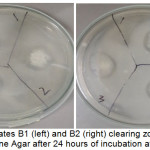 |
Figure 1: Isolates B1 (left) and B2 (right) clearing zone in Gelatin Peptone Agar after 24 hours of incubation at 37˚C. Click here to View figure |
Isolate B1, Enterococcus faecalis are known to possess proteolyic system and lipolytic activity that contributes to development of special flavour once incorporated in manufacturing of cheese.24 Tetragenococcus muriaticus isolated from fermented products had been found to contribute in lowering pH and reduce risk of putrefaction to hasten the degradation of the fermented fish product with the aid of the endogenous enzyme present in the fish.26,29 Lactobacillus delbrueckii commonly use in dairy products that aid as thickener, stabilizer and gelling agent produced exopolysaccharide for that function thus; they have amylase activity.28 Carnobacterium spp. is well known to produce tyramine a natural derivative of amino acid tyrosine.31
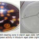 |
Figure 2: Isolate B3 clearing zone in starch agar plate (left) and Isolate B4 lipase activity in tributyrin agar plate (right). Click here to View figure |
Table 4 and Fig. 3 shows the antimicrobial activity and zone of inhibition of bacterial isolates against amoxicillin, chloramphenicol, cephalexin and erythromycin. Susceptibility of the isolates against the antibiotic agents was confirmed using the Clinical Laboratory Standards Institute Manual for Antimicrobial Susceptibility Testing. For amoxicillin, all the four isolates was susceptible with zone of activity of 44mm for the first isolate, 33mm for the second isolate, 35 mm for the third isolate and 28 mm for the fourth isolate. For chloramphenicol, three isolates were susceptible and one had no reaction with zone of activity of 30 mm for the first isolate, 34 mm for the second isolate and 30 mm for the fifth isolate. For cephalexin, only two isolates were susceptible and two others had no reaction, the zone of activity for the first isolate was 35 mm and 38 mm for the second isolate. For erythromycin, only the first isolate was susceptible with 21 mm zone of activity while the other three isolates had no reaction.
Table 4: Antimicrobial activity and zone of inhibition of bacterial isolates against amoxicillin, chloramphenicol, cephalexin and erythromycin.
| Isolate | Antibiotics | |||||||
| Amoxicillin | Chloramphenicol | Cephalexin | Erythromycin | |||||
| Zone of activity (mm) | Antimicrobial activity | Zone of activity (mm) | Antimicrobial activity | Zone of activity (mm) | Antimicrobial activity | Zone of activity (mm) | Antimicrobial activity | |
| B1 | 44mm | S | 30mm | S | 35mm | S | 21mm | S |
| B2 | 33mm | S | 34mm | S | 38mm | S | – | – |
| B3 | 35mm | S | – | – | – | – | – | – |
| B5 | 28mm | S | 30mm | S | – | – | – | – |
Legend: S, susceptible
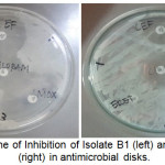 |
Figure 3: Zone of Inhibition of Isolate B1 (left) and Isolate B2 (right) in antimicrobial disks. Click here to View figure |
As can be observed in Fig. 4, all the isolates against Escherichia coli, Bacillus subtilis and Staphylococcus aurues had no inhibition zone. It had only reacted against amoxicillin that serve as control against the three pathogenic microorganisms. Lactic acid bacteria are known to produce variety of antimicrobial compounds such as ethanol, formic acid, acetone, hydrogen peroxide, diacetyl and bacteriocins that contributes for its ability to preserve foods and are natural competitive known to overcome other microorganisms sharing the same niche.5 The antagonistic and antimicrobial properties of lactic acid bacteria are due to the competition for nutrients and the production of one or more active metabolites such as organic acids (lactic and acetic acid), hydrogen peroxide and antimicrobial peptides such as bacteriocins.8
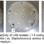 |
Figure 4: Antagonistic activity of LAB isolates (1-5 coding corresponds to B1-B5) to Bacillus subtilis (a), Staphylococcus aureus (b) and Escherichia coli (c) in the MH agar medium. Click here to View figure |
Isolation and characterization of bacteria are important in determining the microbial flora of food products it further distinguish the beneficial and harmful effects of microbiota in certain samples. This test requires a lot of time and effort to end up with essential result, thus it is important to properly choose the exact test required for characterization of certain organisms. Lactic acid bacteria in food products had long been associated to good factors as food preservatives and with added fermentation metabolites. They only require the right environmental factors to grow and proliferate in food products. Bacterial isolation of this organisms are very much promising industry especially in developing countries like the Philippines because population are not yet so aware of this organisms and the benefits that can be derived through their consumption.
Acknowledgments
The authors would like to acknowledge Department of Science and Technology-Accelerated Science and Technology Human Resources Development Program (DOST-ASTHRDP) and University of the Philippines Visayas-College of Fisheries and Marine Sciences for supporting this study.
Conflict of Interest
There is no conflict of interest.
References
- Majumdar R., Basu and Nayak, B. Assessment of Nutritional Quality of ‘shidal’ a Fermented Fish Product of Northeast India. Journal of Indian Fish Association: 36: 25-34: (2009).
- Thwe Moe N. K., Thwe S. M., Shirai T., Terahara T., Imada C. and Kobayashi, T. Characterization of Lactic Acid Bacteria Distributed in Small Fish Fermented with Boiled Rice in Myanmar. Fisheries Science: 81: 373-381: (2015).
CrossRef - Olympia M. Fermented Fish Products in the Philippines. Applications of Biotechnology in Traditional Fermented Foods: 131-139: (1992).
- Yap W. G., Villaluz A. C., Soriano M. G. and Santos M. N. Milkfish Production and Processing Technologies in the Philippines. World Fish Center Contribution- Milkfish Project Publication: 2: 1-97: (2007).
- Agrawal N. and Prakashi A. Isolation of Lactic Acid Bacteria from Fermented Milk Products and their Antimicrobial Activity Against Staphylococcus aureus. Journal of Food Safety: 15: 39-42: (2013).
- Fooks L., Fuller R. and Gibson G. Prebiotics, Probiotics and Human Gut Microbiology. International Dairy Journal: 9(1): 53-61: (1999).
CrossRef - Holzapfel E., Haberer P., Geisen R., Bjorkroth J. and Schillinger U. Taxonomy and Important Features of Probiotic Microorganisms in Food and Nutrition. American Journal of Clinical Nutrition: 73(2): 3655-3735: (2001).
CrossRef - Collins B., Hill C. P. and Ross P. Applications of Lactic Acid Bacteria- Produced Bacteriocins. Biotechnology of Lactic Acid Bacteria Novel Applications: 89-109: (2010).
- Felten A., Barreau C., Bizet C. and Lagrange P. H. Lactobacillus Species Identification, H2O2 Production, and Antibiotic Resistance and Correlation with Human Clinical Status. Journal of Clinical Microbiology: 37(3): 729-733: (1999).
- Desmons, S., Krhouz, H., Evrard, P. and Thonart, P. Improvement of Lactic Acid Cell Production. Applied Biochemical and Biotechnology: 70(1): 513-526: (1998).
CrossRef - Banaay, C. G., Balolong, M. and Elegado, F. Lactic Acid Bacteria in Philippine Traditional Fermented Foods. Lactic Acid Bacteria- R & D for Food, Health and Livestock Purposes: 571-588: (2013).
- Akerberg C. and Zacchi G. An Economic Evaluation of the Fermentative Production of Lactic Acid from Wheat Flour. Bioresources Technology: 75(2): 119-126: (2000).
CrossRef - Espejo-Hermes J. Fish Processing Technology in the Tropics. Philippines: Tawid Publications: 101: (1998).
- Bacteriological Analytical Manual (BAM) Online. Rapid Methods for Detecting Foodborne Pathogens. US Food and Drug Administration Manual. 1-14: (2008).
- De Vos P., Garrity G., Jones D., Krieg N., Ludwig W., Rainey F., Schleifer K. H. and Whitman W. Bergey’s Manual of Determinative Bacteriology. The Firmicutes: 2(3): 465-616: (2009).
- Das P., Mandal S., Khan A., Manna S. K., and Ghosh K. Distribution of Extracellular Enzyme-Producing Bacteria in the Digestive Tracts of 4 Brackishwater Fish Species. Turkish Journal of Zoology: 38: 79-88: (2014).
CrossRef - Biemer J. J. Antimicrobial Susceptibility Testing by the Kirby-Bauer Disc Diffusion Method. Annals of Clinical Laboratory Science: 3(2): 135-140: (1973).
- Clinical and Laboratory Standards Institute. Performance Standard for Antimicrobial Susceptibility Testing. 23rd Informational Supplement Document M100-S23: (2013).
- Maturin, L. J. and Peeler, J. T. Aerobic Plate Count. Food and Drug Administration Bacteriological Analytical Manual: 8: (1998).
- Nickelson R. and Finne G. Fish, Crustaceans and Precooked Seafood’s. Compedium of Methods for the Microbiological Examination of Foods: 875-895: (1992).
- Jay M. J. Intrinsic and Extrinsic Parameters of Foods that Affect Microbial Growth. Modern Food Microbiology: 5: 53: (1996).
- Besas J. and Dizon E. Influence of Salt Concentration on Histamine Formation in Fermented Tuna Viscera (Dayok). Food and Nutrition Sciences: 3: 201-206: (2012).
CrossRef - Tyne, D. V., Martin, M. and Gilmore, M. Structure, Function and Biology of the Enterococcus faecalis Toxins: 5: 895-911: (2013).
CrossRef - Gomes B., De Melo Franco B. and De Martinis E. Dualistic Aspects of enterococcus Current Research, Technology and Education Topics in Applied Microbiology and Microbial Technology: 1119-1125: (2010).
- Satomi M., Kimura B., Mizoi M., Sato T. and Fuji T. Tetregenococcus muriaticus nov., a New Moderately Halophilic Lactic Acid Bacterium Isolated from Fermented Squid Liver Sauce. International Journal of Systematic Bacteriology: 47(3): 832-836: (1997).
CrossRef - Kobayashi, T., Kajiwara, M., Wahyuni, M., Hamada-Sato, N., Iwada, C. and Watanabe, E. Effect of Culture Conditions on Lactic Acid Production of Tetragebococcus Journal of Applied Microbiology: 96: 1215-1221: (2004).
- Michaylova M., Minkova S., Kimura K., Sasaki T. and Isawa K. Isolation and Characterization of Lactobacillus delbrueckii bulgaricus and Streptococcus thermophilus from plants in Bulgaria. Federation of European Microbiological Societies: 269: 160-169: (2007).
CrossRef - Nishimura, J. Exopolysaccharides Produced from Lactobacillus delbrueckii bulgaricus. Advances in Microbiology: 4: 1017-1023: (2014).
CrossRef - Francoise L. Occurrence and Role of Lactic Acid Bacteria in Seafood Products. Food Microbiology: 27(6): 698-709: (2010).
CrossRef - Campos, C. A., Castro, M. P., Rivas, F. P. and Schelegueda, L. I. Bacteriocins in Food: Evaluation of the Factors Affecting their Effectiveness in Microbial Pathogens and Strategies for Combating Them. Science, Technology and Education: 994-1004: (2013).
- Brillet, A., Pilet, M., Prevost, H., Cardinal, M. and Leroi, F. Effect of Inoculation of Carnobacterium divergens V41, a Biopreservative Strain Against Listeria monocytogenes risk, on the Microbiological, Chemical and Sensory Quality of Cold-Smoked Salmon. International Journal of Food Microbiology: 104(3): 309-324: (2005).
CrossRef

This work is licensed under a Creative Commons Attribution 4.0 International License.