Introduction
Glucagon like peptide-1 (GLP-1) and glucose dependent insulinotropic polypeptide (GIP) are incretin hormones released from intestinal enteroendocrine L and K cells respectively in response to food intake, especially by carbohydrates and fat rich diet.1,2 Incretin hormones regulating glucose metabolism by inducing insulin hormone production and pancreatic beta cell proliferation. GLP-1 play important role in stimulation of glucose-dependent insulin secretion, augmentation of β-cell mass, inhibition of glucagon release, decrease gastric empting and food intake3. The physiological action of GLP-1 and GIP are very much similar and additive4. Like GLP-1, GIP also have insulinotropic effect, GIP act through by binding to specific GIP receptor present on pancreatic beta cells and enhance exocytosis of insulin containing granules5. GLP-1 and GIP decreases apoptosis and increase proliferation rate of pancreatic beta cells. Wang et al. reported that GIP induces expression of proinsulin gene and enhance the secretion of insulin hormone6. GLP-1 and GIP incretin hormones have very short half life because these are rapidly degraded by dipeptidylpeptide-4 (DPP-4) enzyme7. DPP-4 is located in endothelial and epithelial cells of intestine. Several studies have reported an impaired incretin effect in diabetes patients that attributed defective insulin secretion8. Incretins based therapy are most recently approved class of therapeutic agents for treatment of diabetes, especially via DPP-IV inhibitors and GLP-1 receptor agonist. Sitagliptin and saxagliptin are DPP-IV inhibitors which exert their action through prevention of incretin degradation9. Exendin-4 and liraglutide are GLP-1 receptor agonist having structurally resemblance to GLP-1. Exendin-4 was approved for treatment of diabetes in the U.S. and liraglutide in the Europe in 2005 and 2006, respectively. These drugs are chemically synthesized and not safe for regular and long-term use, having some adverse effects like weight gain, hypoglycaemia, fever, gastrointestinal discomfort and high cost of drugs10. The side effects associated with incretin based drugs necessitated for safer alternatives, including enhancing endogenous production of incretin hormones in diabetic subjects. Many studies supported that probiotic strains have high potential in treatment of diabetes and obesity11,12. It has been shown that probiotic containing foods delay the streptozotacin induced diabetes and protect islets of pancreatic β-cells from damage, delaying the onset of T2DM and prevent complications associated with DM 13. Balakumar et al. reported that probiotics treatment had ability to normalize the level of GLP-1which is impaired in high fat diet induced obesity14. Probiotics are live microorganism which, when administered in adequate amount confer a health benefit to the host15. Probiotics are used as potential modulators of the intestinal microbial flora in a valuable manner. Probiotics have antidiabetic, antimutagenic, anti-inflammatory, antioxidants, immunomodulatory and antiobesity activities and provide a number of health benefits to host through modulation of the gut microbiota. Yadav et al. reported that administration of various combinations of probiotics to obese mice protected from body weight gain, reduced food intake and insulin resistance16. Various metabolic disorders like diabetes and obesity create imbalance of gut microbiota, It is reported that proportion of Firmicutes and Clostridia were higher in the diabetic patients compared to healthy individuals, patients in the pre-diabetes and T2DM groups had significantly increased level of Betaproteo-bacteria compared with healthy group so by administration of adequate amount of probiotic, use to recover intestinal microbiota with beneficial bacteria17. Species of Lactobacillus and Bifidobacterium are most commonly used microbes as probiotics. Zeng et al. reported that strains of Lactobacillus and Bifidobacterium have DPP-4 inhibitory activity. Thus, these probiotics have potential application in management of diabetes18. Our previous study has shown potential hypoglycaemic and antioxidant activity of L. casei and B. bifidium in diabetic animal models 19. Therefore, the present study was designed to investigate the effect of probiotic treatment on incretin hormone secretion in experimental rat models.
Materials and Methods
Materials
Streptozotocin and acarbose were procured from Sigma Aldrich (St. Louis, Missouri, USA). Blood glucose estimation by Accu check one touch glucometer (Johnson & Johnson, Mumbai, India). De Man Rogosa and Sharpe (MRS) agar and broth media were purchased from HiMedia Laboratories (Mumbai, India). Sitagliptin purchased from Merck Serono Co., Ltd, Guanghoou, china. GLP-1 and GIP were estimated by using standard ELISA kit purchased from RayBio®. BioAssay Systems.
Bacterial Strains
LC017 and BB231 probiotic strains those were used in this study, procured from National collection of dairy culture, National Dairy Research Institute, Karnal, India.
Animal Handling
Male Wistar rats, weighted about 170-210 gm were procured from the animal research division, Defence Research & Development Establishment, Gwalior, India. This study has been approved by the Institutional Animal Ethical committee (IAEC) and application to be submitted to the committee for the purpose of control and supervision of experiments on animals (CPCSEA), New Delhi (Reg.No. BU/PHARMA/IAEC/a/16/12). All rats were housed in animal room in plastic cages with husk bedding and cages were cleaned and changed daily. Animal room maintained with 12-h light-dark cycle at 22±2 0C temperature and humidity 55±5%. All rats were provided normal pellet diet and RO water ad libitum. The rats were acclimatised for one week before starting experiments.
Induction of Diabetes
Diabetes was induced in rats by single dose of freshly prepared streptozotocin solution (50 mg/kg body weight) in sterile 0.1 M citrate buffer (pH 4.5) to overnight fasted animal intraperitoneally. Diabetes was verified after 96 hour by evaluating blood glucose level. FBG and PBG were taken regularly till stable hyperglycaemia condition achieve. Rats having blood glucose level 250mg/dl or more were used in this study.
Preparation and Dosing of LC017 and BB231
Lyophilised bacterial strains were revived in MRS broth medium and incubated at 370C in anaerobically in an anaerobic system Mark II (Anaero Gas Pack, LE002.HiMedia, India) for 48 h. After incubation period, 1 mL of inoculum was diluted six times by serial dilution method in sterile distilled water. 100 uL suspension of last successive serial dilution was inoculated on MRS agar plate and after incubation 50-60 colonies appear in each plate. This plate was used to prepare dose for animals. The concentration of last dilution was 56×107 cfu/mL. One colony picked using sterile loop and dissolved in 1 mL sterile water and mixed with sterile micropipette. All the procedure of preparation of bacterial dose took place in laminar air flow. Same method was used to prepare both bacterial strains. Freshly prepared single daily dose of LC017, BB231 alone and combination administered to treated groups of animals for 28 days.
Experimental Design
The animals were divided into six experimental groups each contain 6 rats (n=6).
| Group-1 | Healthy control | Normal rat + single daily dose of sterile distil. water 1mL |
| Group-2 | Untreated diabetic | Single dose of streptozotocin 50mg/kg b.w.+ single daily dose of sterile water 1mL |
| Group-3 | Treated group | Single daily dose of LC017 (~107cfu/mL) |
| Group-4 | Treated group | Single daily dose of BB231(~107cfu/mL) |
| Group-5 | Treated group | Single daily dose of LC017 and BB231 (~107cfu/mL) |
| Group-6 | Standard group | Single daily dose of acarbose (10 mg/kg b.w.) |
The drugs and bacterial doses were administered orally by using feeding needle once daily for 28 days, continuously.
Body Weight Measurement
The body weight was measured of each rat before treatment and every week during and after experiment.
Sucrose Tolerance Test
For carbohydrate tolerance test, the rats were fasted overnight and administered sucrose (2 g/kg b.w.) orally dissolved in 1 mL of distilled water in combination with treatment. Blood was sampled from tail vein at 0, 30, 60, 90, and 120 min after sucrose administration to measure blood glucose level. Blood glucose level was determined by using Accu. check One touch glucometer.
Serum GLP-1 and GIP Estimations
At the end of the experiment (i.e. on 28th day), rats were given glucose load (2 g/kg b.w.) after overnight fasting by gavage. Blood was sampled at 0, 20, 30, 40, 50 and 60 min from tail vein and collected into chilled tubes containing EDTA and DPP-4 inhibitor (sitagliptin) for serum GLP-1 and GIP analysis21. Quantification of GIP and GLP-1 was accomplished by rat ELISA kit (RayBio® rat GLP-1 enzyme immunoassay kit, EIA-GLP-1 and RayBio® rat GIP enzyme immunoassay kit, EIA-GIP) using Thermo Scientific Varioskan Flash Spectral Scanning Multimode Reader. After collection of blood from tail vein, rats were sacrificed under mild ether anaesthesia. Total loss of consciousness was confirmed by toe-pinch. Blood sample was collected by heart puncture and serum was isolated by centrifugation of blood sample at 3000 rpm for 5 min at 40C and stored in -800C until analysis.
Extraction and Estimation of Intestinal GLP-1
Intestinal segments (jejunum, ileum, colon and cecum) were immediately isolated from sacrificed animals and washed with phosphate buffer-saline22. Different segments of intestine were homogenised separately using ethanol/acid (5:1 v/v) solution (5 mL/g tissue) at 40C. Homogenised tissue kept for 24 h at 40C and after that centrifuged at 10,000 g for 20 min, at 40C. The supernatant was transferred and neutralized with 1 mol/l of NaOH and used for intestinal GLP-1 estimations by using ELISA kits (RayBio® rat GLP-1 enzyme immunoassay kit, EIA-GLP-1) according to manufacturer’s instruction.
Statistical Analysis
All the data were presented as mean ±standard error of mean (SEM). The data analyzed by statistical software (statistical package for social sciences, SPSS Version 20.0, IBM) using one-way ANOVA followed by Tukey’s multiple range post hoc tests. The values were considered significantly different at P<0.05.
Result
Effect of Treatment on Body Weight
During 28 days of experiment, diabetic rats were found to have significant (p < 0.001) weight loss, compared to healthy control group. The body weight was significantly (p < 0.001) increased for the acarbose and combination treated groups compared to diabetic group (Table 1). Results indicate that treatment with probiotics affect body weight and could effectively improve weight loss in diabetic animal.
Table 1: Effect of Probiotic Treatment on Change in Body Weight (g) of Normal and Diabetic Rats.
| Groups | Before treatment | Day 1 | Day 7 | Day 14 | Day 21 | Day 28 |
| Normal control | 231.2 ±1.32 | 235.6 ±1.57 | 249.01 ±1.74 | 260.2 ±2.28 | 272.7 ±1.94 | 285.17 ±2.84 |
| Diabetic control | 229.82 ±2.30 | 208.61 ±2.85*** | 199.4 ±4.01*** | 185.11 ±5.19*** | 168.63 ±6.24*** | 163.08 ±6.14*** |
| L. casei treated diabetic | 226.45 ±2.56 | 209.7 ±2.90***,a | 204.05 ±2.61***,a | 197.16 ±1.93***,a | 192.5 ±2.07***,d | 187.38 ±1.48***,d |
| B.bifidium treated diabetic | 225.7 ±2.20 | 210.7 ±1.35***,a | 202.78 ±1.77***,a | 194.05 ±2.09***,a | 187.27 ±2.46***,c | 180.01 ±1.94***,b |
| L. casei and B.bifidium Combination treated | 224.93 ±1.8 | 206.5 ±2.13***,a | 212.4 ±2.12***,b | 221.8 ±1.59***,d | 230.11 ±1.20***,d | 238.1 ±1.53***,d |
| Acarbose | 228.78 ±2.13 | 209.6 ±2.54***,a | 214.6 ±2.65***,c | 223.1 ±2.96***,d | 232.3 ±3.56***,d | 242.4 ±3.15***,d |
The Values are Mean ± SEM (n = 6 animals/group).
*= P < 0.05, **= P < 0.01, ***= P < 0.001compared to positive (healthy) control.
a= P>0.05, b= P < 0.05, c= P < 0.01•= P>0.05, compared to negative (diabetic) control.
Effect of Treatment on Glp-1 Level in Serum
Significant (P < 0.01) decrease in GLP-1 level was observed in non treated diabetic rats as compared to healthy rats. Significant increase (p < 0.01) in GLP-1 level after 28 days of treatment with LC017, BB231 and combination in therapeutic models (Fig.1). Diabetic animal treated with standard drug showed significantly increased and normalized GLP-1 level compared to healthy control rats.
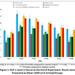 |
Figure 1: GLP-1 Level in Serum at the End of Experiment. Result were Presented as Mean ±SEM (n=6 Animal/Group). |
Effect of Treatment on GIP Level in Serum
Similar to GLP-1, diabetic rats showed a significantly (p< 0.01) decreased level of GIP as compared to nondiabetic healthy rats. Administration of LC017, BB231 and combination therapy significantly (p< 0.01) increased the level of GIP in treated rats as compared to diabetic control (Fig. 2).
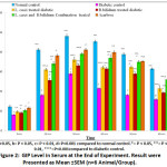 |
Figure 2: GIP Level in Serum at the End of Experiment. Result were Presented as Mean ±SEM (n=6 Animal/Group). |
Effect of Treatment on GLP-1 Level in Intestine
Among different parts of intestine, GLP-1 content was highest in colon region. GLP-1 level was significant decreased (p<0.01) in all animals among different parts of intestine in diabetic rats. After 28 days of treatment, GLP-1 level significantly increased in diabetic rats (Fig. 3).
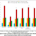 |
Figure 3: GLP-1 Content in Different Part of the Intestinal Tract. Result were Presented as Mean ±SEM (n=6 Animal/Group). |
Effect of Treatment on Sucrose Tolerance
After 28 days of experiment, sucrose tolerance test was performed and we found that maximum blood glucose level reach at 60 min. Blood glucose level of group treated with combination of both LC017 and BB231 were slightly higher than group treated with standard antidiabetic drug acarbose. Oral administration of LC017 and BB231 treatment reduced postprandial blood glucose after sucrose challenge like acarbose. Glucose level significantly (p<0.01) higher in untreated diabetic rat compared to rats treated with probiotcs. (Fig.4). Rats treated with combination of both bacteria showed significantly deceased level of glucose after sucrose tolerance test.
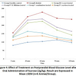 |
Figure 4: Effect of Treatment on Postprandial Blood Glucose Level after Oral Administration of Sucrose (2g/kg). Result are Expressed in Mean ±SEM (n=6 Animal/Group). |
Discussion
Probiotics are the widely used nutritional supplements and therapeutic modality in the management of diabetes and obesity23. Probiotic bacteria are defined as living microorganism which confers health benefits to their host when administered in adequate amounts24. Various studies reported the potential health benefits of Bifidobacterium and lactic acid bacteria such as antitumor activity, antimicrobial activity, improvement of immune system and gastrointestinal microbiota25,26. Manaer et al. reported significant hypoglycaemic effect in diabetic rats through enhancing the release of GLP-1 and improving pancreatic beta cells, by using camel milk fortified with various strains of LAB (Lactic acid bacteria) and yeast27. Our previous study reported that treatment with L. casei and B. bifidum alone and combination (single daily dose 1×10⁷ cfu/mL) ameliorates antioxidant stress and hyperglycemia. So, in the present study we have explored the mechanism by which the probiotic treatment promote hypoglycaemic activity, emphasizing on the effect of probiotic treatment on incretin level. We hypothesized that oral administration of L. casei and B. bifidum alone and in combination might result in change in the level of incretin hormones. To delineate the process we measured the level of incretin hormones especially GLP-1 in serum and intestine that regulate food intake. After 28 days of experiment, we found that hunger reducing hormone GLP-1 was significant decrease in streptozotocin induced diabetic groups as compared to control groups however, oral administration of LC017 and BB231 treatments increase the level of GLP-1 in therapeutic models but interestingly therapeutic group treated with combination of both L. casei and B. bifidum showed significantly increase and normalized GLP-1 level similar to acarbose treated group. These results illustrate that increased the level of GLP-1 correlate with beneficial metabolic effect of LC017 and BB231. Several studies supported that probiotic treatment enhance the secretion of incretin hormone have been correlated to increase the level of insulin hormone by the recovery of pancreatic islets due to antiapoptotic, regeneration activity of GLP-1 and the secretion of GLP-1 associated with modulation of intestinal microbiota28. In the present study, body weight was significantly reduced in diabetic group, this sudden reduction in weight due to degeneration of lipid found in tissue and muscles as lipid involved in gluconeogenesis, in hyperglycaemic condition hence muscles and lipid tissues are important contributor of weight gain29. However, body weight of rats was significantly increased after 28 days treatment with combination of both probiotic strains. The possible mechanism of treatment is associated with increase the level of insulin that in turn improves glycemic control and prevents body weight loss. The observation is supporting previous results30,31.
Conclusion
The result of present study showed that administration of LC017 and BB231 have significant hypoglycaemic potential by increasing the levels of incretin hormones. These incretin hormones improved physiology of islets of pancreatic beta cells and increased the secretion of insulin hormone. The study further supported that combined treatment of L. casei and B. bifidum possesses higher potential as compared to treatment with each one alone.
Acknowledgements
The authors are thankful to Innovation Centre, Bundelkhand University, Jhansi, India for providing facilities and infrastructure for this research.
Financial Support and Sponsorship
The authors received no financial support for the research, authorship, and/or publication of this article.
Conflicts of Interest
The authors declare no conflict of interest.
References
- Seino Y, Yabe D. Glucose-dependent insulinotropic polypeptide and glucagon-like peptide-1: Incretin actions beyond the pancreas. J Diabetes Investig. 2013; 4(2):108-130. doi:10.1111/jdi.12065.
CrossRef - Lim GE, Huang GJ, Flora N, LeRoith D, Rhodes CJ, Brubaker PL. Insulin regulates glucagon-like peptide-1 secretion from the enteroendocrine L cell. Endocrinology. 2009; 150(2):580-591. doi:10.1210/en.2008-0726.
CrossRef - Campbell JE, Drucker DJ. Pharmacology, physiology, and mechanisms of incretin hormone action. Cell Metab. 2013; 17(6):819-837. doi:10.1016/j.cmet.2013.04.008.
CrossRef - Vilsbøll T, Krarup T, Madsbad S, Holst JJ. Both GLP-1 and GIP are insulinotropic at basal and postprandial glucose levels and contribute nearly equally to the incretin effect of a meal in healthy subjects. Regul Pept. 2003;114(2-3):115-121. doi:10.1016/s0167-0115(03)00111-3.
CrossRef - Ding WG, Renström E, Rorsman P, Buschard K, Gromada J. Glucagon-like peptide I and glucose-dependent insulinotropic polypeptide stimulate Ca2+-induced secretion in rat alpha-cells by a protein kinase A-mediated mechanism. Diabetes. 1997;46(5):792-800. DOI: 10.2337/diab.46.5.792.
CrossRef - Wang Y, Montrose-Rafizadeh C, Adams L, Raygada M, Nadiv O, Egan JM. GIP regulates glucose transporters, hexokinases, and glucose-induced insulin secretion in RIN 1046-38 cells. Mol Cell Endocrinol. 1996;116(1):81-87. doi:10.1016/0303-7207(95)03701-2.
CrossRef - Fehmann HC, Göke R, Göke B. Cell and molecular biology of the incretin hormones glucagon-like peptide-I and glucose-dependent insulin releasing polypeptide. Endocr Rev. 1995;16(3):390-410. doi: 10.1210/edrv-16-3-390.
CrossRef - Elahi D, McAloon-Dyke M, Fukagawa NK, et al. The insulinotropic actions of glucose-dependent insulinotropic polypeptide (GIP) and glucagon-like peptide-1 (7-37) in normal and diabetic subjects. Regul Pept. 1994; 51(1):63-74. doi:10.1016/0167-0115(94)90136-8.
CrossRef - Hansotia T, Baggio LL, Delmeire D, et al. Double incretin receptor knockout (DIRKO) mice reveal an essential role for the enteroinsular axis in transducing the glucoregulatory actions of DPP-IV inhibitors. Diabetes. 2004; 53(5):1326-1335. doi:10.2337/diabetes.53.5.1326.
CrossRef - Buse JB, Rosenstock J, Sesti G, et al. Liraglutide once a day versus exenatide twice a day for type 2 diabetes: a 26-week randomised, parallel-group, multinational, open-label trial (LEAD-6). Lancet. 2009; 374(9683):39-47. doi:10.1016/S0140-6736(09)60659-0.
CrossRef - Yadav R, Dey DK, Vij R, et al. Evaluation of anti-diabetic attributes of Lactobacillus rhamnosus MTCC: 5957, Lactobacillus rhamnosus MTCC: 5897 and Lactobacillus fermentum MTCC: 5898 in streptozotocin induced diabetic rats. Microb Pathog. 2018; 125:454-462.
CrossRef - Chaudhari DD, Singh R, Mallappa RH, Rokana N, Kaushik JK, Bajaj R,Batish VK, Grover S. Evaluation of casein & whey protein hydrolysates as well as milk fermentates from Lactobacillus helveticus for expression of gut hormones. Indian J Med Res 2017; 146(3) 409–419.
- Yadav H, Jain S, Sinha PR. Antidiabetic effect of probiotic dahi containing Lactobacillus acidophilus and Lactobacillus casei in high fructose fed rats. Nutrition. 2007; 23(1):62-68. doi:10.1016/j.nut.2006.09.002.
CrossRef - Balakumar M, Prabhu D, Sathishkumar C, et al. Improvement in glucose tolerance and insulin sensitivity by probiotic strains of Indian gut origin in high-fat diet-fed C57BL/6J mice. Eur J Nutr. 2018; 57(1):279-295. doi:10.1007/s00394-016-1317-7.
CrossRef - Food and Agricultural Organization of the United Nations and World Health Organization. Health and nutritional properties of probiotics in food including powder milk with live lactic acid bacteria. World Health Organization [online], http://www.who.int/foodsafety/publications/fs_management/en/probiotics.pdf (2001).
- Yadav H, Lee JH, Lloyd J, Walter P, Rane SG. Beneficial metabolic effects of a probiotic via butyrate-induced GLP-1 hormone secretion. J Biol Chem. 2013; 288(35):25088-25097. doi:10.1074/jbc.M113.452516.
CrossRef - Grigorescu I, Dumitrascu DL. IMPLICATION OF GUT MICROBIOTA IN DIABETES MELLITUS AND OBESITY. Acta Endocrinol (Buchar). 2016;12(2):206-214. doi:10.4183/aeb.2016.206.
CrossRef - Zeng Z, Luo J, Zuo F, Zhang Y, Ma H, Chen S .Screening for potential novel probiotic Lactobacillus strains based on high dipeptidyl peptidase IV and α-glucosidase inhibitory activity. Funct. Foods.2016;20: 486–495.
CrossRef - Sharma P, Bhardwaj P, Singh R. Administration of Lactobacillus casei and Bifidobacterium bifidum Ameliorated Hyperglycemia, Dyslipidemia, and Oxidative Stress in Diabetic Rats. Int J Prev Med. 2016; 7:102. Published 2016 Aug 22. doi:10.4103/2008-7802.188870.
CrossRef - Liu L, Yu YL, Yang JS, et al. Berberine suppresses intestinal disaccharidases with beneficial metabolic effects in diabetic states, evidences from in vivo and in vitro study. Naunyn Schmiedebergs Arch Pharmacol. 2010; 381(4):371-381. doi:10.1007/s00210-010-0502-0.
CrossRef - Artinian SB, Al Lafi SM, Boutary SS, Bitar KM, Zwainy NS, Bikhazi AB. Assessment of glucagon-like peptide-1 analogue and renin inhibitor on the binding and regulation of GLP-1 receptor in type 1 diabetic rat hearts. Exp Diabetes Res. 2011: 489708. doi:10.1155/2011/489708.
CrossRef - Kuhre RE, Albrechtsen NW, Windeløv JA, Svendsen B, Hartmann B, Holst JJ. GLP-1 amidation efficiency along the length of the intestine in mice, rats and pigs and in GLP-1 secreting cell lines. Peptides. 2014; 55:52-57. doi:10.1016/j.peptides.2014.01.020.
CrossRef - Singh S, Sharma RK, Malhotra S, Pothuraju R, Shandilya UK. Lactobacillus rhamnosus NCDC17 ameliorates type-2 diabetes by improving gut function, oxidative stress and inflammation in high-fat-diet fed and streptozotocintreated rats. Benef Microbes. 2017;8(2):243-255. doi:10.3920/BM2016.0090.
CrossRef - Food and Agriculture Organization of the United Nations/World Health Organization (FAO/WHO), 2001. Evaluation of health and nutritional properties of powder milk and live lactic acid bacteria. Available at: http://www.who.int/foodsafety/publications/ fs_management/en/probiotics.pdf.
- Minic R, Gavrovic-Jankulovic M, Petrusic V, et al. Effects of orally applied Fes p1-displaying L. plantarum WCFS1 on Fes p1 induced allergy in mice. J Biotechnol. 2015; 199:23-28. doi:10.1016/j.jbiotec.2015.01.028.
CrossRef - Fasseas MK, Fasseas C, Mountzouris KC, Syntichaki P. Effects of Lactobacillus salivarius, Lactobacillus reuteri, and Pediococcus acidilactici on the nematode Caenorhabditis elegans includes possible antitumor activity. Appl Microbiol Biotechnol. 2013;97(5):2109-2118. doi:10.1007/s00253-012-4357-9.
CrossRef - Manaer T, Yu L, Zhang Y, Xiao XJ, Nabi XH. Anti-diabetic effects of shubat in type 2 diabetic rats induced by combination of high-glucose-fat diet and low-dose streptozotocin. J Ethnopharmacol. 2015; 169:269-274. doi:10.1016/j.jep.2015.04.032.
CrossRef - Lin HV, Frassetto A, Kowalik EJ Jr, et al. Butyrate and propionate protect against diet-induced obesity and regulate gut hormones via free fatty acid receptor 3-independent mechanisms. PLoS One. 2012; 7(4):e35240. doi:10.1371/journal.pone.0035240.
CrossRef - Cani PD, Lecourt E, Dewulf EM, et al. Gut microbiota fermentation of prebiotics increases satietogenic and incretin gut peptide production with consequences for appetite sensation and glucose response after a meal. Am J Clin Nutr. 2009;90(5):1236-1243. doi:10.3945/ajcn.2009.28095.
CrossRef - Mohammed A, Koorbanally NA, Islam MS. Anti-diabetic effect of Xylopia aethiopica (Dunal) A. Rich. (Annonaceae) fruit acetone fraction in a type 2 diabetes model of rats. J Ethnopharmacol. 2016; 180:131-139. doi:10.1016/j.jep.2016.01.009.
CrossRef - Kumar N, Tomar SK, Thakur K and Singh AK. The ameliorative effects of probiotic Lactobacillus fermentum strain RS-2 on alloxan induced diabetic rats. J Funct Foods. 2016; 28: 275-284. doi:10.1016/j.jff.2016.11.027.
CrossRef

This work is licensed under a Creative Commons Attribution 4.0 International License.