Pectin Based Colorimetric Film for Monitoring Food Freshness
Meenambiga Setti Sudharsan*
, Lakshaya Kumar, Vivek Pazhamalai and Sowmya Hari Department of Bioengineering, School of Engineering, Vels Institute of Science Technology and Advanced Studies (VISTAS), Chennai, India.
Corresponding Author E-mail: meenambiga.se@velsuniv.ac.in
DOI : http://dx.doi.org/10.12944/CRNFSJ.11.3.03
Download this article as:
![]()
The colours of anthocyanins are sensitive to variations in the pH of the environment. The field of food engineering is seeing a rise in the utilisation of anthocyanins obtained from plants to produce new and active packaging film. A pH-sensitive colorimetric film was developed using anthocyanin recovered from the peel of Phaseolus vulgaris (dark red kidney bean) in conjunction with composite pectin derived from citron peel. Additionally, Anthocyanins could be employed as colorimetric markers to identify food degradation, due to their sensitivity towards pH alterations and the production of ammonia gas. Solvent casting process was used to produce the film which changed from pink to brownish with rising pH due to the ammonia vapour that was produced. The antioxidant and antibacterial abilities of the anthocyanin containing smart films were investigated, and both Escherichia coli and Staphylococcus aureus growth was successfully inhibited. The developed film was characterized for its physical properties such as water solubility, moisture content and swelling index. To examine the structure of the films' surface, scanning electron microscopy (SEM) investigation has been carried out. The film indicator active response to pH fluctuation was demonstrated through tests on samples of chicken meat under various settings, allowing for the real-time monitoring of spoiled foods. This makes monitoring perishable goods, easy, affordable, environmentally friendly and biodegradable pH-sensitive indicator with visible colour change.
KEYWORDS:Anthocyanin; Citron; Colorimetric pH indicator; Dark red kidney bean; Pectin
Introduction
Due to urgent issues like environmental deterioration and global warming, there is a greater demand for sustainable, environmentally friendly products. Since biodegradability is directly related to the chemical make-up of the materials rather than their source, European Bioplastics mandates biopolymers to be biodegradable1. Customers may now instantly view and understand information about the grade and quality of packaged food goods because of smart packaging, a recent invention in the food sector. Consumers could judge the quality of their food by looking for chromatic shift in the case of microbial spoilage or chemical alterations to packaged food. In recent years, natural materials, which are more environmentally friendly and highly biodegradable than synthetic ones, have gradually taken their position in the food packaging industry.2,3
A smart film is a cheap, efficient, and user-friendly tool for tracking food spoiling in real time from production to consumption. To accomplish this, a variety of indicators and sensors, including time-temperature integrators and freshness indicators, have been researched for smart packaging4. Numerous intelligent packaging strategies have been developed up to this point, including harmful bacteria biosensors, oxygen and carbon dioxide sensors, pH indicators, moisture and time-temperature detectors, and pH indicators5. Freshness indicators will reveal the extent of variation in quality brought on by existent bacteria or enzymes in food products instantaneously, depending upon the colour shift of pH-responsive pigments with in indicator and the total volatile basic-nitrogen (TVB-N) released by packed items. These indicators are composed of a natural dye that can detect pH and a dye carrier. Both substances must be safe for consumption, non-toxic, and pH-stable6.
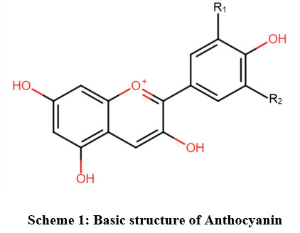
Plants get their red, purple, orange, pink, and blue colour from anthocyanins pigments, that are naturally occurring, non-toxic and soluble in water. Due to the structural changes in anthocyanins associated with colour change as a function of pH, they may be suitable for colorimetric indicators used in intelligent food packaging.7,8 These most astonishing pigments may be found in a variety of natural sources, including fruits and vegetables like red roses, pomegranate seeds, purple carrots, and purple cabbage, as well as flowers like butterfly peas and red roses. Anthocyanins are large subclasses of polyphenols, which are secondary metabolites of plants. The flavylium cation structure serves as their foundation. In nature, there are more than 600 different types of anthocyanins9. Figure 1.1 demonstrates the anthocyanin’s basic structure.
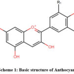 |
Scheme 1: Basic structure of Anthocyanin. |
The six anthocyanidins (in the form of aglycones) that are most frequently found are cyanidin, pelargonidin, delphinidin, petunidin, peonidin, and malvidin. One of the most prevalent anthocyanins is cyanidin-3-glucoside, which is found in black beans, black rice, and several berries10. Numerous pH indicators have been researched from various sources of anthocyanins. To identify the freshness of pork, Choi et al., in 2017, built a smart packaging film employing anthocyanin from purple potatoes as a pH indicator. The consumption of legumes is widespread across the world11. The kidney beans (Phaseolus vulgaris L.), a kind of legume, are gaining popularity due to its superior dietary antioxidant content. They are excellent suppliers of functional ingredients like fibres and polyphenols in addition to being abundant sources of nutritional ingredients like protein and vitamins12. Hence black kidney peels were valorised for extracting anthocyanins in our study.
The second component of the smart film is the support or carrier of the dye. Aside from being non-toxic and insoluble in aqueous solutions, the ideal indicator dye carrier must also be capable of entrapping the indicator13. The food industry frequently uses pectin, a common complex carbohydrate produced from several plants, as a thickening, colloidal stabiliser, and emulsifying agent. Outstanding physicochemical qualities of pectin make it appropriate for a variety of uses, including food packaging. Since it is biocompatible, biodegradable, and non-toxic, it has only lately gained recognition as a promising biopolymer for the production of sustainable bio-based packaging films. It also makes an excellent solid substrate for pH-sensitive dyes14,15. Pectin exhibits antimicrobial property to an extent due to the presence of uronic acid. It can be improved with modification in its structure or by incorporating other biopolymers, thereby enhancing its physical properties. Citrus peel (mostly lemon, orange, and lime) is the most often used commercial source for industrial pectin extraction, followed by pomace from apples and sugar beetroot pulp.16,17. Thus, citron peel was utilized for film formation.
Meat is high in protein and fat, making it vulnerable to oxidation and contamination by microbes during storage. Because of this, a substantial quantity of meat is wasted and lost each year, resulting in significant financial losses18,19. Traditional methods of assessing freshness of food, such as various physicochemical identification, microbial characteristics, and visual or gustatory perception have drawbacks like labour-intensive operations, protracted detection times, and inaccurate evaluation. This makes it challenging to achieve precise real time data about meat freshness eventually contributing to food waste. Our study was focused on utilizing dark red kidney bean peel and citron fruit peel to create a smart film that could detect meat freshness and substantially address the concerns associated with it. Extraction of anthocyanin from dark red kidney bean peel and incorporation of anthocyanin in pectin extracted from citrus fruit peel is less explored. Studies on extraction of anthocyanin from red flowers and fruits are done extensively when compared to peels of kidney bean which is rich in anthocyanin, pelargonidin 20.
Materials and Methods
Extraction of Anthocyanin from dark red kidney bean
A technique described by Francis et al., (1982) was used for anthocyanin extraction and quantification. A scalpel was used to hand remove the seed coats of Phaseolus vulgaris (dark red kidney bean), which were then dried for up to two hours at 400 °C. A sample of the dark red kidney bean seed cover was placed in falcon tubes, and the tubes were heated at 40 °C while being filled with a 30 ml acidified ethanol solution (85 ml ethanol in 15 ml of 1.5 mol L-1 HCl). After that, it underwent an hour of centrifugation at 4000 g while being continuously stirred. The sample was subjected to steady stirring as the supernatant was manually separated, with subsequent addition of 20mL of acidified ethanol once again. The solvent components were pooled after the extraction. Extracts of anthocyanins were found in the clear supernatant and was filtered out.
Characterisation of Anthocyanin
UV/Visible spectra analysis
The anthocyanins isolated from black bean seed covers were characterised by their mass spectra fragmentation profiles UV/Vis (500–550 nm), and the results are validated with information from the database. Using the absorption spectrum and its concentration, anthocyanins in the samples were measured.
pH analysis
A series of 2 ml pH testing solutions; Hydrochloric acid (HCL), water (H2O), and sodium chloride (NaCl) were taken in three different test tubes. By transferring 2 ml of (BKBP) extract into each test tube containing 2 ml of a different pH solution, colour changes in the samples were detected.
FTIR analysis
The presence of phenolic contents and anthocyanins in the dark red kidney bean extract was examined using a Fourier transform infrared spectrometer. Throughout the measurement, the sample was maintained in position on the diamond crystal. A typical 128 scans with 4 cm-1 of spectral resolution made up each spectrum. The 4000 to 400 cm-1 spectral measuring range was used.
Extraction of Pectin from Citron Peel
The Citron peel was washed, cut into small pieces in order to boil fast. Inner rind or pericarp of citron peel was weighed to 25 g and mixed with 100ml distilled water kept on low flame and boiled. To that, 2 ml of dilute HCl was added and stirred continuously to distribute acid well. The mixture was boiled for 30 mins and ethanol was gently poured at the centre of the beaker to form a gel like substance. It was then filtered with wet muslin cloth to obtain pure gel substance rich in pectin. The degree of esterification was determined based on the method described by Ma et al., 2022.21
Fabrication of Colorimetric film
The films were created using the solution casting technique. The pectin solution (6 g/l) combined with glycerol (18 g/l) were added in to distilled water and stirred with a magnetic stirrer at 300 rpm for 30 minutes at 80 °C 22. Glycerol acts as as a humectant and the solution was transferred to a Petridish, where it was dried for 24 hours at a fixed temperature and relative humidity (RH) of 40%. The film was removed and left at room temperature for upcoming tests.
Characterisation of colorimetric Film
Moisture content test
The samples of dimensions 2.5 × 2.5 cm, were weighed to determine their initial mass before calculating its moisture content. After being stored for 24 hours at 105 °C, the film’s final mass was determined23. The film’s moisture content (MC) was determined using the formula (Eq.1)
where, W and D represents the film initial and final weights respectively.
Water solubility and swelling index
According to Erna et al., (2022) the swelling index and water solubility of the indicator film were measured23. The samples were sliced into 2.5 cm × 2.5 cm square film fragments, which were subsequently soaked in 25 mL of distilled water and remained at room temperature for a day. The samples were taken after a 24-hour period and weighed right afterwards (M0). The final mass of the film (W1) was computed after drying the remaining film for 24 hours at 105 °C. To calculate the swelling index, a circle of the film that was 1.5 cm (S0) in diameter was submerged in 25 mL of distilled water and remained at room temperature for 24 hours. The size of the film (S1) was then determined right away. The swelling index (SI) and water solubility (WS) were calculated using the formulas listed in Eq. 2 and Eq.3.
Barrier properties of the film
An ultraviolet spectrophotometer was used to determine the films’ UV-visible absorption spectra. A film (0.6 cm 0.4 cm) was loaded inside a quartz cuvette, and an absorption spectrum scan was then carried out using 1 nm as the sample interval, at wavelengths ranging from 300 to 1100 nm.
Water vapour permeability (WVP) test
The water vapour permeability of the film was tested using the American Society for Testing and Materials (ASTM) methodology. Six ml of distilled water was added to the test cup, and a rubber seal was placed over the film sample. The temperature and relative humidity (RH) of the cups were kept under control at 50 and 23 degrees Celsius, respectively. For nine hours, at one-hour intervals, the test cups were weighed. The WVP was calculated using the formula as shown in Eq. 4

Film Surface Morphological Analysis
The microscopic structure of the pectin layer was examined using a scanning electron microscope. Because the film cannot conduct electricity on its own, gold must be sprayed onto it a little before taking measurements to coat the layer adequately with Au/Pb. At a 5 kV accelerating voltage, the section was magnified 200 times and the outer layer 5000 times.
Film Antimicrobial Activity Test
The examination of anti-bacterial ability of the film was carried out using the agar well diffusion technique. With the use of a puncher, samples from the acquired films were sliced into 8 mm discs. On a sterile operating table, UV radiation was used for two hours to sterilize the samples and plates. The agar plate medium was prepared in a Petri dish that is 90 mm wide. Then, 100μl stock culture of E. coli and of S. aureus respectively, were equally distributed throughout the agar medium. The film sample was then spread throughout the agar plate and stored at 37 °C in an incubator with a continuous temperature for 24 hours. The bacteriostatic zone’s circumference (mm) was measured.
Table 1: Film Antimicrobial activity test
|
Food borne pathogen |
Positive control |
Negative control |
Testing sample |
|
Escherichia coli |
Filter paper soaked with Amoxicillin |
PVA film |
Anthocyanin coated pectin film |
|
Staphylococcus aureus |
Filter paper soaked with Amoxicillin |
PVA film |
Anthocyanin coated pectin film |
Sterility Test
In order to determine if a biofilm is sterile, a sample was taken and examined for the presence of live bacteria. In order to encourage the growth of microorganisms, the sample was placed in a nutrient-rich media. The growth of microorganisms in the medium was then observed overnight.
Colorimetric Film Evaluation on Meat Products
As pH intelligent indicators to identify food spoiling, anthocyanin coated pectin-based film was tested in real systems. The anthocyanin films were utilized to keep track of the freshness of chicken flesh in order to evaluate their effectiveness as a pH-detecting indication. Chicken flesh is placed in the beaker and colorimetric indicator are embedded in the back of the beaker. After placing it in room temperature, the pH and colour shift of the film was observed till 8 days.
Results and Discussion
Pectin yield from citron peel
Similarly, according to Cortes et al., (2017), pectin was extracted from various citrus peels like mango peels, potato pulp, and apple peels etc24. However, in our tests, citron peel pectin extraction produced high yields. 300 g of pectin were found through our trails. Obtained pectin is shown in the figure 1. Degree of esterification (DE) of pectin is one of the major characteristics which is related to the gelation property of pectin. The DE of pectin yield was found to be 65.8% indicating its ability to form a stable gel.
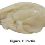 |
Figure 1: Pectin |
Anthocyanin confirmatory test
Spectrophotometer analysis
Anthocyanin extract sample was loaded in a UV-spectrophotometer to analyse the UV spectral peak range of anthocyanin. In UV-spectrophotometer the anthocyanin absorbs the spectral peak range of 520 nm. Figure 2 shows the UV-vis spectral peak obtained for anthocyanin.
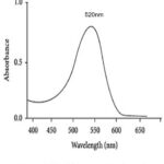 |
Figure 2: UV- visible spectra of anthocyanin |
pH Confirmation test for anthocyanin
Anthocyanins have a dark purple colour at acidic pH ranges (2-5) due to the flavylium cation form, which changes to a lighter shade of purple at neutral pH ranges (6-7). The coloration changed to yellow-brown with a further rise in pH (8–12), showing its quinoidal base form. Figure 3 displays the observations of colour change.
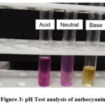 |
Figure 3: pH Test analysis of anthocyanin |
FTIR analysis test of anthocyanin
Identification of functional groups is accomplished through the use of FTIR studies. Since each functional group absorbs light at a specific frequency in the infrared spectrum, anthocyanin compounds can be distinguished in FTIR spectra by the presence of specific functional groups25. Concentrated extracts of BKBP anthocyanins were analysed using FTIR spectroscopy, revealing prominent absorption bands. The observed values of FTIR spectra are listed in table 2. Aromatic rings may be detected by their absorption band, which ranges from 600 to 1000 cm-1. The absorbance at 1043 cm-1 is caused by the stretching vibrations of C-O-C esters, while the stretching of the pyran rings causes absorbance at 1224 cm-1, a feature of flavonoid compounds. The C=C groups in aromatic rings are represented by bands at 1632 cm-1, whereas bands at 1725 cm-1 show the C=O distortion of phenols. The phenol O-H groups and sugar vibrations are responsible for the wide band at 3342 cm-1. Therefore, the spectral examination of the concentrated extract confirmed the existence of functional groups like C-O-C, C=O, C=C, and O-H, which were the distinguishing characteristics of anthocyanins. Figure 4 and 5 represents the standard FTIR curve and observed FTIR curve for anthocyanin respectively.
Table 2: FTIR analysis of anthocyanin sample
|
Spectrum Wavelength |
Literature |
Bands |
Functional groups |
|
3342 cm-1 |
3700- 3100 cm-1 |
Broaden |
O-H |
|
1725 cm– 1 |
1705-1725 cm-1 |
Sharp |
C=O |
|
1632 cm-1 |
1500-1600 cm-1 |
Medium |
C=C |
|
1224 cm-1 |
1080- 1300 cm-1 |
Sharp |
C-O-C |
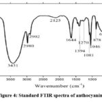 |
Figure 4: Standard FTIR spectra of anthocyanin |
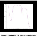 |
Figure 5: Obtained FTIR spectra of anthocyanin. |
Characterisation of colorimetric film
Based on the solution casting process, anthocyanins from the dark red kidney bean peel (BKBP) were mixed with composite pectin from the citron peel (CP) to create a pH-sensitive colorimetric film. Pectin and anthocyanins enhance the materials’ mechanical, moisture-resistance, and UV-visibility screening qualities. Additionally, because the anthocyanins are sensitive to pH changes and the formation of ammonia gas, they might be employed as colorimetric markers to identify food deterioration26. Figure 6 shows the anthocyanin coated pectin film.
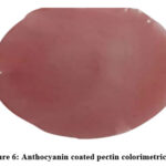 |
Figure 6: Anthocyanin coated pectin colorimetric film |
Physical Properties of colorimetric film
We tested the films’ moisture level, solubility in water, and swelling index.17. The hydrophilic nature of the indicator film determines how strongly it interacts with water molecules, which can lead to delays or inaccurate results when determining the freshness of meat. According to the indicator film’s moisture content, there was little change. Due to the interaction of hydrophilic moieties and water as well as the crosslinking of anthocyanin and pectin, the films had lesser moisture content over 117.5. This shows that the pectin-based film has a lot of hydroxyl groups and may take in moisture from the air around it. In the past, it was found that when the anthocyanin concentration increased, the moisture content of polyvinyl alcohol and starch films with roselle anthocyanin declined significantly, with no detectable variation in the water solubility of the indicator films27. The indicator films were less soluble in water with a solubility of 2.50, caused by crosslinking and stronger molecular bond formation between pectin and anthocyanin. Hence, Anthocyanins help reduce pectin’s hydrophilicity and solubility in the layer created by water. Our pectin-film has a 0.405 swelling index which is higher than starch due to their repulsion between adjacent amylopectin chains’ negatively charged phosphate groups, which inhibits the bonding of hydrogen between chains and promotes fast hydration and swelling.
Barrier properties of film
The anthocyanin-coated pectin films had the highest light transmission because they lacked groups that could efficiently absorb UV-vis light, which may be because the anthocyanins in the films have UV-vis radiation-absorbing properties. It was observed that the film’s light transmittance remained almost constant at zero between the wavelengths of 300 and 800 nm, and that the film absorbs UV-visible light transmission at 452 nm, indicating that it can effectively shield food from UV radiation. Our findings indicated that the incorporation of anthocyanins significantly increased the film’s UV-light barrier property (Figure 7).
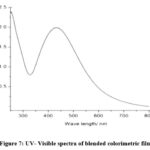 |
Figure 7: UV- Visible spectra of blended colorimetric film |
Water Vapour Permeability Test
Another important quality to consider is the water vapour permeability of the films being used for packing, as this affects how much moisture food products retain or lose while being stored. The WVP value obtained for the film was 0.073. The results for the anthocyanins loaded pectin films differed significantly. This suggested how the inclusion of the polysaccharide seemed to have a considerable effect upon the water molecule diffusion across the films. The anthocyanins had zero effect on size of the holes within the biopolymer network, but they did change how the chains of the biopolymer interacted with one another. According to other study, incorporating anthocyanin pigments did modify the WVP characteristics of biopolymer films, although the impacts varied depending on the anthocyanin and biopolymer types. As an example, adding anthocyanins to pectin films made them more permeable to water vapour.
Film microstructure SEM Analysis
The microscopic structure of colorimetric films was examined using SEM to look for cracks as well as holes that would affect the films’ mechanical and barrier properties. The films’ sectional view also clearly depicts that the anthocyanin addition improved its thickness28. In figure, 8 to 11 the SEM outcomes are displayed. The pectin film’s 3D-micrograph revealed a smooth surface. Valleys and peaks became more numerous.as a result of the anthocyanin addition29. Giancone et al., (2011) further noted that the pectin films can be identified by their tightly packed clusters30. Compared to those from a traditional SEM, the circumstances in the current investigation enable the visualisation of more minute details from sample surfaces. A substantially smoother surface was produced using pectin-based colorimetric film that included anthocyanin polyphenols at the maximum concentration (12%). In comparison to the anthocyanin-incorporated film with a lesser concentration, there was minimal sign of clustering, and the thickness was considerably more consistent. In comparison to that of the film exhibiting a higher concentration, the cross-section of this one revealed a more broken framework. Pectin has a strong capacity for building films since the surface of the films lacked any cavities or fissures. SEM images (Figure 8a) also showed that anthocyanin was evenly distributed in pectin-based films. The films’ roughness and unevenness (Figure 8b and 8c) can be due to less interaction between the anthocyanin compounds and the components of starch and glycerol. Pinho starch and jabuticaba flour were used to create films by Luchese et al., (2015), who also noticed films with roughened surfaces31. However, they claimed that the pH-dependent colour fluctuation of the film was unaffected by the surface roughness.
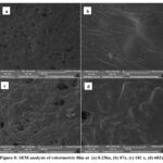 |
Figure 8: SEM analysis of colorimetric film at (a) 8.23kx, (b) 87x, (c) 182 x, (d) 682x |
Film antimicrobial activity test
To reduce microbial contamination, food packaging sheets must have strong antibacterial activity32. They claimed that various antibacterial properties were demonstrated by anthocyanins isolated from dark red kidney beans. At the same time, anthocyanins demonstrated good antibacterial efficacy against Staphylococcus aureus and Escherichia coli. In contrast, films made of anthocyanins produced clear inhibitory zones in S. aureus and E. coli. It can alter the potential and permeability of bacterial cell membranes, causing cell harm. As a result, the cytoplasm is lost and the cellular structures are damaged, which causes the bacteria to die quickly. E. coli and S. aureus culture had inhibitory zones with maximum diameters of 20.65 mm and 37.30 mm, respectively (Table 3). The structural variations between these two kinds of bacteria may be the cause of this. While E. coli is Gram-negative bacteria with an outer protective lipopolysaccharide membrane, S. aureus is Gram-positive bacteria lacking such membrane in its cell wall. The performance of the films’ antibacterial properties against these two foodborne pathogens is shown in the figure 9.
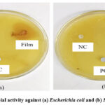 |
Figure 9: Antimicrobial activity against (a) Escherichia coli and (b) Staphylococcus aureus |
Table 3: Zone of inhibition of film against foodborne pathogens
|
Food borne Pathogens |
Zone of inhibitions (mm) |
||
|
Positive control |
Negative control |
Colorimetric film |
|
|
Escherichia coli |
23.65 |
Nil |
21 |
|
Staphylococcus aureus |
26 |
Nil |
24 |
Film sterility test
A sample of film was tested for the presence of live bacteria by encouraging the growth of microorganisms in a nutrient-rich media overnight. As from day-1 to day-2 there was no appearance of contamination around the film, which may be due to anthocyanin content present in the film. The test shows no signs of living microorganisms and therefore the film proved to be sterile. Addition of anthocyanin has improved the film’s antimicrobial activity, so it plays vital role against microorganisms (Figure 10).
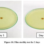 |
Figure 10: Film sterility test for 2 days |
Colorimetric Film Tested on Meat Products
During the meat storage process, the breakdown of proteins and other macromolecules that contain nitrogen releases ammonia and amines, two basic volatile nitrogen compounds, raising the pH level in the packing systems. To determine if anthocyanin coated pectin films were effective, fresh chicken meat was used as a pH detecting indicator. When stored at 4°C for eight days, the colorimetric film, in contrast, had a significant colour shift from pink to brownness (Figure 11).
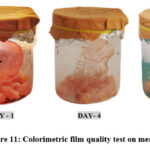 |
Figure 11: Colorimetric film quality test on meat sample. |
Conclusion
This study’s main objective is to significantly improve society. Based on anthocyanin recovered from dark red kidney bean peel mixed with composite pectin extracted from citron peel, a pH-sensitive colorimetric film was created. Solvent casting was used to make the film. Using scanning electron microscopy, the morphological structure of the film was investigated. The findings showed that the pectin isolated from BKBP had good film-forming capacity and that the anthocyanins were evenly distributed throughout the pectin matrix. When the anthocyanin-containing smart films’ antibacterial abilities were examined, it was discovered that they have a strong ability to inhibit bacterial growth of Escherichia coli and Staphylococcus aureus. The film’s physiological characteristics, such as its moisture content, water solubility, and swelling index, were also described. This makes tracking perishable foods simple, inexpensive, and safe thanks to our environmentally friendly, biodegradable pH-sensitive indicator. The major limitation of the study is the degradation parameters of the film and the degardation studies of the film is yet to be analysed in future . It has the potential to increase public health, decrease food waste, avoid foodborne infections, and open new avenues for innovation and economic development.
Acknowledgement
The authors would like to thank the management of Vels Institute of Science Technology and Advanced Studies, Chennai for providing necessary lab requirements in completing this research work.
Conflict of Interest
The authors declare no conflict of interest.
Funding sources
The research, writing, and/or publishing of this work were all done without any financial assistance from the authors.
References
- Siracusa V, Rocculi P, Romani S, Rosa MD. Biodegradable polymers for food packaging: a review. Trends in Food Science & Technology. 2008;19(12):634-643. doi:https://doi.org/10.1016/j.tifs.2008.07.003
CrossRef - Choi I, Lee JY, Lacroix M, Han J. Intelligent pH indicator film composed of agar/potato starch and anthocyanin extracts from purple sweet potato. Food Chemistry. 2017;218:122-128. doi:https://doi.org/10.1016/ j.foodchem.2016.09.050
CrossRef - Musso YS, Salgado PR, Mauri AN. Smart gelatin films prepared using red cabbage (Brassica oleracea L.) extracts as solvent. Food Hydrocolloids. 2019;89:674-681. doi:https://doi.org/10.1016/ j.foodhyd.2018.11.036
CrossRef - Park YW, Kim SM, Lee JY, Jang W. Application of biosensors in smart packaging. Molecular & Cellular Toxicology. 2015;11(3):277-285. doi:https://doi.org/10.1007/s13273-015-0027-1
CrossRef - Kalpana S, Priyadarshini SR, Maria Leena M, Moses JA, Anandharamakrishnan C. Intelligent packaging: Trends and applications in food systems. Trends in Food Science & Technology. 2019;93:145-157. doi:https://doi.org/10.1016/j.tifs.2019.09.008
CrossRef - Halász K, Csóka L. Black chokeberry (Aronia melanocarpa) pomace extract immobilized in chitosan for colorimetric pH indicator film application. Food Packaging and Shelf Life. 2018;16:185-193. doi:https://doi.org/10.1016/j.fpsl.2018.03.002
CrossRef - Timberlake CF, Bridle P. Flavylium salts, anthocyanidins and anthocyanins. I.—Structural transformations in acid solutions. Journal of the Science of Food and Agriculture. 1967;18(10):473-478. doi:https://doi.org/10.1002/jsfa.2740181008
CrossRef - Iacobucci GA, Sweeny JG. The chemistry of anthocyanins, anthocyanidins and related flavylium salts. Tetrahedron. 1983;39(19):3005-3038. doi:https://doi.org/10.1016/s0040-4020(01)91542-x
CrossRef - Li D, Wang P, Luo Y, Zhao M, Chen F. Health benefits of anthocyanins and molecular mechanisms: Update from recent decade. Critical Reviews in Food Science and Nutrition. 2015;57(8):1729-1741. doi:https://doi.org/10.1080/10408398.2015.1030064
CrossRef - Clifford MN. Anthocyanins – nature, occurrence and dietary burden. Journal of the Science of Food and Agriculture. 2000;80(7):1063-1072. doi:https://doi.org/10.1002/(sici)1097-0010(20000515)80:7%3C1063::aid-jsfa605%3E3.0.co;2-q
CrossRef - Dueñas M, Sarmento T, Aguilera Y, et al. Impact of cooking and germination on phenolic composition and dietary fibre fractions in dark beans (Phaseolus vulgaris L.) and lentils (Lens culinaris L.). LWT – Food Science and Technology. 2016;66:72-78. doi:https://doi.org/ 10.1016/j.lwt.2015.10.025
CrossRef - Halász K, Csóka L. Black chokeberry (Aronia melanocarpa) pomace extract immobilized in chitosan for colorimetric pH indicator film application. Food Packaging and Shelf Life. 2018;16:185-193. doi:https://doi.org/10.1016/j.fpsl.2018.03.002
CrossRef - Roy S, Priyadarshi R, Łopusiewicz Ł, Biswas D, Chandel V, Rhim JW. Recent progress in pectin extraction, characterization, and pectin-based films for active food packaging applications: A review. International Journal of Biological Macromolecules. 2023;239:124248. doi:https://doi.org/10.1016/j.ijbiomac.2023.124248
CrossRef - Guo Z, Ge X, Li W, Yang L, Han L, Yu Q. Active-intelligent film based on pectin from watermelon peel containing beetroot extract to monitor the freshness of packaged chilled beef. Food Hydrocolloids. 2021;119:106751. doi:https://doi.org/10.1016/j.foodhyd.2021.106751
CrossRef - Sucheta, Rai SK, Chaturvedi K, Yadav SK. Evaluation of structural integrity and functionality of commercial pectin based edible films incorporated with corn flour, beetroot, orange peel, muesli and rice flour. Food Hydrocolloids. 2019;91:127-135. doi:https://doi.org/10.1016/j.foodhyd.2019.01.022
CrossRef - Huang J, Hu Z, Hu L, Li G, Yao Q, Hu Y. Pectin-based active packaging: A critical review on preparation, physical properties and novel application in food preservation. Trends in Food Science & Technology. 2021;118:167-178. doi:https://doi.org/10.1016/j.tifs.2021.09.026
CrossRef - Anas M, Ahmad S, Malik A. Microbial Escalation in Meat and Meat Products and Its Consequences. Health and Safety Aspects of Food Processing Technologies. Published online 2019:29-49. doi:https://doi.org/10.1007/978-3-030-24903-8_3
CrossRef - Khaled AY, Parrish CA, Adedeji A. Emerging nondestructive approaches for meat quality and safety evaluation—A review. Comprehensive Reviews in Food Science and Food Safety. 2021;20(4):3438-3463. doi:https://doi.org/10.1111/1541-4337.12781
CrossRef - Zhao L, Liu Y, Zhao L, Wang Y. Anthocyanin-based pH-sensitive smart packaging films for monitoring food freshness. Journal of Agriculture and Food Research. 2022;9, p.100340. https://doi.org/ 10.1016/ j.jafr.2022.100340.
CrossRef - Ma J, Tong P, Chen Y, Wang Y, Ren H, Gao Z, Yue T, Long F. The inhibition of pectin oligosaccharides on degranulation of RBL-2H3 cells from apple pectin with high hydrostatic pressure assisted enzyme treatment. Food Chemistry. 2022; 371, p.131097. DOI: 10.1016/j.foodchem.2021.131097.
CrossRef - Merz B, Capello C, Leandro GC, Moritz DE, Monteiro AR, Valencia GA. A novel colorimetric indicator film based on chitosan, polyvinyl alcohol and anthocyanins from jambolan (Syzygium cumini) fruit for monitoring shrimp freshness. International Journal of Biological Macromolecules. 2020;153:625-632. doi:https://doi.org/10.1016/ j.ijbiomac.2020.03.048
CrossRef - Erna KH, Felicia WXL, Vonnie JM, Rovina K, Yin KW, Nur’Aqilah MN. Synthesis and Physicochemical Characterization of Polymer Film-Based Anthocyanin and Starch. Biosensors. 2022;12(4):211. doi:https://doi.org/10.3390/bios12040211
CrossRef - Cortez R, Luna-Vital DA, Margulis D, Gonzalez de Mejia E. Natural Pigments: Stabilization Methods of Anthocyanins for Food Applications. Comprehensive Reviews in Food Science and Food Safety. 2016;16(1):180-198. doi:https://doi.org/10.1111/1541-4337.12244
CrossRef - Swer TL, Mukhim C, Bashir K, Chauhan K. Optimization of enzyme aided extraction of anthocyanins from Prunus nepalensis L. LWT. 2018;91:382-390. doi:https://doi.org/10.1016/j.lwt.2018.01.043
CrossRef - Sani MA, Tavassoli M, Hamishehkar H, McClements DJ. Carbohydrate-based films containing pH-sensitive red barberry anthocyanins: Application as biodegradable smart food packaging materials. Carbohydrate Polymers. 2021;255:117488. doi:https://doi.org/10.1016/j.carbpol.2020.117488
CrossRef - Zeng P, Chen X, Qin YR, et al. Preparation and characterization of a novel colorimetric indicator film based on gelatin/polyvinyl alcohol incorporating mulberry anthocyanin extracts for monitoring fish freshness. Food Research International. 2019;126:108604. doi:https://doi.org/10.1016/j.foodres.2019.108604
CrossRef - Yun YH, Lee CM, Kim YS, Yoon SD. Preparation of chitosan/polyvinyl alcohol blended films containing sulfosuccinic acid as the crosslinking agent using UV curing process. Food Research International. 2017;100:377-386. doi:https://doi.org/10.1016/j.foodres.2017.07.030
CrossRef - Ahmad M, Hani NM, Nirmal NP, Fazial FF, Mohtar NF, Romli SR. Optical and thermo-mechanical properties of composite films based on fish gelatin/rice flour fabricated by casting technique. Progress in Organic Coatings. 2015;84:115-127. doi:https://doi.org/10.1016/ j.porgcoat.2015.02.016
CrossRef - Tiziana Giancone, Torrieri E, Próspero Di Pierro, Cavella S, Giosafatto VL, Masi P. Effect of Surface Density on the Engineering Properties of High Methoxyl Pectin-Based Edible Films. Food and Bioprocess Technology. 2009;4(7):1228-1236. doi:https://doi.org/10.1007/s11947-009-0208-9
CrossRef - Luchese CL, Frick JM, Patzer VL, Spada JC, Tessaro IC. Synthesis and characterization of biofilms using native and modified pinhão starch. Food Hydrocolloids. 2015;45:203-210. doi:https://doi.org/10.1016/j.foodhyd.2014.11.015
CrossRef - Li N, Zhou Z, Wu F, et al. Development of pH-Indicative and Antimicrobial Films Based on Polyvinyl Alcohol/Starch Incorporated with Ethyl Lauroyl Arginate and Mulberry Anthocyanin for Active Packaging. Coatings. 2022;12(10):1392. doi:https://doi.org/10.3390/ coatings12101392
CrossRef
Accepted on: 22 Dec 2023
Second Review by: Misha Patel
Final Approval by: Dr. Krešimir Mastanjević


Web of Science Coverage
Emerging Sources Citation Index (ESCI)
2024 Journal Impact Factor: 1.1
Scopus Journal Metrics
CiteScore 2025: 2.6
CiteScore Details
Sustainable Nutrition: Food Systems, Nutrient Retention, and Public Health Impact
![]()
This journal is a member of, and subscribes to the principles of, the Committee on Publication Ethics (COPE)


