Introduction
The International Agency for Research on Cancer, reported a continuing rise in the number of cancer patients. According to GLOBOCAN figures for 2020, nearly 10.0 million people died from cancer, and there were 19.3 million new occurrences of the disease. According to reports, breast, lung, colorectal, prostate, and stomach cancers are the main killers of people with cancer 1. Cancer cells develop when healthy cells lose their ability to control the cell cycle, proliferation, and apoptosis 2,3. As a result, these cells multiply continually and quickly without restriction, which is a hallmark of the growth of cancer 4. Surgery, radiation, and chemotherapy are the most popular and extensively used cancer treatments, but they are expensive and have a lot of adverse effects 5. The use of natural ingredients as anticancer agents offers a solution to these issues. Previous studies have shown that a variety of compounds derived from medicinal plants have anticancer activity by modifying cell cycle and/or apoptosis 6, therefore it can be said that this is a highly effective method for treating cancer. Additionally, it has been demonstrated that reducing oxidative stress early on with exogenous antioxidants—most commonly found in plants or herbs like curcumin, resveratrol, catechin, and genistein—as well as eating meals high in vitamins and antioxidants—can help prevent cancer7.
One of functional food which has a lot of attention to be used as an alternative for cancer treatment is pigmented rice. A blackish-purple pigmented rice, Keunnunjami, has anticancer effect against cervical and gastric cell lines due to the high antioxidant activity8. However, one the most popular pigmented rice which is rich in pharmaceutical value is black rice. Black rice is an antioxidant-rich food, and a prior study we conducted in mice with diabetes model indicated that this extract has anti-inflammatory properties 9. The high value of antioxidant activity in black rice become one the most concern that link to the anticancer effect8. Besides, anthocyanins, phenols, flavonoids, -oryzanols, and tocol are all present in black rice 10–12. According to Chen et al13, anthocyanin from black rice extract has been shown to inhibit liver cancer cells as well as a breast cancer metastasis-promoting molecule both in vitro and in vivo14. Zhou et al. (2017) also observed that black rice anthocyanin inhibited the phosphorylation of molecules that promote metastasis in cell lines for breast cancer in humans MCF-7 as well as MDA-MB-453 through FAK, cSrc, and p130Cas. However the comparison effect of black rice in different human cancer cell lines and which pathway that influence the most in the anti-cancer mechanism of this extract are still remain unclear. So that, the objective of the study is to examine the anti-cancer potential of aqueous black rice extract on the cancer cell lines in humans such as HeLa, T47D, and U2OS.
Materials and Methods
Black Rice Extraction
Wojalaka variety of black rice (Oryza sativa L.) from Kepanjen, Malang, East Java, Indonesia, was used in this study. Black rice powder was pounded and mashed using less than 80 mesh size according to the American Standard Test Sieve Series (ASTM). The grounding process affected the simplicial or powder form characteristic which must be considered15. After then, Simplicia was macerated with Aquadest (1:3, m:v) for an hour while being stirred numerous times. The sample was then filtered to separate the filtrate from the pulp. After that, 4 percent maltodextrin was added, and the filtrate was freeze-dried at -40 °C. The yield was then calculated by weighing the extracted material.
Total Phenolic and Flavonoid Content Estimation
Based on Christina et al (2021)16 the total phenolic content was assessed using the Folin-Ciocalteu colorimetric test. 20 µl of BR extract or standard (gallic acid, SigmaÒ, Germany) were combined and put into a 100 µl batch of freshly prepared 100 % Folin-Ciocalteu reagent (SigmaÒ, Germany). The combination was given a five-minute incubation period at room temperature before being neutralized with 75 µl of saturated sodium carbonate (MerckÒ, Germany). It was then given another two hours at room temperature without exposure to light. Next, a wavelength of 740 nm was used to measure the absorbance following a 2-hour reaction.
The measurement of the flavonoid content was based on Christina et al (2021)16 Quercetin (SigmaÒ, Germany) was used as the standard, and 50 µl of BR extract was added along with 70 µl of distilled water, 15 µl solution of sodium nitrate (MerckÒ, Germany) (5%) and incubated for 5 min. After incubating for 5 min, the sample received 15 µl of 10% (m/v) AlCl3 (MerckÒ, Germany), which was then added. After that, 100 µl of sodium hydroxide solution (MerckÒ, Germany) was added to the mixture (1 M). The sample absorbance was then compared to the blank using a 510 nm wavelength analysis.
DPPH Scavenging Ability Assay
The 2, 2-Diphenyl-1-picrylhydrazyl (DPPH, SigmaÒ, Germany) test was done according to Ablat et al (2014)17 with a slight modification, the effect of BR extract free radical scavenging was investigated. In a nutshell, ethanol was made using a series of BR extract concentrations. The same volume of a 1 ml DPPH solution (0.004 %) in ethanol was then combined with the extract or ascorbic acid (MerckÒ, Germany) as the standard. The combination was then allowed to stand for 30 minutes at room temperature in the dark. The absorbance value of the scavenging activity was then computed using the following formula after the absorbance at 517 nm was determined;
![]()
Where At denoted the absorbance of the sample, and Ac denoted the absorbance of the control (ethanol and DPPH reaction). The IC50 or 50% of scavenging activity was determined trhough the graph plotted against percentage inhibition and standard.
Ferric-reducing/antioxidant Power
The FRAP assay was done to assess antioxidant activity according to Fernandes et al (2016)18 with slight modification . In a nutshell, the TPTZ (2,4,6-tripyridyl-S-triazine, SigmaÒ, Germany) and 1.0 mol/l (10 volumes) acetate bauffer (SigmaÒ, Germany) were combined with 1 volume of 20 mmol/l ferric chloride (CDHÒ, India) and 40 mmol/l hydrochloric acid (MerckÒ, Germany) to create the FRAP reagent. Then, the sample was mixed with 3 ml of FRAP reagent, 100 µl of sample, 300 µl of deionized water, and let to sit for 8 minutes.With varied concentrations of FeSO4.7H2O (MerckÒ, Germany) (100–1,000 µmol/l), the absorbance was measured and a standard curve was created.
Total Antioxidant Capacity Determination
The phosphomolybdate technique was done according to Jafri et al (2017)19 and used to determine the total antioxidant capacity (TAC). In a nutshell, 2.0 ml of reagent solution was combined with 0.2 ml (0.5 mg/ml) of BR extract (600 mM sulfuric acid (MerckÒ, Germany), 28 mM sodium phosphate (MerckÒ, Germany), and 4 mM ammonium molybdate (MerckÒ, Germany)). The mixture was then heated for 60 minutes at 95°C. The absorbance was measured at 695 nm and compared to a control (2 ml reagent solution). Ascorbic acid’s antioxidant activity served as a standard, and its equivalent expression was used.
Cell Culture and Flow Cytometry Analysis
Five dosages of an anticancer test were performed on selected samples; Control (C; 0 µg/mL), dosage I (BR1; 100 µg/mL), dose II (BR2; 200 µg/mL), dose III (BR3; 300 µg/mL), dose IV (BR4; 400 µg/mL), dose V (BR5; 500 µg/mL), and Cisplatin (PT. Ferron Pharmaceuticals, Indonesia) (Cis; 1.5 µg/mL). Several cancer cell lines, including human breast cancer (T47D), sarcoma (U2OS) cells, and servical cancer (HeLa), were used in in vitro anti-cancer tests. Besides, this study also used human peripheral blood mononuclear cells (PBMC) as normal or control cell which was obtained from healthy female donor aged 45 years old who was fulfilled the inform consent before conducting the study. The protocol was approved by the ethics commission of University of Brawijaya with ethical clearance number 474-KEP-UB. The PBMC isolation from whole blood was done according to Puleo et al (2017)20 with modification. The blood was collected and mixed with PBS (1:1) (Biowest) and put in 15 ml centrifuge tube filled with Ficoll-Hypaque 1077. The sample was then formed 2 separated layer and centrifuged at 1500 rpm 20° C for 30 min. The sample was then formed 3 layers and PBMC in interphase part was collected and washed with PBS 5 ml in centrifuge tube. The PBC was then centrifuged again for 5 min at 2500 rpm and 10 °C and the pellet was collected as PBMC. In addition, U2OS cells were grown in DMEM complete medium (Gibco®, USA) whereas PBMC, HeLa and T47D cells were grown in RPMI 1640 complete medium (Gibco®, USA). The cells were kept alive in a cell culture incubator with 5% CO2 and temperature 37 °C. A 10% fetal bovine serum/FBS (Gibco®, USA) and 1% penicillin-streptomycin (Gibco®, USA) were added to each basal medium, DMEM and RPMI. After one day of cell culture, the viability, apoptosis, and cell cycle of the treated cells were examined. The cells were then treated with various doses of BR extract.
Cell Viability Analysis
The cell viability analysis was done using WST-1 reagents (Roche Diagnostics GmbH, Germany) according to Christina et al (2022)21 with modification. The PBMC, T47D, U2OS, and HeLa cells were cultured in 96-well plate and several doses of BR extract (BR1, BR2, BR3, BR4, BR5) were given and compared to cisplatin-treated cells (100 μl). Then, the treatment medium was dicarded and added by 100 μl medium containing 5 μl WST-1 reagent in the dark. After 30 min incubation, a wavelenght 450 nm used to measure the absorbance.
Cell Cycle Analysis
The cell cycle analysis was done by flow cytometry analysis using PI staining (BioLegend®, USA). With a slight modification, the cell cycle analysis was refered to Pumiputavon et al (2017)22. The administration of BR extract and cisplatin was done as the cell viability and apoptosis analysis. After treatment, the cells were harvested using trypsin solution (Gibco®, USA) . After trypsination, the cells were added with the same volume of culture medium and centrifuged at 2500 rpm, 10 ºC and 5 min. The pellet was then stained with 50 μl PI antibody and incubated in 4 ºC for 30 min. After staining, the flow cytometry analysis was done.
Cell Apoptosis Analysis
The analysis of cell apoptosis was done through flow cytometry analysis with Annexin V/PI staining22 (BioLegend®, USA). The antibody (50 μl) was added after the cells were harvested and incubated for 30 min. After incubation, the samples were analyzed using flow cytometer.
Statistical Data Analysis
The flow cytometry data was analyzed using BD CellQuest ProTM. The data showed as the mean + SD results where the statistical data were analyzed using One-way ANOVA using SPSS software ver. 16 for windows.
Results
According to this investigation, the BR extract contained 11.12 mg QE/g of total flavonoids and 66.42 mg GAE/g of total phenolic content. The extract contained higher level of phenolic than flavonoid which strongly correlated to the antioxidant activity. This study analyzed the antioxidant activity through DPPH, FRAP, and TAA assays. The analysis showed the antioxidant value based on the IC50 of DPPH assay was 53.19 µg/ml, while the value based on FRAP and TAA assay were 49.86 mg/ml and 96.70 mg/ml, respectively (Table 1).
Table 1: Yield percentage (%), water content (%), total phenol content (TPC as mg gallic acid/g extract), total flavonoid content (TFC as mg of quercetin/g extract), DPPH % inhibition (IC50), FRAP % inhibition, and TAA % inhibition of BR extract.
| Yield in percentage (%) | Water Content (%) | TPC(mg/g) | TFC(mg/g) | DPPH (μg/ml) | FRAP (mg/g) | TAC (mg/g) |
| 69.35 | 5 | 66.42 ± 0.23 | 11.12 ± 0.19 | 53.19 | 49.86 ± 0.04 | 96.70 ± 0.30 |
Using the WST-1 reagent, the viability of cancer cells (HeLa, T47D, and U2OS) was assessed in comparison to peripheral blood mononuclear cell (PBMC) as normal or non-cancer cells. The findings demonstrated that BR extract decreased cell viability specifically in cancer cells (Tab. 1). Following the administration of cisplatin (31.35 %), the viability of PBMC dramatically decreased (p<0.05), however it had grown along with BR extract. The study also demonstrated that, in a dose dependent manner, BR extract dramatically decreased the viability of HeLa, T47D, and U2OS cells (p<0.05). When compared to BR extract, cisplatin therapy, however, had the lowest effect on cancer cell viability.
According to the study, BR extract significantly and dose-dependently increased apoptosis in HeLa and T47D cells (p<0.05). In addition, U2OS cells saw a substantial increase in apoptosis in a dose-independent manner (p<0.05) when exposed to the effects of BR extract. BR extract was more capable of inducing T47D cell apoptosis than any other cancer cell lines. Compared to cisplatin, the number T47D apoptosis cells of BR extract groups were similar to low dose of cisplatin treatment (1.5 µg/mL). Cisplatin stimulated T47D cells apoptosis to a level of 95.15 %, whereas BR extract was 93.64 % (BR5). However, untreated cells group displayed 0.82, 1.19, and 1.21 % of cell death (HeLa, T47D, and U2OS, respectively).
Table 2: BR extract effect on several cancer cell lines viability
| Group | Cell viability (%) | |||
| PBMC | HeLa | T47D | U2OS | |
| Untreated | 61.91 ± 0.28b | 100 ± 0g | 100 ± 0g | 100 ± 0f |
| BR1 (100 µg/mL) | 65.366 ± 0.55c | 68.55 ± 0.55f | 71.47 ± 0.61f | 61.43 ± 0.54e |
| BR2 (200 µg/mL) | 76.21 ± 0.61e | 63.58 ± 0.53e | 67.57 ± 0.54e | 60.26 ± 0.72e |
| BR3 (300 µg/mL) | 69.5 ± 0.5d | 57.75 ± 0.68d | 62.3 ± 0.16d | 46.61 ± 0.26d |
| BR4 (400 µg/mL) | 85.49 ± 0.45f | 50.3 ± 0.31c | 55.56 ± 0.57c | 47.9 ± 0.27c |
| BR5 (500 µg/mL) | 89.22 ± 0.25g | 37.83 ± 0.61b | 41.76 ± 0.28b | 45.35 ± 0.48b |
| Cis (1.5 µg/mL) | 31.35 ± 0.47a | 14.83 ± 0.50a | 15.19 ± 0.3a | 25.73 ± 0.19a |
*Black rice extrat treatment on several cancer cell lines. Untreated group as control cells without BR extract treatment. BR1 to BR5 (Black rice extract dose 1 to dose 5), and Cis group as cells group treated with Cisplatin. Value was the mean of data ± sd. Superscript notation showed a significantly difference by Tukey test (p<0.05).
Through the use of PI staining and flow cytometry analysis, the cancer cell cycle was identified. The graph’s orange and grey lines, which correspond to the S and G2/M phases, respectively, and the blue line, which represents the G0-G1 phase (Fig. 2). On HeLa cells, BR extract dramatically inhibited G2/M in a dose-dependent manner (p<0.05) and significantly inhibited S phase in a dose-independent manner (p<0.05). Additionally, the study showed that BR extract strongly promotes cell cycle arrest to the G0/G1 phase, with BR5 showing the highest percentage of cells in this phase (73.58%). Cisplatin treatment suppressed the cells in the G0/G1, S, and G2/M phases; we hypothesized that this was because the medication mostly triggered apoptosis in the cells (Fig. 1).
Table 3: BR extract effect on several cancer cell lines apoptotis
| Group | HeLa | T47D | U2OS |
| Untreated | 0.82 ± 0.032a | 1.196 ± 0.135a | 1.21 ± 0.149a |
| BR1 (100 µg/mL) | 20.66 ± 0.085b | 83.29 ± 0.305b | 56.12 ± 0.2f |
| BR2 (200 µg/mL) | 24.06 ± 0.16c | 90.83 ± 0.745c | 42.86 ± 0.29e |
| BR3 (300 µg/mL) | 24.57 ± 0.6c | 90.17 ± 0.244c | 39.79 ± 0.9d |
| BR4 (400 µg/mL) | 30.64 ± 0.52d | 93.11 ± 0.615d | 35.15 ± 0.8c |
| BR5 (500 µg/mL) | 36.33 ± 0.53e | 93.64 ± 0.449d | 30.74 ± 0.42b |
| Cis (1.5 µg/mL) | 50.5 ± 0.087f | 95.19 ± 0.871e | 67.1 ± 0.19g |
*Black rice extrat treatment on several cancer cell lines. Untreated group as control cells without BR extract treatment. BR1 to BR5 (Black rice extract dose 1 to dose 5), and Cis group as cells group treated with Cisplatin. Value was the mean of data ± sd. Superscript notation showed a significantly difference by Tukey test (p<0.05).
On the other hand, the percentage of untreated T47D cells in S and G2/M phase have been relatively low. BR extract maintained the cell to not increase the S and G2/M phase that showed by the flat of organge and grey line on the graph. But the G0/G1 phase increased along with the treatment of BR extract aignificantly in dose independent manner (p<0.05). The treatment of BR extract induced G0/G1 phase of U2OS cells significantly (p<0.05) and inhibited S and G2/M phase significantly in dose dependent manner (p<0.05). BR extract and cisplatin treatment showed the same inhibition of cell cycle to S and G2/M phase.
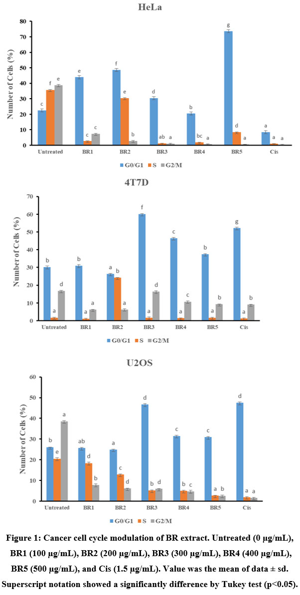 |
Figure 1: Cancer cell cycle modulation of BR extract. Untreated (0 µg/mL), BR1 (100 µg/mL), BR2 (200 µg/mL), BR3 (300 µg/mL), BR4 (400 µg/mL),BR5 (500 µg/mL), and Cis (1.5 µg/mL). |
Discussion
Numerous bioactive substances found in black rice which has been througly documented, such as phenolic and flavonoid content. Phenolic substances have the potential to treat cancer and other oxidative stress-related disorders. Phenolic compounds are frequently claimed to have anti-carcinogenic potential due to a variety of properties including cell cycle arrest induction, cell proliferation suppression, ROS level modulation, protein tumor suppressor modulation, and cell repair augmentation23. Promising substances such as flavonoids are utilized to control reactive oxygen species (ROS), induce cell cycle arrest and death, autophagy, and reduce the growth and invasiveness of cancer cells24.
Based on this study, an active value of antioxidant activity (50–100 µg/ml) was assigned to the BR extract’s DPPH assay which was categorized as a strong class of antioxidant activity25. The antioxidant activity of black rice extract was the highest when compared to red and brown rice. In agreement with that, red and brown rice extracts were contrasted with BR extract, which showed the greatest antiproliferative action, with IC50 values of 148.6 and 119.2 mg/mL against the MCF-7 and MDA-MB-231 cell lines, respectively26. The presence of phenolic and flavonoid chemicals, which are responsible for the suppression of cancer growth, contributes to the strong antioxidant activity of black rice extract.
In line with Hui et al (2010)27, BR extract has a reductive effect on the viability of cancer cell types. Black rice extract high in anthocyanins decreased the viability of the cell lines MCF-7, MDA-MB-231, and MDA-MB-453 from human breast cancer and activated the caspase cascade to cause cell death in MDA-MB-453 cells. When compared to T47D (BR5; 41.76 percent) and U2OS, the cytotoxic activity of BR extract was shown to be most effective on HeLa cells, reaching up to 37.38 percent viable cells on BR5 (BR5; 45.35 %). However, there was inconsistent data between viability and apoptosis cells. The study found BR5 induced the T47D cells for 94% cells, where the viability cells in previous analysis showed 41.76%. This was possible because the were a lot of factors can influence the cells health, including passage number, medium, incubator, etc. Nevertheles, the BR extract’s ability to induce apoptosis was consistent with that of Pratiwi et al28. Because cyanidin 3-glucoside and peondin 3-glucoside were present, it was believed that the black rice brand fraction caused apoptosis in HeLa cells. Pratiwi discovered that the apoptosis-induced capability of black rice fraction extract was superior to doxorubicin in terms of effectiveness. Compared to doxorubicin, an Indonesian black rice methanolic extract was observed to increase apoptosis in T47D cells29. The study discovered that T47D cells were more sensitive to the BR extract’s ability to induce apoptosis than HeLa or U2OS cells, which is similar with Pratiwi et al29 research.
Unregulated cell cycle and unchecked cell proliferation are characteristics of cancer. Controlling cell proliferation and growth is mostly governed by the cell cycle and cyclin-dependent kinases (CDKs) and cyclin30. Unchecked proliferation is caused by dysregulation of genes involved in the cell cycle machinery, which is frequently detected in cancer. According to the study, HeLa, T47D, and U2OS cells showed a significant percentage of cells in the S and G2/M phases when left untreated. G0/G1 were elevated along with the BR extract, however S and G2/M were considerably decreased. One promising method of treating cancer was the triggering of cell cycle arrest.
Conclusion
This study reported that the total phenolic and flavonoid content of BR extract were 66.42 and 11.12 mg/ml. Besides, the antioxidant activity of BR extract based on different analysis such as DPPH, FRAP, and TAC analysis were strong with value of 53.19 µg/ml, 49.86 mg/ml, and 96.70 mg/ml, respectively. According to the findings of an in vitro research, the high antioxidant content of the BR extract was expected to display anti-cancer action. Human cancer cells’ viability was selectively decreased by BR extract. However, T47D showed the greatest sensitivity to BR extract-induced apoptosis in range of 83 to 95%. In cell cycle analysis, it was discovered that BR extract caused HeLa, T47D, and U2OS cells to enter the G0/G1 phase of the cell cycle.
Acknowledgement
Direktorat Riset dan Pengabdian kepada Masyarakat (Directorate of Research and Community Service) of Ministry of Research and Technology Indonesia and we are thankful to Wirdatun Nafisah for assiting this research.
Conflict of Interest
Authors declare that there is no conflict of interest in this study.
Funding sources
This study is funded by Direktorat Riset dan Pengabdian kepada Masyarakat (Directorate of Research and Community Service) of Ministry of Research and Technology Indonesia with a grant no, (8/E1/KPT/2020.
References
- Sung H, Ferlay J, Siegel RL, et al. Global Cancer Statistics 2020: GLOBOCAN Estimates of Incidence and Mortality Worldwide for 36 Cancers in 185 Countries. CA Cancer J Clin. 2021;71(3):209-249. doi:10.3322/caac.21660
CrossRef - Hatzold, Julia; Conradt B. Control of Apoptosis by Asymetric Cell Division. Published online 2008:e84. doi:10.1371/journal.pbio.0060084
CrossRef - Mair W. pbio.1000423 1..2 _ Enhanced Reader.pdf. Published online 2010:8. doi:10.1371/journal.pbio.1000423
CrossRef - Frank SA, Iwasa Y, Nowak MA. Patterns of cell division and the risk of cancer. Genetics. 2003;163(4):1527-1532. doi:10.1093/genetics/163.4.1527
CrossRef - Frank SA. Cancer the whole story.pdf. Published online 2011:e1001044. doi:10.1371/journal.pbio.1001044
CrossRef - Mondal S, Bandyopadhyay S, K. Ghosh M, Mukhopadhyay S, Roy S, Mandal C. Natural Products: Promising Resources for Cancer Drug Discovery. Anticancer Agents Med Chem. 2012;12(1):49-75. doi:10.2174/187152012798764697
CrossRef - Vanden Berghe W. Epigenetic impact of dietary polyphenols in cancer chemoprevention: Lifelong remodeling of our epigenomes. Pharmacol Res. 2012;65(6):565-576. doi:10.1016/j.phrs.2012.03.007
CrossRef - Chung SI, Lee SC, Yi SJ, Kang MY. Antioxidative and antiproliferative activities of ethanol extracts from pigmented giant embryo rice (Oryza sativa L. cv. Keunnunjami) before and after germination. Nutr Res Pract. 2018;12(5):365-370. doi:10.4162/nrp.2018.12.5.365
CrossRef - Hartati FK, Widjanarko SB, Widyaningsih TD, Rifa’i M. Anti-Inflammatory evaluation of black rice extract inhibits TNF-α, IFN-γ and IL-6 cytokines produced by immunocompetent cells. Food Agric Immunol. 2017;28(6). doi:10.1080/09540105.2017.1332006
CrossRef - Shao Y, Xu F, Sun X, Bao J, Beta T. Identification and quantification of phenolic acids and anthocyanins as antioxidants in bran, embryo and endosperm of white, red and black rice kernels (Oryza sativa L.). J Cereal Sci. 2014;59(2):211-218. doi:10.1016/j.jcs.2014.01.004
CrossRef - Hou F, Zhang R, Zhang M, et al. Hepatoprotective and antioxidant activity of anthocyanins in black rice bran on carbon tetrachloride-induced liver injury in mice. J Funct Foods. 2013;5(4):1705-1713. doi:10.1016/j.jff.2013.07.015
CrossRef - Walter M, Marchesan E, Massoni PFS, da Silva LP, Sartori GMS, Ferreira RB. Antioxidant properties of rice grains with light brown, red and black pericarp colors and the effect of processing. Food Res Int. 2013;50(2):698-703. doi:10.1016/j.foodres.2011.09.002
CrossRef - Chen XQ, Nagao N, Itani T, Irifune K. Anti-oxidative analysis, and identification and quantification of anthocyanin pigments in different coloured rice. Food Chem. 2012;135(4):2783-2788. doi:10.1016/j.foodchem.2012.06.098
CrossRef - Luo LP, Han B, Yu XP, et al. Anti-metastasis activity of black rice anthocyanins against breast cancer: Analyses using an ErbB2 positive breast cancer cell line and tumoral xenograft model. Asian Pacific J Cancer Prev. 2014;15(15):6219-6225. doi:10.7314/APJCP.2014.15.15.6219
CrossRef - Mohite AM, Mishra A, Sharma N. Effect of Different Grinding Processes on Powder Characteristics of Tamarind Seeds. Agric Res. 2020;9(2):262-269. doi:10.1007/s40003-019-00431-9
CrossRef - Christina, Yuyun Ika; Nafisah, Wirdatun; Widodo; Rifa’i, Muhaimin; Djati MS. In vitro antioxidant and anticancer activity of crude ethanol extract of Mahkota Dewa (Phaleria macrocarpa) leaves. AIP Conf Proc. 2021;2353(May):030015.
CrossRef - Ablat A, Mohamad J, Awang K, Shilpi JA, Arya A. Evaluation of antidiabetic and antioxidant properties of Brucea javanica seed. Sci World J. 2014;2014. doi:10.1155/2014/786130
CrossRef - Fernandes RPP, Trindade MA, Tonin FG, et al. Evaluation of antioxidant capacity of 13 plant extracts by three different methods: cluster analyses applied for selection of the natural extracts with higher antioxidant capacity to replace synthetic antioxidant in lamb burgers. J Food Sci Technol. 2016;53(1):451-460. doi:10.1007/s13197-015-1994-x
CrossRef - Jafri L, Saleem S, Ihsan-ul-Haq, Ullah N, Mirza B. In vitro assessment of antioxidant potential and determination of polyphenolic compounds of Hedera nepalensis K. Koch. Arab J Chem. 2017;10:S3699-S3706. doi:10.1016/j.arabjc.2014.05.002
CrossRef - Puleo A, Carroll C, Maecker H, Gupta R. Isolation of PBMCs Using Vacutainer® Cellular Preparation Tubes (CPTTM). Bio-Protocol. 2017;7(2):1-6. doi:10.21769/bioprotoc.2103
CrossRef - Christina YI, Rifa’I M, Widodo N, Djati MS. Comparative Study of Antiproliferative Activity in Different Plant Parts of Phaleria macrocarpa and the Underlying Mechanism of Action. Sci World J. 2022;2022. doi:10.1155/2022/3992660
CrossRef - Pumiputavon K, Chaowasku T, Saenjum C, et al. Cell cycle arrest and apoptosis induction by methanolic leaves extracts of four Annonaceae plants. BMC Complement Altern Med. 2017;17(1):1-11. doi:10.1186/s12906-017-1811-3
CrossRef - Anantharaju PG, Gowda PC, Vimalambike MG, Madhunapantula S V. An overview on the role of dietary phenolics for the treatment of cancers. Nutr J. 2016;15(1):1-16. doi:10.1186/s12937-016-0217-2
CrossRef - Kopustinskiene DM, Jakstas V, Savickas A, Bernatoniene J. Flavonoids as anticancer agents. Nutrients. 2020;12(2):1-25. doi:10.3390/nu12020457
CrossRef - Riyadi PH, Atho’illah MF, Tanod WA, Rahmawati IS. Tilapia viscera hydrolysate extract alleviates oxidative stress and renal damage in deoxycorticosterone acetate-salt-induced hypertension rats. Vet World. 2020;13(11):2477-2483. doi:10.14202/VETWORLD.2020.2477-2483
CrossRef - Ghasemzadeh A, Karbalaii MT, Jaafar HZE, Rahmat A. Phytochemical constituents, antioxidant activity, and antiproliferative properties of black, red, and brown rice bran. Chem Cent J. 2018;12(1):1-13. doi:10.1186/s13065-018-0382-9
CrossRef - Hui C, Bin Y, Xiaoping Y, et al. Anticancer activities of an anthocyanin-rich extract from black rice against breast cancer cells in vitro and in vivo. Nutr Cancer. 2010;62(8):1128-1136. doi:10.1080/01635581.2010.494821
CrossRef - Pratiwi R, Tunjung WAS, Rumiyati R, Amalia AR. Black Rice Bran Extracts and Fractions Containing Cyanidin 3-glucoside and Peonidin 3-glucoside Induce Apoptosis in Human Cervical Cancer Cells. Indones J Biotechnol. 2016;20(1):69. doi:10.22146/ijbiotech.15271
CrossRef - Pratiwi R, Amalia AR, Anindito Sri Tunjung W, Rumiyati. Active fractions of black rice bran cv cempo ireng inducing apoptosis and S-phase cell cycle arrest in T47D breast cancer cells. J Math Fundam Sci. 2019;51(1):47-59. doi:10.5614/j.math.fund.sci.2019.51.1.4
CrossRef - Schwartz GK, Shah MA. Targeting the cell cycle: A new approach to cancer therapy. J Clin Oncol. 2005;23(36):9408-9421. doi:10.1200/JCO.2005.01.5594
CrossRef

This work is licensed under a Creative Commons Attribution 4.0 International License.