Prevention of Higher Triglycerides, Malondialdehyde, and Fatty Liver Disease using the Ethanolic Extract of Sea Lettuce (Ulva Lactuca) in Male Wistar Rats (Rattus Norvegicus)
Putu Austin Widyasari Wijaya1
and Luh Putu Ratna Sundari3* 1Biomedic Science Postgraduate Program, Faculty of Medicine, Udayana University, Bali, Indonesia.
2Department of Biochemistry, Faculty of Medicine, Udayana University, Bali, Indonesia.
3Department of Physiology, Faculty of Medicine, Udayana University, Bali, Indonesia.
Corresponding Author Email: luhputu_ratnafk@unud.ac.id
DOI : http://dx.doi.org/10.12944/CRNFSJ.10.1.23
Download this article as:
![]()
Fatty liver disease is caused by high-calorie intake and the prevalence is currently increased due to lack of definite treatment. However, antioxidants are used as a preventive measure, and some exist as bioactive compounds in the Ulva lactuca extract used. These compounds include alpha-tocopherol, vitamin C, and polyphenols, which has an anti-hyperlipidemic and antioxidant effect. Therefore, this study aims to analyze the Ulva lactuca extract in preventing higher triglyceride, malondialdehyde (MDA), and fatty liver disease. This experiment was a randomized control with a post-test only group design using 36 male Wistar rats. The animals were given high fat and fructose diet, and divided randomly into 2 groups, those were: control group who were given a placebo; and treatment group were given ethanol extract of Ulva lactuca 200mg / kg-weight for 42 days. Data between groups were analyzed by Independent-t test. The results showed that Ulva lactuca extract can prevent higher triglyceride and MDA levels in treatment group significantly, which is p less than 0.001 and p=0.003 respectively (p less than 0.05). Furthermore, histological examination showed the infiltration of fat vacuoles in control group. In conclusion, Ulva lactuca extract could be an alternative prevention for fatty liver disease due to its ability to prevent higher triglyceride and mda level in male wistar rats.
KEYWORDS:Antioxidant; Fatty Liver; Sea lettuce; Triglyceride; Ulva Lactuca
Introduction
Sedentary lifestyle, as a practical diet and rich in calories causes excess body energy in the form of fat. Accumulation of fat in the body triggers various metabolic diseases such as obesity, diabetes mellitus, metabolic syndrome, and asymptomatic fatty liver.
Fatty liver or Non-alcoholic fatty liver disease (NAFLD) is a condition of the accumulation of fat in the liver without alcohol consumption. High levels of fat in the blood are stored in fat tissue (adipose) or stored in the liver in the form of triacylglycerols or triglyceride (TG).1 Research by Chatrath et al. (2012) stated that hypertriglyceridemia in fatty liver is triggered by overproduction of very low-density lipoprotein (VLDL) which is rich in triglycerides due to dysregulation of fat metabolism in the liver.2 In addition, several studies showed that hypertriglycerides are closely related to fatty liver.1,2,3 The prevalence of Non-Alcoholic Fatty Liver Disease increases every year asymptomatically. It was estimated to be around 25% on a global scale and was 7.9% at the medical checkup population of Charitas Palembang Hospital in 2013. Furthermore, the prevalence at the Dr. Kariadi Semarang General Hospital increased by 4% to 7% from 2005 to 2009 4,5 even with an uncertain NAFLD management. Since the condition is caused by the productions of free radicals from liver fat peroxidation, the best method of treatment is to consume food or drinks containing high quantity of antioxidants. One of the local food ingredients that is useful as an antioxidant is sea lettuce (Ulva lactuca). It contains bioactive compounds in the form of vitamin C, polyphenols, and α-tocopherol, namely 35.64 mg / 100g, 694.57 mgGAE / 100g, and 308.54 mg/100g.6
The content of Ulva lactuca which acts as anti-hyperlipidemic are vitamin E or alpha-tocopherol and its polyphenols.7,8 Vitamin E is known to modify lipid metabolism through PPARα activation which will increase β-oxidation of fatty acids.9 Polyphenols as anti-hyperlipidemic through CPT1A1 activation which will also induce β-fatty acid oxidation.8,10 Apart from being anti-hyperlipidemic, Ulva lactuca also has potential as anti-inflammatory with the ability to inhibit free radicals thus preventing the activation of inflammatory cytokines and chemokines such as TNF-α, IL-1, IL-2, IL-4, IL-6, and IL-8.9,10 A study by Sathivel et al. (2014) showed that Ulva lactuca had a hepatoprotective effect on D-Galactosamine induction. Ulva has strong activity in inhibiting superoxide formation and decreasing intracellular ROS. 7 This supports the ability of Ulva lactuca as an antioxidant. Several studies have also shown the existence of the antioxidative effect of Ulva using the malondialdehyde (MDA) marker. Giving Ulva lactuca extract was found to prevent the increase in MDA and increase the work of antioxidant enzymes in vitro and in vivo.8
Apart from a high-fat diet, fructose can also cause fatty liver through the de novo lipogenesis mechanism.11 This study used a sample with a high-fat and fructose diet to induce fatty liver. Then analyzed the effect of the ethanol extract of Ulva lactuca on fatty liver in rats fed a high fat and fructose diet through examination of blood triglycerides, malondialdehyde (MDA).
Although some studies have shown that Ulva lactuca has antioxidative and hepatoprotective effect 8,9,10, there has been no study of its effect on triglyceride and oxidative biomarker MDA in rats fed a high fat and high fructose diet (fatty liver model). This study was objected to determine the antioxidant activity of Ulva lactuca, its effect on triglyceride, lipid peroxidation and fatty liver.
Material and Methods
Research Design
This is an experimental study with the Post Test Control Group Design method. It was conducted and treated for 6 weeks. Furthermore, the independent and the dependent variables in the form of ethanol extract of sea lettuce (Ulva lactuca) and blood triglycerides, malondialdehyde (MDA), and fatty liver were tested respectively.
Experimental Animals
The sample used consisted of male Wistar (Rattus norvegicus) rats aged 12-14 weeks with a weight of 180-200 grams and in good health. The number of samples was calculated using Federer’s formula to divide 36 animals into treatment and control groups. The treatment group received a high fat and fructose diet with ethanol extract of Ulva lactuca, while the control received the same diet and placebo for 42 days. In vivo rat experiment was carried out at the Experimental Animal Care Unit, Laboratory of Pharmacology and Therapy, Faculty of Medicine Udayana University from 1st November 2019 – 21 th December 2019. This research has been approved by the Animal Ethics Committee, of the Faculty of Veterinary, Udayana University No. 3181/UN.14.2.9/PD/2019.
High Fat Diet and High Fructose Diet
High fat diet is composed of 200 grams standard diet (CP-594), 100 grams wheat flour, 8 grams cholesterol, 40 ml pork tallow and 50 ml water. These components were mixed until homogeneous. The mixture is formed into pellets and the pellets were dried in an oven at 1500C for 4 hours.13 The pellet of high-fat diet contains of 30% fat, 55% carbohydrate, 13% protein and 2% cholesterol.
The high-fructose diet is a 30% fructose solution (30ml fructose / 100ml water) added to the drinking water of rats every day.11 Food and water were given ad libitum.
Preparation of Ulva lactuca Extract and Determination of the Dosage
The material used is sea lettuce (Ulva lactuca), and the extract was made using the Soxhletation method with 90% ethanol solvent. The extraction was conducted for 5 hours, and the dosage was consistent with the study by Sathivel et al. (2014) at 200mg / kgBW. 12 This study used Ulva lactuca extract with a dose of 200mg / kgBW once a day for 42 days.
Administration of Ulva lactuca Extract
Thirty-six male Wistar rats 12-14 weeks with weight 180-200 grams which are randomly distributed into 2 groups of treatment. Control group were given high fat, high fructose diet and placebo solution (aquadest) and treatment group were given high fat, high fructose diet and extract of Ulva lactuca. Food and water were given ad libitum, placebo solution and extract Ulva lactuca were given once daily with a dose of 200 mg/kgBW for 42 days. At the end of 6 weeks of administration, the animal were sacrificed, and 3 ml blood samples were taken from retro-orbital plexus and centrifuged at 4000 rpm for 15 minutes to obtain the serum. The serum was stored frozen until analyses.
Blood Triglyceride Examination
Triglyceride examination used the enzymatic colorimetric GPO-PAP (Dia-Sys method), according to manufacturer instructions (Triglycerides, Cobas Integra, Roche, Germany), and the normal range in male rats aged 8-16 weeks was 20-114 mg / dL. 14
Malondialdehyde (MDA) Examination
The MDA examination used the ELISA method with the Rat Malondialdehyde Kit, according to manufacturer instructions (Bioassay, Shanghai, China), and the levels were measured using Optical Density (OD value).
Liver Histology Examination
The liver histological examination was conducted using a Hematoxylin Eosin (HE) streak. Liver preparations were histopathologically assessed to find NAFLD-specific lesions such as steatosis. This complication is characterized by infiltration or accumulation of fat vacuoles on hepatocytes based on the NASH CRN score. 15,16
Statistical Analysis
In this study, descriptive analysis was conducted for data characteristics with a confidence level of 95%. In addition, the normality, homogeneity, and comparative tests were conducted using the Shapiro-Wilk, Levene, and the Independent T-test respectively.
Result
In each group, there was an increase in body weight (Table 1) before and after treatment, because of high fat and high fructose diet, but after treatment there were not differ significantly, whereas p value = 0.163 (p>0.05). It indicated that extract of Ulva lactuca has no effect to bodyweight. Our result was similar to Widyaningsih et al., (2015) who also found no difference in body weight rat which given high-fat diet and extract of Ulva lactuca.17
Table 1: The Mean Comparison of Rats’ Weight between Groups Before and After Treatment of Ulva lactuca.
| Group n | Control Bodyweight (g) + SD | Treatment Bodyweight (g) + SD | p-value |
| Before 18 | 195.72 + 7.217 | 197.61+ 6.251 | 0.437 |
| After 18 | 244.94 + 18.928 | 236.33 + 12.58 | 0.163 |
p significance (p<0.05)
Table 2 showed that the mean of triglyceride and MDA levels were 164.72 mg / dL and 124.44 mg / dL, and 0.407 nmol/ml, and 0.297 nmol/ml respectively for control and treatment group. The independent t-test on the triglyceride and the MDA variables obtained p-value <0.001, and p-value = 0.003 respectively. Therefore, the mean levels of triglycerides and MDA between the two groups after treatment differed significantly (p <0.05).
Table 2: The Mean Comparison Results of the Research Variables after Treatment.
| Variable | Group | n | Mean | SD | p |
| Triglycerides (mg/dL) |
Control | 18 | 164.72 | 7.813 | <0,001 |
| Treatment | 18 | 124.44 | 15.901 | ||
| MDA (nmol/ml) | Control | 18 | 0,407 | 0,130 | 0,003 |
| Treatment | 18 | 0,297 | 0,048 |
p significance (p<0.05)
Liver Histology Examination
The liver histology examination was conducted on four pairs of randomized samples each from the control and treatment group, based on the Lambertz et al. (2017) study.18 The results obtained showed that the hepatocytes of only two of the four samples from the control group were infiltrated with the fat vacuole known as steatosis. Meanwhile, in the treatment group, they did not show steatosis signs, which was assessed based on the NASH CRN score. In this study, the control group showed a ± 5% steatosis. Therefore, there was a fatty liver process at an early stage in the control group (grade 1 steatosis). The results of the liver histology examination are shown in Figure 1.
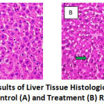 |
Figure 1: The Results of Liver Tissue Histological Examination in Control (A) and Treatment (B) Rats. |
Description
| Figure A (Control) | Figure B (Treatment) |
| § Blue arrow: fat vacuoles infiltrate in hepatocyte cells | § There is no infiltration of fat vacuoles |
| § Green arrow: widening of the sinusoid vessels | § Green arrow> widening of the sinusoid vessels |
Discussion
The results showed that the ethanol extract of Ulva lactuca decreased the level of triglyceride in the treatment group (Table 2). This is in line with previous studies conducted by Kammoun et al. (2017), where a decrease in triglyceride levels after the administration of Ulva lactuca extract for 30 days was reported. The role of vitamin C and E contained in the ethanolic extract was also mentioned. Furthermore, the study by Hassan et al. (2011) showed that administering Ulva lactuca extract for 21 days may reduce triglyceride levels by 66%. 7,8
Alpha-tocopherol in the extract acts as anti-hyperlipidemic on adiponectin (suppresses fatty acid synthesis) and the peroxisome proliferator-activated receptor (PPAR) mechanism. PPAR modifies β-oxidation of free fatty acids and regulates lipogenesis-related genes, as well as triggers a decrease in serum lipid levels. 8,9,19,20 Kim et al. (2013) reported that the administration of α-tocopherol supplements will increase the PPAR-α expression and β-oxidation of fatty acids in the liver, as well as decrease serum triglyceride levels in rats fed with a high-fat diet.9
Polyphenol compounds in the extract also show protective effects against mitochondria and are antisteatotic by increasing the carnitine palmitoyltransferase 1 (CPT1A1) enzyme expression. It plays a role in β-oxidation and decreases the fatty acid synthase complex (FAS) expression. 20 The MDA levels was also decreased in the treatment group and the bioactive compounds acted as antioxidants in vitro and in vivo. 8,22,23,24,25 The activity was measured using the MDA value as a lipid peroxidation marker in vivo. This result is in line with the study conducted by Hassan et al. (2011) where the administration of Ulva extract significantly prevented oxidative damage by minimizing lipid peroxidation. 8 Other studies by Widyaningsih et al. (2014) and Kammoun et al. (2018) showed a similar result, where the polyphenol and flavonoid content significantly reduce MDA levels.10,25
Polyphenol donates hydrogen ions to reduce oxidants, while vitamin E or α-tocopherol acts as an oxidant chain breaker by donating the atoms to its phenolic hydroxyl groups. It can also suppress inflammation by inhibiting the transforming growth factor-beta (TNF-β) expression. In contrast, vitamin C neutralizes reactive oxygen species in the aqueous phase before lipid peroxidation. 6,20
The antioxidant effect and reduction of triglyceride levels by Ulva lactuca also in line with the result of research with positive control by Widyaningsih et.al (2016). 26 In that study, the effect of Ulva lactuca was compared with the drug of choice lipid lowering agent, namely simvastatin (statin) which is known to have anti-oxidative and anti-inflammatory effects. Ulva lactuca (200mg / kgBW) was found to have the same effect (with statins), even better in lowering blood cholesterol levels on the 56th day of administration. This is because Ulva lactuca contains various compounds that work as antioxidants and anti-inflammatory, such as chlorophyll, phytomelatonin, and polyphenols.7,20,26
In addition, it was reported that the ethanol extract of Ulva lactuca prevented the fatty acid accumulation in the rats’ liver tissue. The fat vacuole accumulation occurred in only two of the four samples from the control group examined, and this is because of their age. Due to this reason, they can adapt to the excess fat and fructose consumption. These experimental animals were 12-14 weeks, or 20-22 years old in humans, while the highest prevalence of fatty liver is at the age of 40-65 weeks.27 This study shows the occurrence of early stages of fatty liver with a high-fat and fructose diet, where there is ± 5% steatosis. Therefore, it is included in grade 1 steatosis on the NASH CRN score for the fatty liver.
Similarly, previous studies showed that Ulva extract has an antioxidant and hepatoprotective activity that prevents fat deposition, lipotoxic lipids, and inflammation in the liver. 7,12,23
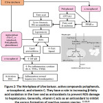 |
Figure 2: The Workplace of Ulva lactuca, Active Compounds: Polyphenols, α-tocopherol, and Vitamin C. |
Conclusion
From the results, it is reasonable to conclude that ethanol extract of Ulva lactuca can prevent higher triglycerides and MDA levels, as well as the fat vacuoles accumulation in male Wistar rats (Rattus norvegicus) when treated with a high fat and fructose diet.
Funding Sources
The authors received no financial support for the research, authorship, and/or publication of this article.
Conflict of Interest
The authors declare no conflict of interest.
References
- Chatrath, H., MD., Vuppalanchi R., MD., Chalasani N., MD. Dyslipidemia in Patients with Non-alcoholic Fatty Liver Disease. NIH Public Access. 2012; 32 (1): 22-29.
CrossRef - Tracer, K.F., dan D. Rozman. Non-alcoholic Fatty Liver Disease: Focus on Lipoprotein and Lipid Deregulation. Journal of Lipid in Hindawi Publishing Corporation. 2011; 1-14.
CrossRef - Tomizawa, M., Y. Kawanabe, F. Shinozaki, S. Sato, Y. Matoyashi, T. Sugiyama, S. Yamamoto, M. Sueishi. Triglyceride is Strongly Associated with Non- alcoholic Fatty Liver Disease among Markers of Hyperlipidemia and Diabetes. Biomedical reports. 2014; 2: 633-636.
CrossRef - Suharjo, B.C., P. Bayupurnama, N. Ratnasari, S. Maduseno, and S. Murdjanah. Ultrasound-Diagnosed Non-Alcoholic Fatty Liver Disease among Medical Checkup Patients. The Indonesian Journal of Gastroenterology, Hepatology and Digestive Endoscopy. 2013; 14 (3), 145-9.
- Sari, G.A.C., H.D. Purnomo and E. Sudijanto. Non-alcoholic Fatty Liver Disease in Adult Metabolic Syndrome: clinical features and the relationship between the number of components of the metabolic syndrome are disturbed and the degree of ultrasound. Jurnal Media Medika Muda. 2012; 1-12.
- Yunita, D., L.P. Wrasiati, & L. Suhendra. Characteristics of Bioactive Compounds Extract of Sea Lettuce (Ulva lactuca L.) on Ethanol Solvent Concentration and Extraction Time. Jurnal Rekayasa dan Manajemen Agroindustri. 2018; 6 (3), 189- 195.
CrossRef - Kammoun, I., H.B. Salah, H.B. Saad, B. Cherif, M. Droguet, C. Magne, C. Kallel, O. Boudawara, A. Hakim, N. Gharsallah, and I.B. Amara. Hypolipidemic and cardioprotective effects of Ulva lactuca ethanolic extract in hypercholesterolemic mice. Archives of Physiology and Biochemistry. 2018; 124 (4): 313-325.
CrossRef - Hassan, S., A. El-Twab, M. Hetta, dan B. Mahmoud. Improvement of Lipid Profile and Antioxidant of Hypercholesterolemic Albino Rats by Polysaccharides Extracted from Green Alga Ulva lactuca Linnaeus. Saudi Journal of Biological Sciences.2011; 18: 333-340.
CrossRef - Kim, Y., J. Kim, H.J. Ham, dan R. Choue. Effects of d-α-tocopherol Supplements on Lipid Metabolism in a High-Fat Diet-Fed Animal Model. Nutrition Research and Practice. 2013; 7 (6): 481-487.
CrossRef - Widyaningsih W., R. Sativa, dan I. Primardiana. Antioxidant Effect of Green Algae Ethanol Extract (Ulva lactuca L.) on Malondialdehyde (MDA) Levels and Superoxide Dismutase (SOD) Enzyme Activity of CCl4-Induced Rats. Media Farmasi. 2015; 12 (2); 163-175.
CrossRef - Jensen, T., M. F. Abdelmalek, S. Sullivan, K. J. Nadeau, M. Green, C. Rocal, T, et al. Fructose and Sugar: A Major Mediator of Nonalcoholic Fatty Liver J Hepatol. 2018; 68 (5): 1063-1075.
CrossRef - Sathivel, A. Balavanayagamani, B. R. H. Rao, dan T. Devaki. Sulated Polysaccharide isolated from Ulva lactuca attenuates D-galactosamine induced DNA fragmentation and Necrosis during Liver Damage in Rats. Pharmaceutical Biology. 2014; 52 (4). 498-505.
CrossRef - Marques, C., M. Meireles, S. Norberto, J. Leite, J. Freitas, D. Pestana, A. Faria, and C. Calhau. High-fat diet-induced obesity Rat model: a comparison between Wistar and Sprague-Dawley Rat. Adipocyte, 2016; 5(1): 11-21.
CrossRef - Giknis, M. L. A., dan C. B. Clifford. Clinical Laboratory Parameters for Crl:Wl (rats). Charles River Laboratories. 2008; 1-14.
- Puri, P. dan A. J. Sanyal. Nonalcoholic Fatty Liver Disease: Definitions, Risk Factors, Workup. Clinical Liver Disease. 2012; 1 (4), 99-103.
CrossRef - Krisnansari, D., H. Sulistyo, V. R. B. Ati. Effect of Propolis on the Liver Function and Fatty Liver Disease of White Rat (Rattus norvegicus) Hypercholesterolemia Model. Penel Gizi Makan. 2014; 37 (1): 77-85.
- Widyaningsih, Wahyu & Salamah, Nina. The Effect of Green Algae (Ulva Lactuca L) Ethanol Extract on Body Weight and Feed Consumption of Male Rats Given a High Fat Diet. Pharmaciana.2015. 10.12928/pharmaciana.v5i2.2438.
CrossRef - Lambertz, J., T. Berger, T.W. Mak, J. Heiden, dan R. Weiskirchen. Lipocalin- 2 in Fructose-Induced Fatty Liver Disease. Frontiers in Physiology. 2017. 8(964): 1-16.
CrossRef - Nan, Y.M., W.J. Wu, N. Fu, B.L. Liang, R.Q. Wang, L.X. Li, S.X. Zhao, J.M Zhao, dan J. Yu. Antioxidants Vitamin E and 1-aminobenzotriazole Prevent Experimental Non-alcoholic Steatohepatitis in Mice. J. Gastroenterol. 2009; 44: 1121-1131.
CrossRef - Perumpail, B.J., A. A. Li, N. John, S. Sallam, N.D. Shah, W. Kwong, G. Cholankeril, D. Kim, dan A. Ahmed. The Role of Vitamin E in the Treatment of NAFLD. Diseases. 2018; 6, 86.
CrossRef - Rafiei, H., K. Omidain, dan B. Bandy. Dietary Polyphenol Protect Against Oleic Acid-Induced Steatosis in an in Vivo Model of NAFLD by Modulating Lipid Metabolism and Improving Mitochondrial Function. Nutrients. 2019; 11: 541.
CrossRef - Mahmud, I., R. Pertiwi, N.R. Azis, dan D.N. Reviana. 2014. Utilization of the Potential of Green algae (Ulva lactuca) as a Natural Antioxidant in the Prevention of Acute Myocardial Infarction. E-Proceeding PIMNAS PKM-P Ditjen Dikti Kmdikbud RI.[Online]PKM-P2014. Available at: http://artikel.dikti.go.id/index.php/PKM-P/issue/view/35 [downloaded: 21 March 2018].
- Kumar, S. A., M. Magnusson, L. C. Ward, N. A. Paul, dan L. Brown. Seaweed Supplements Normalise Metabolic, Cardiovascular and Liver Responses in High-Carbohydrate, High-Fat Fed Rats. Marine Drugs. 2015; 13:788-805.
CrossRef - Sundari, L.P.R., N. Adiputra, dan I.M.K. Dinata. Supplementation of Vitamin E 400 IU Decreases Malondialdehyde Level of Obese Women Staff at School of Medicine Udayana University. Asian Jounal of Pharmaceutical and Clinical Research. 2017; 10 (9): 61-63.
CrossRef - Kammoun, I., H.B Salah, H. B. Saad, B. Cherif, M. Droguet, C. Magne, C. Kallel, O. Boudawara, A. Hakim, N. Gharsallah, dan I.B. Amara. Archives of Physiology and Biochemistry. 2018; 124 (4): 313-325.
CrossRef - Widyaningsih, W., N. Salamah, F.Q. Maulida. The effects of ethanolic extract of green algae (Ulva lactuca l.) on blood cholesterol levels in male rats induced by a high-fat diet. Indonesian Journal of Medicine and Health. 2016; 7(5):181-186.
CrossRef - Chalasani, N., Z. Younossi, J.E. Lavine, M. Charlton, K. Cusi, M. Rinella, S.A, et al. The Diagnosis and Management of Nonalcoholic Fatty Liver Disease: Practice Guidance from the American Association for the Study of Liver Diseases. Hepatology. 2018; 67 (1), 328-357.
CrossRef
Accepted on: 13 Sept 2021
Second Review by: Dina Keumala Sari Indonesia
Final Approval by: Prof. Jiwan S. Sidhu


Web of Science Coverage
Emerging Sources Citation Index (ESCI)
2024 Journal Impact Factor: 1.1
Scopus Journal Metrics
CiteScore 2025: 2.6
CiteScore Details
Sustainable Nutrition: Food Systems, Nutrient Retention, and Public Health Impact
![]()
This journal is a member of, and subscribes to the principles of, the Committee on Publication Ethics (COPE)


