Chlorophyll Determination in Green Pepper Using two Different Extraction Methods
1Dept. of Crop Production Technological Educational Institute of Peloponnese, School of Agricultural Technology, Food Technology and Nutrition
2Department of Food Technology,Antikalamos 24100 Kalamata, Greece.
Corresponding Author Email: theovarzakas@yahoo.gr
DOI : http://dx.doi.org/10.12944/CRNFSJ.4.Special-Issue1.05
Download this article as:
![]()
The aim of this paper is the comparison of the classic Arnon method with the DMSO method regarding the determination of chlorophyll content in California Wonder peppers stored for 25 days at 5, 10 and 20°C. The results suggest that the factors affecting chlorophyll degradation are temperature and storage time, as well as their interaction. There is a linear relationship between changes in chlorophyll content and storage temperature. The statistical analysis indicates that the two chlorophyll extraction methods considerably differ, as the chlorophyll contents obtained through the Arnon method were, in all cases, lower than those obtained through DMSO. It has to be stressed that the results greatly depend on both the selected solvent and the chlorophyll extraction method.
KEYWORDS:Chlorophyll; DMSO; Arnon method; Pepper
Introduction
Color plays a key role in the commercial value of plant organs and along with texture determine the freshness of most vegetables.1 The perception of color is due to the presence of pigments. Fruits and vegetables are rich in pigments and those attract consumers. Plant pigments can be divided into categories according to their chemical composition. Hence, chlorophyll, carotenoids, anthocyanins and flavonoids can be distinguished.
Chlorophyll is a natural dye derived from green plants and algae. Absorbs energy from the sun that is used to synthesize carbohydrates from CO2 via photosynthesis.2 In the chloroplasts of higher plants two kinds of chlorophyll are found, chlorophyll a (blue-green) and chlorophyll b (yellow-green), which differ in the substituent of a pyrrole ring II. Besides its photosynthetic role it has other applications and is used as a natural pigment in food and cosmetics.3 The chlorophyll degradation during curing, processing or during aging of plant tissues results in a colour change. The factors that affect the degradation of chlorophyll are: water stress, light, temperature, ethylene or their combination.4,5 In the case of pepper6,7 Minguez-Mosquera and Méndoz (1994 a, b) reported that following harvest carotenogenesis was observed which was affected by light and temperature. Hence, ripe fruits are rich in carotenoids which affects their color.
Cantwell et al. (1998),8 report that temperature is one of the major factors that determine the postharvest quality of the green vegetables. High temperature of preservation accelerates deterioration and reduces storage time whereas low temperature increases shelf life of most fresh vegetables9 delaying degradation of chlorophyll.10 The reduction of the intensity of green colour in vegetables is associated with aging, the reduction in the nutritional value and in general their quality.9
Pepper is one of the most popular flavors and is used internationally due to its characteristic spicy flavor and the aroma characteristic. It is a rich source of vitamin A and C, antioxidants, zeaxanthin, components that play an important role in the human diet.11
The purpose of this study was to compare the classical method of Arnon with the DMSO method in the determination of chlorophyll in green pepper fruits preserved at 0,10 and 20° C since few studies on the comparison of these two methods have been conducted.12
Materials and Methods
Raw material
California Wonder peppers were cut at maturity level from a farm at Plati Messinia. Following harvest, peppers were sorted according to size and colour and then divided into three groups each one of which was stored under a different temperature. Storage conditions of peppers were as follows. 5, 10 and 20°C, relative humidity 90%, and storage period of 25 days.
Chlorophyll determination
A wide range of solvents has been proposed for chlorophyll determination13 (Castle et al., 2011). Acetone and DMSO are two popular and characteristic methods used widely in ecophysiological research.12 For this reason in this study for chlorophyll determination two methods were employed i.e. the classical method of Αrnon, (1949)14 and DMSO method.15,16,17,18
Αrnon method
Chlorophyll extraction was carried out with a mixture of acetone and water at a ratio of 80% -20% (v/v). 2 g of pepper tissue was homogenized with 25 ml acetone solution 80%, using a laboratory blender (BLENDER 8010E, MODEL 38BL40) for 2 min. Filtration was followed (MN G15¼ 125mm), and the filtrate transferred to a volumetric flask of 100 ml covered with aluminum foil to avoid oxidation of chlorophyll from light and filled to the top with acetone solution 80%. Absorption was measured at 663 and 645 nm using a HITACHI spectrophotometer U-2000.
Concentration of chlorophyll (a, b, total) was expressed as mg /g fresh weight. Determination of chlorophyll a, b and total was carried out using the formula by Arnon:14
Chlorophyll a (mg/g F.W) = (12.7 A663 -2.69 A645) x X/1000x n (1)
Chlorophyll b (mg/g F.W) = (22.9 A645 – 4.68 A663) x X/1000x n (2)
Total chlorophyll (mg/g F.W) = (20.2 A645 + 8.02 A663) x X/1000x n (3)
where: A645 = absorption value at 645 nm, , A663= absorption value at 663 nm, Χ = total volume of filtrate, n = tissue weight.
DMSO method
Extraction has been carried out with DMSO solvent which has amphiphilic properties.19 0.1 g of pepper tissue were cut in smaller pieces and placed in test tubes containing 10 ml of solvent. Test tubes were incubated in a water bath at 60-65ºC for an hour. From preliminary studies this time was judged satisfactory for the full decolorisation of tissues. Cooling at room temperature was followed for 30 min, filtration and absorption measured at 665 nm and 648 nm being the final stages. Blank determination was carried out with DMSO. Absorption measurement was carried out with a HITACHI Spectrophotometer U-2000.
Chlorophyll concentration (a, b and total) was expressed as mg /g fresh weight and determined by the following formulae16 (Barnes et al., 1992).
Chlorophyll a (mg/g F.W) = (14.85 A665 -5.14 A648) (1)
Chlorophyll b (mg/g F.W) = (25.48 A665 – 7.36 A648) (2)
Total chlorophyll (mg/g F.W) = (7.49 A665 + 20.34 A648) (3)
where: A665 = absorption value at 665 nm, A648= absorption value at 648 nm.
Chlorophyll determination was carried out at 0, 3rd, 6th,10th,13th, 17th, 20th and 25th day in 6 samples per temperature.
Results were statistically analysed by statistical package Statgraphics Plus (5.1). Comparison of the means was carried out with the (LSD) at confidence level of p= 0, 05.
Results and Discussion
Chlorophyll determination with Αrnon method
The concentration of pepper tissue in chlorophyll a, b and total during storage at 5, 10 and 20° C is shown in Figure 1. From Figure 1 it can be shown that at the end of storage at 5°C, the total chlorophyll shows an increase of 50%, which can be attributed to damage caused by the low storage temperature (cold injury) in tissues. At 10° C the total chlorophyll shows little variation (12%) and kept almost to baseline levels by the end of storage, whereas at 20°C, after the 15th day, the total chlorophyll shows a reduction which at the end of storage (day 25) reaches 62%. Chlorophyll a shows greater rates compared to chlorophyll b. Based on the literature20 chlorophyll a is the predominant pigment and chlorophyll b is complementary, and their ratio is 3: 1. In this study the ratio of both chlorophylls is of the order of 2.25 / 1. Deviations from the ratio of 1.3 have been observed in other plant tissues, such as spinach, where the ratio is 4.02 / 1,21 and okra wherein the ratio is 1.2 /1.20 The ratio of chlorophyll a / chlorophyll b varies depending on the type of fruit, the variety, environmental conditions (including agronomic practices) and growth stage.22 During storage the ratio changes because chlorophyll a decomposes faster than chlorophyll b21 which agrees with our results because at the end of storage the ratio chlorophyll a / chlorophyll b at 5°C is 1,1/1, at 10°C 1,4/1 and at 20 °C 0,6/1.
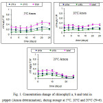 |
Figure 1: Concentration change of chlorophyll a, b and total in pepper (Αrnon determination), during storage at 5°C, 10°C and 20°C (Ν=6). |
Chlorophyll determination with DMSO method.
Concentration in chlorophyll a, b and total in pepper during storage at 5, 10 and 20 °C, as determined by DMSO method is presented in Figure 2. From this figure it is evident that at the end of storage (25th day), total chlorophyll remained very close to the initial levels of 5°C and 10°C (reduction of 0.8% and 13%, respectively) whereas at 20°C the reduction was more intense and came up to 27%.
The ratio of chlorophyll a to chlorophyll b was 3.2/1 which agrees with the literature20 whereas at the end of storage this ratio changed and reached to 1,6/1 at 5°C, 2,3/1 at 10°C and 2,6/1 at 20°C.
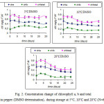 |
Figure 2: Concentration change of chlorophyll a, b and total in pepper (DMSO determination), during storage at 5°C, 10°C and 20°C (Ν=6). |
Effect of temperature on chlorophyll change
To study the effect of temperature on chlorophyll change, the change of total chlorophyll at temperatures of 5, 10 και 20°C will be presented as determined by Arnon and DMSO methods.
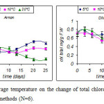 |
Figure 3: Effect of storage temperature on the change of total chlorophyll as determined by Arnon and DMSO methods (Ν=6). Click here to View figure |
Statistical analysis (ANOVA) showed that the factors affecting the degradation of chlorophyll are: storage temperature, storage life and their interaction (p = 0.05).
From Figure 3 it appears that in the case of the determination of chlorophyll by Arnon method no statistically significant difference between the three storage temperatures was observed until day 13, after which a marked decrease is observed at 20° C. At the end of storage (day 25) the higher total chlorophyll content was shown by the fruits stored at 5° C, the intermediate by fruits stored at 10° C and the smallest by fruits stored at 20° C. The difference is statistically significant (p = 0.05).
In the case of chlorophyll determination by the DMSO method, up to day 10 no statistically significant difference was observed between the three storage temperatures, but then the fruits stored at 5° C showed higher rates (statistically significant) compared to fruits stored at 10 and 20 ° C which showed no significant difference between them until the end of storage (day 25).
In order to show the change in total chlorophyll as an effect of temperature storage the change of chl / chlo factor at 3 temperatures was used in both assays.
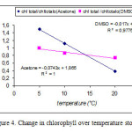 |
Figure 4: Change in chlorophyll over temperature storage |
Figure 4 shows that chlorophyll change is linearly correlated with temperature with a correlation coefficient R2= 1 in the case of acetone and 0.98 in the case of DMSO.
It is known that the temperature plays an important role in the degradation of chlorophyll. Generally, the high temperature stimulates the enzymatic degradation whereas the low temperature delays it.5 The characteristic green color of immature fruit is due to the presence of chlorophylls and carotenoids.23 With the onset of ripening many complex biochemical changes occur which include the change of the composition of colouring agents and the color change. During ripening the chloroplasts are transformed into chromoplasts containing only carotenoids. In the case of non-climacteric fruits such as pepper, the degradation of chlorophyll biosynthesis of carotenoids in general is very slow.24 When chlorophyll is in the chloroplast it is a component with moderate stability. But when chloroplasts are disorganized chlorophyll is very unstable and prone to a wide range of structural changes caused by factors such as temperature, acidic or basic environment, the activity of the enzymes, the molecular O2, light and other post harvest operations.23
Acceleration of ripening of climacteric fruit or degreening non climacteric fruit is accelerated by the temperature and the action of ethylene. The temperature range to be used for degreening depends on the species and variety. The optimum temperature for the biosynthesis of carotenoids depends on the type of carotenoid and the type of fruit and usually ranges between 15-25° C.
Comparison of the two methods.
For comparison of the two methods of chlorophyll determination the change in total chlorophyll will be determined per temperature and method.
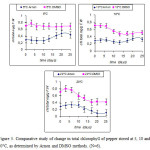 |
Figure 5: Comparative study of change in total chlorophyll of pepper stored at 5, 10 and 20°C, as determined by Arnon and DMSO methods. (Ν=6). Click here to View figure |
Statistical analysis (ANOVA) showed that there is a statistically significant difference (p = 0.05) between the two methods for chlorophyll determination. From Figure 5 we can conclude that at all storage temperatures of pepper, determination of chlorophyll in pepper using the DMSO method showed higher values, whereas the Arnon procedure gave smaller values which are consistent with other researchers.25,12,26 Table 1 shows the relationship between the assay methods of chlorophyll determination at both the start of storage and the end of storage per temperature.
Table 1: Relation of total chlorophyll between two methods of determination.
| Methods | Start of storage |
End of storage |
||
|
5°C |
10°C |
20°C |
||
| DMSO/Arnon |
2,41 |
1,6 |
1,6 |
3,76 |
Table 1 shows that at the start of storage chlorophyll determined by the Arnon method was 2.4 times lower than that determined by the method DMSO. At the end of storage at 5° C and 10° C the ratio was 1.6 and at 20°C 3,76. According to Porra (2002)27 and Wright et al., (1997)28 DMSO was more effective than acetone at low dye concentrations, as in this case at 20°C due to degradation of chlorophyll. The chlorophyll is insoluble in water but soluble in organic solvents. It follows clearly that there is a significant effect of the solvent and the method used to extract chlorophyll. According to Leung (1998),29 Makeen et al., (2007),30 breaking the tissue, filtration and the use of large amounts of solvent (Arnon method) results in low yield of dye compared to other methods, in this case with the use of the solvent DMSO.
The method of Arnon is widely used by many researchers despite the fact that it has some errors, and the mixture of solvents used has disadvantages.31 The main problem is that the mixture of acetone and water (80% v / v) may account for the incomplete extraction of chlorophyll, and that we can not determine the amount of the acetone evaporated at the breakage of tissues, centrifugation, filtration and measurement with the spectrophotometer.
The changes of the concentration of acetone to water is an important error factor as the specific absorption coefficient of chlorophyll a and b varies with the acetone content (e.g. 79% of this is slightly different to that of 80%).31 According to Sumanta et al., (2014),32 acetone is not the ideal solvent for the determination of chlorophyll, because besides the small yield in chlorophyll determination, it is volatile, flammable, irritating the skin and affecting the plastic wells used for spectroscopic measurement. The use of alternative solvents for the spectrophotometric determination of pigments has been requested for various reasons such as the precise determination of the rates of calculation of equations for the different behavior of the various types of plant tissues at different extraction agents. Thus, there are extraction means (e.g. 80% acetone) which are ineffective, others which are effective but require wetting, breakage of the tissue, clarification, centrifugation and generally large preparation time and others only require soaking and shaking to extract the pigments.31
DMSO is a solvent with amphiphilic properties19 which has been used successfully for the extraction of chlorophyll of algae,33,34 lichen16,35 and leaves of higher plants.15,16,17,18 It penetrates the membranes and denatures the protein by replacing or displacing the water around them. It is considered superior to acetone for determination of chlorophyll in higher plants35 (Ronen and Galun, 1984). The method is simple, the extraction is complete and is based on the immersion of tissue fragments in a given quantity of DMSO and incubation at a temperature of 60-65° C. After incubation the solvent is drained and the absorbance is measured at appropriate wavelengths which absorb in the photosynthetic pigments. Advantageous over other methods (methanol, acetone, ethanol) in the fact that the extraction of chlorophyll is easy and fast since no grinding required and centrifugation or filtration.36 For this reason it is suitable for application even in field conditions17 and allows for the preparation and analysis of large numbers of samples in a short time.
The stability of the chlorophyll extracted by DMSO during storage is better than that of the acetone method.15,16 The drawback that the method shows is that the extraction of the pigments is based on diffusion of DMSO in the photosynthetic tissues, and not mechanical disruption of the cells. Therefore, the incubation time is not constant but depends on the particular anatomical and morphological characteristics of the tissues of each plant species.15,16 This practically means that the samples are incubated in DMSO until completely discolored tissues turn up. In some cases, the complete extraction of chlorophyll with DMSO requires many hours of incubation.37 It should be noted that there may be health problems at routine analysis with DMSO which need to be taken into account.
Conclusions
The procedure of chlorophyll extraction from plant tissues is quite composite. We can conclude from our experimental findings that chlorophyll concentration in tissues has been affected by the storage temperature. The extraction quantity of pigment has been affected significantly by the method of determination. DMSO method was capable to extract a higher percentage of chlorophyll compared to the classical method, and was fast and did not require high quantities of solvent.
References
- Kader, A., 2002. Maturation and maturity index. Postharvest Technology of Horticultural crops. (ed. A. Kader), University of California Agr and Natural Resources Pub.3311 pp. 55-62.
- Raven, P.H., Evert, R.F., Eichhorn, S.E., 2005. In: Photosynthesis, Light, and Life. Biology of Plants, seventh ed. W.H. Freeman, San Francisco, CA, pp. 119–127.
- Humphery, A.M., 2004. Chlorophyll as a color and functional ingredient. J. Food Sci. 69, C422–C425.
CrossRef - Heaton, W.J and Marangoni, G.A., 1996. Chlorophyll degradation in processed foods and senescent plant tissues. Trends in Food Science and Technology, V.7: 8-15.
CrossRef - Yang, X., Zhang, Z., Joyce, D., Huang,X., Xu, L., Pang, X., 2009. Characterization of chlorophyll degradation in banana and plantain during ripening at high temperature. Food Chemistry, 114:383-390.
CrossRef - Minguez-Mosquera MI and Hornero-Mendez D., 1994a. Formation and transformation of pigments during the fruit ripening of Capsicum annuum cv. Bola and Agridulce,J. Agric. Food Chem., 42, 38–44.
CrossRef - Minguez-Mosquera MI and Hornero-Mendez D., 1994b. Changes in carotenoid esterification during the fruit ripening of Capsicum annuum cv. Bola. J. Agric. Food Chem., 42, 640–644.
CrossRef - Cantwell, M., J. Rovelo, X. Nie and V. Rubatzky, 1998. Specialty salad greens:Postharvest physiology and shelf-life. Acta Hort., 467:371-377.
CrossRef - Cantwell, .M and Kasmire, F.R., 2002. Postharvest handling systems: flower, leafy and stem vegetables. In: Postharvest Technology of Horticultural crops. (ed. A. Kader), University of California Agr and Natural Resources Pub.3311,pp.423-433.
- Pogson, B.J., and Morris, S.C., 1997. Consequences of cool storage of broccoli on physiological and biochemical changes and subsequent senescence at 20°C. J. Amer. Soc. Hort. Sci. 122:553-558.
- Raffo, A., BaimonteI. & Paoletti F., 2008. Changes in antioxidants and taste related compounds content during cold storage of fresh–cut red sweet peppers. European Food Research Technology 226(5), 1167–1174.
CrossRef - Wang, Wen-Jie, He Hai-Sheng, Guan, Yu Li, Wen-Xin Zhang, Zhong-Hua Zu, Yuan-Gang, 2009. Methodological Comparison of Chlorophyll and Carotenoids Contents of Plant Species Measured by DMSO and Acetone-extraction Methods Bulletin of Botanical research V29 (2) www.cnki.com.cn.
- Castle, S. C., Conor D. Morrison, Nichole N. Barger, 2011. Extraction of chlorophyll a from biological soil crusts: A comparison of solvents for spectrophotometric determination. Soil Biology & Biochemistry, 43,853-856.
CrossRef - Arnon, D. 1949. Copper enzymes in isolated chloroplasts polyphenoloxidase in Beta Vulgaris. Plant Physiology, V.24 (1), pp. 1-15.
CrossRef - Hiscox JD, Israelstam GF, 1979. A method for the extraction of chlorophyll from leaf tissue without maceration. Can. J. Bot.57:1332-1334.
CrossRef - Barnes JD, Balaguer L, Manrique E, Elvira S, Davison AW, 1992. A reappraisal of the use of DMSO for the extraction and determination of chlorophylls a and b in lichens and higher plants. Envir. Exp. Bot. 32:85-100.
CrossRef - Tait MA, Hik DS., 2003. Is dimethylsylfoxide a reliable solvent for extracting chlorophyll under field conditions? Photos. Res. 78:87-91.
CrossRef - Richardson AD, Duigan SP, Berlyn GP., 2002. An evaluation of noninvasive methods to estimate foliar chlorophyll content. New Phytol.153: 185-194.
CrossRef - Notman R, Noro M, Ο;Malley B, Anwar J, 2006. Molecular basis for dimethylsulfoxide (DMSO) action on lipid membranes. J. Am. Chem. Soc.128: 13982-13983.
- Gross J, 1991. Pigments in Vegetables, Chlorophylls and Carotenoids, Van Nostrand Reinhold, New York.
CrossRef - Goodwin, T.W., 1965. Chemistry and Biochemistry of plant pigments. Academic Press, New York .Gross J,1987. Chlorophylls. In: Pigments in fruits. Academic Press Inc. London, UK: 1-55.
- Gross J, 1987. Chlorophylls. In: Pigments in fruits. Academic Press Inc. London, UK: 1-55.
- Artés,F., Mínguez, M.I and Hornero, D., 2002. Analysing changes in fruit pigments. In: Colour in Food, Improving quality, McDougall (ed), CRC Press, Boca Raton, Boston, New YorkWashington, pp. 249-282.
CrossRef - Eaks, I.L., 1977. Physiology of degreening-summary and discussion of related topics. Proc. Int. Soc. Citriculture, 1:223-226.
- Seo, K.Y., Yi, M.J., Son, Y.H., Koo, J.W., Noh, N.J., 2005. Extraction and Determination of Chlorophyll Contents of Korean Pine Needles Using Acetone and DMSO (Dimethylsulfoxide). Journal of Korea Forest Society V94(4) agris.fao.org/aos/records/KR2006013490 (accessed 20-10-2015).
- Lan, S., Li Wu, Delu Zhang, Chunxiang Hu, Yongding Liu, 2011. Ethanol outperforms multiple solvents in the extraction of chlorophyll-a from biological soil crusts. Soil Biology & Biochemistry, 43, 857-861.
CrossRef - Porra, R. J., 2002 The chequered history of the development and use of simultaneous equations for the accurate determination of chlorophylls a and b, Photosynth. Res., 73,149–156.
CrossRef - Wright S. W., Jeffrey S. W. and Mantoura F. R. C., 1997.Evaluation of methods and solvents for pigment analysis. In: Phytoplankton pigments in oceanography: guidelines to modern methods, UNESCO Publ., Paris, 261–282.
- Leung, A.Y., 1998. Methods and techniques for study of biochemistry of crop plants. J. Amer. Oil Chem. Soc., 50:407-410.
- Makeen, K., Babu, G.S., Lavanya, G.R., Abraham, G., 2007. Studies of chlorophyll content by different methods in black gram. Intern. J. Agric. Res. 2(7):651-654.
CrossRef - Wellburn, A.R., 1994. The spectral determination of chlorophylls a and b, as well as total carotenoids, using various solvents with spectophotometers of different resolution. J. Plant Physiol. V.144: 307-313.
CrossRef - Sumanta Nayek, Choudhury Imranul Haque, Jaishee Nishika and Roy Suprakash, 2014. Spectrophotometric Analysis of Chlorophylls and Carotenoids from Commonly Grown Fern Species by Using Various Extracting Solvents. Research Journal of Chemical Sciences, V4(9),63-69.
- Burnison BK, 1980. Modified dimethylsulfoxide (DMSO) extraction for chlorophyll analysis of phytoplankton. Can . J. Fish. Aquat. Sci.37:729-732.
CrossRef - Shoaf WT, Lium BW., 1976. Improved extraction of chlorophyll a and b from algae using dimethylsulfoxide. Limnol. Oceanog.21:926-928.
CrossRef - Ronen R, Galun M., 1984. Pigment extraction from lichens with dimethylsulfoxide (DMSO) and estimation of chlorophyll degradation. Envir. Exp. Bot. 24:239-245.
CrossRef - Devesa R., Moldes A., Diaz-Fierros F., Barral M.T., 2007. Extraction study of algal pigments in river bed sediments by applying factorial designs. Talanta 72:1546-1551.
CrossRef - Shinano T, Lai TT, Kawamukai T, Inoue MT, Koike T, Tadano T., 1996. Dimethylsulfoxide method for the extraction of chlorophylls a and b from the leaves of wheat, field bean, dwarf bamboo and oak. Photosynthetica 32:409-415.


Web of Science Coverage
Emerging Sources Citation Index (ESCI)
2024 Journal Impact Factor: 1.1
Scopus Journal Metrics
CiteScore 2025: 2.6
CiteScore Details
Sustainable Nutrition: Food Systems, Nutrient Retention, and Public Health Impact
![]()
This journal is a member of, and subscribes to the principles of, the Committee on Publication Ethics (COPE)


