
Introduction
In the past decades, non-communicable diseases (NCD), such as diabetes mellitus, hyperuricemia, and obesity have become serious concern, due to their various serious complications and the risks for cardiovascular diseases. The prevalence of diabetes mellitus, hyperuricemia, and obesity were projected to increase worldwide in the next decades.1 The growing prevalence in metabolic diseases indicates that other alternative approach is required in conjunction with current management of these diseases, such as the use of key enzyme inhibitors. In addition, some of the drugs currently available in the market have been known to cause undesirable side effects.2, 3
Enzyme inhibition has become important alternative therapeutic strategy in designing new drugs.4 Even though enzymes are crucial for numerous biochemical processes in the body system, excessive enzymes activities may lead to over production and accumulation of metabolites that induce pathologies, such as those in metabolic syndrome. Therefore, enzyme inhibition is essential for the prevention and treatment of metabolic disorders, such as diabetes mellitus, hyperuricemia, and obesity.
Epidemiology studies have shown positive correlation between high consumption of fruits and vegetables and reduced risk of metabolic syndrome.5, 6 Such health properties of fruits and vegetables may partially be attributed to various important micronutrients, such as vitamin C, vitamin E, flavonoids, phenolics, all of which are crucial in maintaining human health. Screening for new drugs or supplements to help combat metabolic disorders has become one of the major research interests. Plants are potential source for screening natural enzyme inhibitors for metabolic related diseases, such as α-glucosidase and α-amylase,7 xanthine oxidase,8 lipase,9 and protease.10 One of the plants that has gained growing interest recently is Muntingia calabura.
Muntingia calabura is the only species in the genus Muntingia (family Elaeocarpaceae). It is widely distributed in the tropical and subtropical areas, including India, South and Central America (where it is known as Jamaican cherry) and Southeast Asia, such as Indonesia (local name: Kersen or Talok) and Malaysia (Kerukup Siam). The trees can grow up to 7 to 12 meters, with horizontal spreading of branches that make them ideal for shade trees. The fruit is spherical, around 1.0 cm in diameter. The ripe fruit is red, sweet and juicy, containing numerous tiny yellowish seeds. However, the fruit is not yet promoted as marketable fruit.
M.calabura is used in folk medicine to treat a number of illnesses, such as headache and gastric disorders.11 Different parts of M.calabura have been studied and found to have pharmacological properties. Most studies reported bioactivities of the leaves and stem bark, such as antibacterial,12 antioxidant,13 antinociceptive,14 gastroprotective,15 antidiabetic,16 and cardioprotective17 activities. However, limited studies were reported on the medicinal properties of the fruit, such as antioxidant,13, 18 and anti-inflammatory19 activities. In domestic context, the juice is extracted from the fruit and the fruit pulp is generally discarded.
This study was conducted to investigate the potential inhibition activity of both the fruit juice and extracts of the fruit pulp of M.calabura on enzymes (α-glucosidase, α-amylase, xanthine oxidase, lipase, and protease) involved in some metabolic diseases. The antioxidant activity of the juice and extract was also investigated.
Materials and Methods
Chemicals
All solvents and chemicals used in the experiments were of analytical grade. Folin & Ciocalteu’s phenol reagent, α-glucosidase from Saccharomyces cerevisiae (EC 3.2.1.20), p-nitrophenyl-α-D-glucopyranoside, α-amylase from porcine pancreas (EC 3.2.1.1), xanthine oxidase from bovine milk (EC1.17.3.2.), acarbose, allopurinol, azocasein, 2,2-diphenyl-1-picryl-hydrazyl (DPPH), and 3,5-di-tert-butyl-4- hydoxytoluene (BHT), protease from Streptomyces griceus (EC 3.4.23.6), orlistat, and sodium diclofenac were purchased from Sigma-Aldrich (St. Louis, USA). Gallic acid was obtained from Santa Cruz Biotechnology (Dallas, USA). Sodium carbonate (Na2CO3) and starch soluable were purchased from Merck (Darmstadt, Germany). All remaining reagents were of the highest purity available (>98%).
Spectrophotometer measurements were carried out using a Biochrom Libra-S22 (Cambridge, UK).
Plant Materials
Muntingia calabura fruits were collected from Sentani district, Jayapura, Papua province, Indonesia in August 2018.
Preparation of Fruit Juice (FJ) and Crude Extract of Fruit Pulp (FP)
After washing the fruits thoroughly, they were crushed using food extractor. The fruit juice of M. calabura (FJ) obtained from the extractor was used directly for analysis. FJ stock solution was prepared by dissolving FJ in dimethylsulfoxide (DMSO). The fruit pulp of M. calabura (FP) left in the juice extractor which consisted of seeds, peels and fibre were freeze-dried (Biobase Bk-Fd10P, Shandong, China). The dried pulp was macerated with ethanol for 3 days at room temperature and the filtrate was evaporated under reduced pressure (Buchi R3, Flawil, Switzerland). The stock solution of dried FP was prepared by dissolving the sample in DMSO. All stock solutions were stored in the dark at -20 oC prior to analysis.
Total Phenolic Content Estimation Assay
The total phenolic content of FJ and FP of M.calabura was estimated using a Folin-Ciocalteu method with slight modification.20 In brief, sample (0.5 mL) was mixed with Folin-Ciocalteu reagent (2.5 mL, 10%, v/v in water) and left to stand for 10 mins. Thereafter, Na2CO3 (2.5 mL, 75 g/L) was added and the reaction mixture was incubated for 2 hours at ambient temperature. The absorbance was measured using a spectrophotometer at 765 nm. Gallic acid (13 – 200 µg/mL) was used to generate a calibration curve (R2 = 0.9976, y = 0.008x + 0.034). Results were expressed as mgGAE/mL.
α-Glucosidase Inhibition Assay
The inhibition activity of FJ and FP on α-glucosidase was determined based on our reported method 21. The method was based on the release of p-nitrophenol resulting from catalytic hydrolysis of the substrate p-nitrophenyl-α-D-glucopyranoside by α-glucosidase which was monitored spectrophotometrically at 405 nm. Different concentrations of the samples were mixed with α-glucosidase and p-nitrophenyl-α-D-glucopyranoside as a substrate. Briefly, into each diluted sample of different concentrations (50 µL), phosphate buffer (50 µL, 0.05 M pH 6.8) and α-glucosidase (50 µL, 0.5 U/mL) were added. Concentration range was selected such that 50% inhibition would be included in the range. The mixture was preincubated for 5 mins at 37±0.5 °C. Thereafter, p-nitrophenyl-α-D-glucopyranoside (100 µL, 1 mM) was added to initiate the reaction and the reaction was allowed to proceed for 20 mins at 37±0.5 °C. A solution of Na2CO3 (750 µL, 0.1 M) was added to stop the reaction and the absorbance was read at 405 nm using a spectrophotometer. The inhibition percentage was calculated based on: (Ac – As)/Ac x 100, where Ac was absorbance of control solution and As was absorbance of sample. Acarbose was used as a positive control. The inhibition activity was calculated as IC50 values, which is the concentration of the extract causing 50% of enzyme inhibition. The IC50 values were compared with that of acarbose.
α-Amylase Inhibition Assay
The inhibition activity of FJ and FP on α-amylase was determined using a reported method.22 Different concentrations of the samples were prepared, each was mixed with porcine pancreatic α-amylase and starch as a substrate. Concentration range was determined in which 50% enzyme inhibition would be included in the range. In brief, a mixture of sample (100 µL), 100 µL phosphate buffer (0.02 M, pH 6.9, ionic strength NaCl 6 mM), and α-amylase (100 µL, 0.5 mg/mL) was preincubated for 5 mins at 37±0.5 °C. Into this mixture, starch (200 µL, 1 % w/v) was added to start the reaction. The mixture was incubated for 10 mins at 37±0.5 °C, thereafter DNS reagent (500 µL) was added to stop the reaction. DNS reagent consisted of 3,5-dinitrosalicylic acid (1%, w/v), phenol (1.1%, w/v), Rochelle salt (K-Na-tartarate) 22% w/v, and NaOH (1.5%, w/v) in aqueous solution. The reaction mixture was incubated in a boiled water bath for 5 mins. Afterwards, the mixture was diluted by the addition of water (14 mL). The absorbance was read at 540 nm. Acarbose was used as a positive control. The inhibition activity was expressed as IC50 values and com
Xanthine Oxidase Inhibition Assay
The inhibition activity of FJ and FP of M.calabura on xanthine oxidase was determined based a reported method23 with slight modification.24 Sample (100 µL) was added with xanthine oxidase from bovine milk (100 µL, 0.2 U/mL) and phosphate buffer (100 µL, 0.05 M pH 7.4). The mixture was incubated for 5 mins at 37±0.5 °C. Xanthine (200 µL, 0.3 mM) was added to start the reaction, and the reaction mixture was further incubated for 30 mins at 37±0.5 °C. A solution of HCl (200 µL, 0.1 M) was added to stop the reaction and the absorbance was read at 290 nm. The absorbance was corrected against blank sample solution containing all components except for the enzyme which was replaced by phosphate buffer. Allopurinol (2.75 – 6.87 µg/mL), which is a standard inhibitor for xanthine oxidase, was used to generate a calibration curve (y = – 0.0142x + 0.9365, R2 = 0.8851). The inhibition activity of FJ and FP was presented as standard compound equivalent25 of mg allopurinol equivalent/mL as a unit.
Lipase Inhibition Assay
The inhibition activity of FJ and FP of M.calabura on lipase was assessed based on a qualitative method from previously reported method26 with some modifications. A phenol red agar was prepared consisting of agar (2%, w/v), olive oil (1%, v/v) and phenol red as an indicator (0.01%, v/v). A circular well was made in each quadrant of the plate. Extracts or orlistat (reference inhibitor) was mixed with pancreatic porcine lipase (200 U/mL in 0.05 M tris buffer pH 8 and NaCl 0.03 M) in a 1:1 ratio. A 50 uL of this mixture was suspended into each well and the reaction was left to proceed for 10 min at 37±0.5 °C. Catalytic degradation of olive oil releases fatty acid which changes colour of the indicator from yellow to red. Lipase inhibition activity was observed based on the decrease in zone diameter in the presence of lipase inhibitors. Blank well containing only olive oil was also tested.
Protease Inhibition Assay
The inhibition activity of FJ and FP of M.calabura on protease was determined based on a method described previously 27, 28 with some modifications. Azocasein was used as a substrate. Extracts of different concentrations (50 µL) was well-mixed with 50 μL protease (0.08 U/mL) and 100 uL azocasein (5 mg/mL). The concentrations were selected to include 50% enzyme inhibition. The reaction mixture was incubated for 90 mins at 37±0.5 °C. Afterwards, 400 μL trichloroacetic acid (TCA) (5% v/v, water) was added. After centrifugation at 15,000 rpm for 10 mins, the supernatant was retained and added with 700 μL NaOH (0.56 M). Absorbance was measured at 442 nm and the inhibition percentage was calculated using the following formula: (Ac – As)/Ac x 100, where Ac was absorbance of control solution and As was absorbance of sample. Anti protease activity was reported as IC50 and activities were compared with that of Na-diclofenac. .
2,2-Diphenyl-1-Picryl-Hydrazyl (DPPH) Radical Scavenging Assay
The antioxidant activity of FJ and FP was evaluated based on their scavenging activity on DPPH radicals, according to the method described previously29 with slight modifications.30 Briefly, into each diluted sample of different concentrations (3 mL) was added DPPH solution in ethanol (1 mL, 0.6 mM). Sample concentrations were selected to include 50% inhibition. The reaction mixture was protected from light and incubated for 30 mins at room temperature. The absorbance was read at 517 nm. Results were expressed as IC50 values. BHT and ascorbic acid was used as reference antioxidants.
Statistical Analysis
Experiments were performed in triplicates. The results were expressed as mean ± standard deviation. Students’ t-test and one-way ANOVA with Tukey test (SPSS software version 23 for Windows) were used to analyse the difference, and P < 0.05 was considered to be statistically significant.
Results
Estimation of Phenolic Compounds
The total phenolic content in FJ (2.34 ± 0.19 mgGAE/mL) was significantly higher (p=0.045) than in FP (0.27 ± 0.01 mgGAE/mL). The juice contained higher content of phenolics, more than 8 times the amount in FP.
In vitro Antidiabetic Activity
Antidiabetic activity of FJ and FP was evaluated by two different in vitro procedures; the α-glucosidase and α-amylase inhibition assays. α-Glucosidase catalyses hydrolysis of the α (1-4)-glycosidic links in polysaccharide or oligosaccharide liberating glucose molecules. As can be seen on Table 1, FJ and FP were able to inhibit α-glucosidase in a concentration dependent manner. FP exhibited a stronger inhibition than FJ (P = 0.001), with IC50 value almost twice lower than FJ. It is worth noting that FP showed a more potent inhibition activity on α-glucosidase compared to acarbose.
Table 1: α-Glucosidase inhibitory activity of the juice and pulp of M. calabura fruit and acarbose.
| Concentration(μg/mL) | Inhibition(%) | IC50(μg/mL) | |
| FJ | 73.75147.50221.25295.00 | 9.84 ± 1.1159.09 ± 8.7576.21 ± 11.7585.25 ± 0.98 | 162.00 ± 9.46 |
| FP | 13.7534.3855.00137.50 | 11.62 ± 10.1220.27 ± 0.5536.80 ± 1.1377.94 ± 0.88 | 84.70 ± 2.70 |
| Acarbose | 13.0026.0052.00104.00 | 7.64 ± 2.6619.55 ± 3.0030.18 ± 2.7750.15 ± 2.43 | 130.66 ± 5.94 |
Similarly, FJ and FP exhibited significant α-amylase inhibition which were comparable with the standard acarbose. Table 2 shows that for both extracts, inhibition activity on α-amylase increased with increasing concentration. Similar to the results on α-glucosidase, a significant difference in the inhibition activity was observed for FJ and FP (P = 0.001), with FP exhibited stronger inhibition activity with IC50 values 5 times lower than FJ.
Table 2: α-Amylase inhibitory activity of the juice and pulp of M. calabura fruit and acarbose.
| Concentration(μg/mL) | Inhibition(%) | IC50(μg/mL) | |
| FJ | 53.13106.25212.50425.00 | 20.35 ± 13.4725.41 ± 0.3138.67 ± 0.2051.34 ± 5.55 | 422.34 ± 13.47 |
| FP* | 1.152.2918.3336.67 | 8.93 ± 1.5311.29 ± 2.3819.66 ± 3.1129.34 ± 10.02 | 80.46 ± 33.67 |
| Acarbose | 20.0040.0080.00150.00200.00 | 5.72 ± 1.3516.99 ± 0.3532.30 ± 0.4854.06 ± 0.9459.39 ±2.52 | 152.46 ± 8.43 |
*IC50 of the fruit pulp was calculated from linier regression equation, thus extrapolated from the graph
Anti-Hyperuricemia Activity
Anti-hyperuricemia activity of FJ and FP was investigated based on an in vitro enzyme inhibition method, by studying the inhibitory effect of FJ and FP on xanthine oxidase. Inhibition on xanthine oxidase was estimated using allopurinol standard curve. Results showed that both FJ and FP had inhibition activity on xanthine oxidase. However, a significant difference (P = 0.000) was observed between FJ and FP, in which FP exhibited a much stronger inhibition on xanthine oxidase (2.78 mg allopurinol equivalent/mL) when compared with FJ (0.02 mg allopurinol equivalent/mL).
Anti-Lipase Activity
In the present study, an agar plate test was used to investigate anti-lipase activity of FJ and FP and the results are shown in Figure 1. Lipolytic enzyme lipase hydrolyses olive oil which was used as a substrate, and the release of free fatty acids change the colour of phenol red indicator from yellow to red. Well C contained olive oil without lipase and inhibitors, thus no formation of free fatty acid was observed. Well E was lipase and olive oil without inhibitor. As expected, the largest red zone was observed, indicating no inhibition of lipase. Well A, B, and D contained the lipase-olive oil test samples in the presence of orlistat, FP and FJ, respectively. In each case, the presence of lipase inhibitors decreased the halo diameter, indicating that hydrolytic reaction on olive oil by lipase was inhibited.
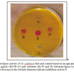 |
Figure 1. Anti-lipase activity of M. calabura fruit and control based on an agar plate assay: (A) orlistat (500 μg/mL) (B) FP (C) only substrate (D) FJ and (E) Substrate-lipase system without inhibitor. The decrease in the red halo diameter indicates inhibition activity.Click here to View figure |
Antiprotease Activity
The current study screened antiprotease activity of FJ and FP and results are presented in Table 3. Both extracts were able to modulate protease activity in a concentration dependent manner. The IC50 values were calculated to compare inhibition activity. The antiprotease activity of FP was much stronger than FJ, observing almost twice stronger than FJ.
Table 3: Antiproteases activity of M. calabura L. fruit and standard.
| Concentration(mg/mL) | Inhibition(%) | IC50(mg/mL) | |
| FJ | 0.0310.0610.1230.2460.492 | 22.75 ± 0.8127.95 ± 1.1632.87 ± 4,3047.34 ± 0,5659.06 ± 3,38 | 0.35 ± 0,02 |
| FP | 0.0290.0570.1150.2290.458 | 3.98 ± 0.639.36 ± 1.1112.64 ± 1.3922.50 ± 2.4932.63 ± 1.50 | 0.75 ± 0,04 |
| Na-Diclofenac | 0.0080.0200.0830.167 | 6.75 ± 0.0516.10 ± 0.04963.57 ± 0.4887.03 ± 0.28 | 0.04 ± 0,00 |
Antioxidant Activity
Antioxidant activity of FJ and FP was determined based on the effect of both extracts on DPPH radicals. As can be seen on Table 4, both extracts were able to scavenge DPPH radicals. As the concentration of the extracts increased, the % of scavenging activity was also incrementally increased. The IC50 of FJ was significantly weaker than FP (P = 0.000). However, when compared with BHT and ascorbic acid (antioxidant standards), the scavenging activity (IC50) of both FJ and FP were much lower, indicating a weak radical scavenging potential.
Table 4: DPPH radical scavenging activity of M. calabura L. and standards.
| Concentration(μg/mL) | Inhibition(%) | IC50(μg/mL) | ||||
| FJ | 239.00478.00717.00956.001195.001434.00 | 22.56 ± 1.8134.47 ± 0.0348.96 ± 1.1059.82 ± 0.1172.34 ± 0.0877.79 ± 0.26 | 780.80 ± 4.15 | |||
| FP | 330.00440.00660.00880.001100.00 | 32.62 ± 0.2044.34 ± 4.1665.56 ± 4.4086.65 ± 5.3898.08 ± 3.33 | 532.37 ± 46.41 | |||
| Ascorbic acid | 10.0020.0040.0080.00 | 8.56 ± 0.1115.29 ± 0.1140.48 ± 1.2775.96 ± 1.28 | 53.24 ± 0.82 | |||
| BHT | 3.336.6713.3326.67 | 12.66 ± 1.0323.15 ± 0.2040.99 ± 0.5956.28 ± 1.86 | 21.36 ± 0.80 | |||
Discussion
Phenolic compounds have been associated with various plants bioactivities, including antienzymes and antioxidant activities.31, 32 The current study found that phenolic compounds were identified not only in the fruit juice, but also in the fruit pulp, suggesting that the fruit pulp may still have nutritional benefit. This observation is similar with previous studies regarding the presence of phenolics in the residue or pulp of various fruits.33 Previously, phenolic contents were reported for M. calabura fruit, partitioned using various organic solvents of different polarity. Results reported previously found that methanol was the best solvent to extract phenolic compounds from fruits, as indicated by the highest amount of phenolics found.34
Type 2 diabetes mellitus (T2DM) is characterised by chronic hyperglycaemia, which may be due to a defect in insulin secretion, or insulin action, or a combination of both.35 Controlling the level of glucose in blood plasma is considered a practical and effective therapy for type 2 DM. In consequence, inhibition of α-glucosidase and α-amylase can delay the release of glucose from dietary carbohydrates and its absorption. In this way, inhibitors that target these enzymes can be an important therapeutic strategy in the management of type 2 DM. Acarbose, the drug of choice prescribed for T2DM, succeeds in controlling glucose level in plasma, however it has been associated with several adverse side effects.2 Strong inhibition of α-amylase and α-glucosidase leads to accumulation of undigested polysaccharides in the gastrointestinal tract (GIT). This cause abdominal disturbances due to bacterial fermentation.36 Other study has reported hepatotoxicity of long-term use of acarbose.37 Therefore, other alternatives are required that have fewer side effects for treating T2DM. In this context, it is necessary to evaluate the antidiabetic activity of fruits and vegetables.
Data obtained from the present study indicate that M. calabura fruit contain some components that are good inhibitors of α-glucosidase and α-amylase. Recently, study by Pereira et al reported the presence of cyanidin-3-O-glucoside and some phenolic compounds (gallic acid and caffeic acid) in M. calabura fruit.13 These compounds that were identified by UHPLC-MS/MS technique have been demonstrated to have inhibition activities against α-glucosidase and α-amylase.38, 39 In addition, the same study identified quercetin and catechin derivatives as the main flavonoids in the fruit, which were also reported to inhibit α-glucosidase and α-amylase.30, 40
For both α-glucosidase and α-amylase inhibition tests, FP showed a better inhibition activity than FJ, despite having lower phenolic content. This suggests that α-glucosidase and α-amylase inhibition activity is not necessarily associated with high phenolic content as other non-phenolic compounds such as alkaloids might be involved in α-glucosidase and α-amylase inhibition.41 The use of ethanol in FP preparation may have extracted more bioactive compounds involved in the enzyme inhibition, compared to FJ which was prepared directly from the fresh juice. Other parts of M. calabura, such as the leaves and stem barks, have been reported to have antidiabetic effects using animal model.16, 17 To the best of our knowledge, the present study is the first to report on the enzymes (α-glucosidase and α-amylase) inhibitory properties of M. calabura fruit.
Xanthine oxidase is responsible for the catalytic oxidation of hypoxanthine to xanthine, which generates superoxide (O2∙−) radicals and uric acid. Previous studies reported that phenolic compounds are considered as antioxidant, not only due to their ability to scavenge free radicals, but also because of its inhibition action on xanthine oxidase.42, 43 In addition, it is well known that the accumulation of uric acid in the joints and kidneys may lead to acute gout arthritis and uric acid nephrolithiasis.44 Allopurinol is a popular drug of choice for the treatment of hyperuricemia. However, its use has been linked to several side effects, such as rash, hypersensitivity.45 Various fruits have been found to have potent xanthine oxidase activity.46, 47 It is also worth noting that several flavonoids identified in M calabura fruit such as catechin derivatives and quercetin have been reported to have xanthine oxidase inhibitory activity.48 The present study found both FJ and FP exhibited inhibition activity on xanthine oxidase, with FP exerted stronger activity as compared with FJ.
Pancreatic lipase is responsible for the hydrolysis of almost 70% of dietary triglycerides. Therefore, inhibition of lipase is one of the effective mechanisms of anti-obesity agents. Orlistat, the first line lipase inhibitor, inhibits catalytic digestion of triglycerides, thus reduces intestinal absorption of free fatty acids. The accumulation of intestinal lipids causes steatorrhea, flatulence, and inconsistent stool.49 The anti-lipase screened in this study indicate that the fruit of M.calabura can be further explored for its anti-lipase activity.
Proteases are an important class of enzymes that involve in numerous biological processes, involving metabolic and physiological processes. This suggests the critical role of proteases in maintaining body homeostasis. Indeed, excessive protease activity has been associated with pathological conditions such as cardiovascular, neurodegenerative, and inflammatory diseases.50 The result obtained in the current study indicates that FJ and FP have anti protease activity, although both were seen to have a weaker activity in comparison with sodium diclofenac. The latter is a potent nonsteroidal anti-inflammatory drug. Gastrointestinal side effects have been reported for long term use of sodium diclofenac.51 Protease inhibition activity observed for FJ and FP may be further explored for a more catalytic specificity of protease inhibition.
Oxidative stress contributes to some of the pathological conditions in the development of metabolic diseases,52 such as in T2DM, obesity, and gouty arthritis. Therefore, antioxidant intake is considered important as part of therapeutic regimen in metabolic diseases. FJ and FP showed weak DPPH radical scavenging activity. Several phenolic and flavonoid compounds such as, gallic acid, caffeic acid, catechin, gallocatechin, naringenin, and quercetin identified in fruits and vegetables have been proven to exert antioxidant activity.13, 53The present study found that FP showed stronger antioxidant activity than FJ. This indicates that the fruit pulp still has nutritional benefit. Stronger DPPH scavenging activity was reported for fruit prepared by maceration and partition in various organic solvent, with strongest activity observed for methanol extract of the fruit of M. calabura.34
Conclusion
Results in the present study clearly demonstrate anti-enzymes (α-glucosidase, α-amylase, xanthine oxidase, lipase, and protease) activities of M. calabura fruit juice and pulp extracts. The study also reports antioxidant (DPPH) activity of the extracts. Significant inhibition of α-glucosidase and α-amylase suggests their potential use as a natural antidiabetic agent. In addition, the fruit was evidenced to have inhibition on xanthine oxidase, an enzyme targeted for the management of hyperuricemia.
Acknowledgement
The authors wish to thank the Faculty of Medicine and Health Sciences, Krida Wacana Christian University for the use of laboratory facilities.
Funding Source
The authors wish to thank The Research Institute of Krida Wacana Christian University for the financial support for the publication of this article.
Conflict of Interest
The authors declare no conflict of interest.
References
- Saklayen M. G. The global epidemic of the metabolic syndrome. Curr Hypertens Rep. 2018;20(2):12.
CrossRef. - Holman R. R., Coleman R. L., Chan J. C. N., Chiasson J. L., Feng H., Ge J., et al. Effects of acarbose on cardiovascular and diabetes outcomes in patients with coronary heart disease and impaired glucose tolerance (ACE): a randomised, double-blind, placebo-controlled trial. The lancet Diabetes & endocrinology. 2017;5(11):877-86.
CrossRef. - Faruque L. I., Ehteshami-Afshar A., Wiebe N., Tjosvold L., Homik J., Tonelli M. A systematic review and meta-analysis on the safety and efficacy of febuxostat versus allopurinol in chronic gout. Seminars in Arthritis and Rheumatism. 2013;43(3):367-75.
CrossRef. - Balbaa M., El Ashry E. Enzyme inhibitors as therapeutic tools. Biochem Physiol. 2012;1(2):1000103.
CrossRef. - Lim M., Kim J. Association between fruit and vegetable consumption and risk of metabolic syndrome determined using the Korean Genome and Epidemiology Study (KoGES). European journal of nutrition. 2019.
CrossRef. - de Oliveira E. P., McLellan K. C. P., Vaz de Arruda Silveira L., Burini R. C. Dietary factors associated with metabolic syndrome in Brazilian adults. Nutr J. 2012;11(1):13.
CrossRef. - Choudhury H., Pandey M., Hua C. K., Mun C. S., Jing J. K., Kong L., et al. An update on natural compounds in the remedy of diabetes mellitus: a systematic review. J Tradit Complement Med. 2018;8(3):361-76.
CrossRef. - Ayyappan P., Nampoothiri S. V. Chapter 13 – Bioactive natural products as potent inhibitors of xanthine oxidase. In: Atta Ur R, editor. Studies in Natural Products Chemistry. 64: Elsevier; 2020. p. 391-416.
CrossRef. - Rajan L., Palaniswamy D., Mohankumar S. K. Targeting obesity with plant-derived pancreatic lipase inhibitors: a comprehensive review. Pharmacological Research. 2020:104681.
CrossRef. - Hellinger R., Gruber C. W. Peptide-based protease inhibitors from plants. Drug Discov Today. 2019;24(9):1877-89.
CrossRef. - Mahmood N., Nasir N., Rofiee M., Tohid S. M., Ching S., Teh L. K., et al. Muntingia calabura: a review of its traditional uses, chemical properties, and pharmacological observations. Pharm Biol. 2014;52(12):1598-623.
CrossRef. - Sufian A. S., Ramasamy K., Ahmat N., Zakaria Z. A., Yusof M. I. M. Isolation and identification of antibacterial and cytotoxic compounds from the leaves of Muntingia calabura L. J Ethnopharmacol. 2013;146(1):198-204.
CrossRef. - Pereira G. A., Arruda H. S., de Morais D. R., Eberlin M. N., Pastore G. M. Carbohydrates, volatile and phenolic compounds composition, and antioxidant activity of calabura (Muntingia calabura L.) fruit. Food research international (Ottawa, Ont). 2018;108:264-73.
CrossRef. - Zakaria Z., Hazalin N. M. N., Zaid S. M., Ghani M. A., Hassan M., Gopalan H., et al. Antinociceptive, anti-inflammatory and antipyretic effects of Muntingia calabura aqueous extract in animal models. J Nat Med. 2007;61(4):443-8.
CrossRef. - Zakaria Z. A., Balan T., Suppaiah V., Ahmad S., Jamaludin F. Mechanism(s) of action involved in the gastroprotective activity of Muntingia calabura. J Ethnopharmacol. 2014;151(3):1184-93.
CrossRef. - Aligita W., Susilawati E., Sukmawati I. K., Holidayanti L., Riswanti J. Antidiabetic Activities of Muntingia calabura L. Leaves Water Extract in Type 2 Diabetes Mellitus Animal Models. Indones Biomed J. 2018;10(2):165-70.
CrossRef. - Yemineni M., Kancharlapalli V. R., Subrahmanyeswari P. Preclinical pharmacological profiling and cardioprotective activity for methanolic extracts of stem bark of Muntingia calabura L against adrenaline induced cardio-toxicity. Res J Pharm Technol. 2019;12(8):3773-80.
CrossRef. - Lin J.-T., Chen Y.-C., Chang Y.-Z., Chen T.-Y., Yang D.-J. Effective compounds in the fruit of Muntingia calabura Linn. cultivated in Taiwan evaluated with scavenging free radicals and suppressing LDL oxidation. Food Func. 2017;8(4):1504-11.
CrossRef. - Preethi K., Premasudha P., Keerthana K. Anti-inflammatory Activity of Muntingia calabura Fruits. Pharmacogn J. 2012;4(30):51-6.
CrossRef. - Simamora A., Santoso A. W., Timotius K. H. Bioactivities of methanol and ethyl acetate mace extracts of Myristica fragrans Houtt. Pharmacognosy Communications. 2018;8(3):103-7.
CrossRef. - Simamora A., Santoso A. W., Timotius K. H. α-Glucosidase inhibitory effect of fermented fruit juice of Morinda citrifolia L and combination effect with acarbose. Current Research in Nutrition and Food Science. 2019;7(1):218-26.
CrossRef. - Adisakwattana S., Ruengsamran T., Kampa P., Sompong W. In vitro inhibitory effects of plant-based foods and their combinations on intestinal α-glucosidase and pancreatic α-amylase. BMC Complementary Altern Med. 2012;12(1):110.
CrossRef. - Azmi S. M. N., Jamal P., Amid A. Xanthine oxidase inhibitory activity from potential Malaysian medicinal plant as remedies for gout. Int Food Res J. 2012;19(1):159-65.
- Aktumsek A., Zengin G., Guler G. O., Cakmak Y. S., Duran A. Antioxidant potentials and anticholinesterase activities of methanolic and aqueous extracts of three endemic Centaurea L. species. Food and Chemical Toxicology. 2013;55:290-6.
CrossRef. - Zengin G., Ceylan R., Katanić J., Mollica A., Aktumsek A., Boroja T., et al. Combining in vitro, in vivo and in silico approaches to evaluate nutraceutical potentials and chemical fingerprints of Moltkia aurea and Moltkia coerulea. Food and Chemical Toxicology. 2017;107:540-53.
CrossRef. - Zubairi N. H. M., Alam M. Z., Salleh M. N., Salleh H. M., Fazil N. A. A new isolate of thermophilic and organic solvent tolerant bacteria for lipase production using basal medium of palm kernel cake. Malays J Microbiol. 2018;14(2):80-7.
- Benitez J. A., Silva A. J., Finkelstein R. A. Environmental signals controlling production of hemagglutinin/protease in Vibrio cholerae. Infect Immun. 2001;69(10):6549-53.
CrossRef. - Coêlho D. F., Saturnino T. P., Fernandes F. F., Mazzola P. G., Silveira E., Tambourgi E. B. Azocasein substrate for determination of proteolytic activity: reexamining a traditional method using bromelain samples. BioMed Res Int. 2016;2016.
CrossRef. - Blois M. S. Antioxidant determinations by the use of a stable free radical. Nature. 1958;181(4617):1199-200.
CrossRef. - Limanto A., Simamora A., Santoso A. W., Timotius K. H. Antioxidant, α-glucosidase inhibitory activity and molecular docking study of gallic acid, quercetin and rutin: a comparative study. Molecular and Cellular Biomedical Sciences. 2019;3(2):67-74.
CrossRef. - Paixão N., Perestrelo R., Marques J. C., Câmara J. S. Relationship between antioxidant capacity and total phenolic content of red, rosé and white wines. Food Chemistry. 2007;105(1):204-14.
CrossRef. - Oboh G., Ademiluyi A. O., Akinyemi A. J., Henle T., Saliu J. A., Schwarzenbolz U. Inhibitory effect of polyphenol-rich extracts of jute leaf (Corchorus olitorius) on key enzyme linked to type 2 diabetes (α-amylase and α-glucosidase) and hypertension (angiotensin I converting) in vitro. J Funct Foods. 2012;4(2):450-8.
CrossRef. - Peschel W., Sánchez-Rabaneda F., Diekmann W., Plescher A., Gartzía I., Jiménez D., et al. An industrial approach in the search of natural antioxidants from vegetable and fruit wastes. Food Chem. 2006;97(1):137-50.
CrossRef. - Preethi K., Vijayalakshmi N., Shamna R., Sasikumar J. In vitro antioxidant activity of extracts from fruits of Muntingia calabura Linn. from India. Pharmacogn J. 2010;2(14):11-8.
CrossRef. - Petersmann A., Müller-Wieland D., Müller U. A., Landgraf R., Nauck M., Freckmann G., et al. Definition, Classification and Diagnosis of Diabetes Mellitus. Experimental and clinical endocrinology & diabetes : official journal, German Society of Endocrinology [and] German Diabetes Association. 2019;127(S 01):S1-s7.
CrossRef. - Derosa G., Maffioli P. Efficacy and safety profile evaluation of acarbose alone and in association with other antidiabetic drugs: a systematic review. Clin Ther. 2012;34(6):1221-36.
CrossRef. - Carrascosa M., Pascual F., Aresti S. Acarbose induced acute severe hepatotoxicity. The Lancet. 1997;349(9053):698-9.
CrossRef. - Akkarachiyasit S., Charoenlertkul P., Yibchok-anun S., Adisakwattana S. Inhibitory activities of cyanidin and its glycosides and synergistic effect with acarbose against intestinal α-glucosidase and pancreatic α-amylase. Int J Mol Sci. 2010;11(9):3387-96.
CrossRef. - Oboh G., Ogunsuyi O. B., Ogunbadejo M. D., Adefegha S. A. Influence of gallic acid on α-amylase and α-glucosidase inhibitory properties of acarbose. J Food Drug Anal. 2016;24(3):627-34.
CrossRef. - Proença C., Freitas M., Ribeiro D., Oliveira E. F., Sousa J. L., Tomé S. M., et al. α-Glucosidase Inhibition by Flavonoids: an In Vitro and In Silico Structure–Activity Relationship Study. J enzyme inhibit med chem. 2017;32(1):1216-28.
CrossRef. - Trinh D. H., Tran P. T., Trinh B. T. D., Nguyen H. T., Nguyen H. D., Ha L. D., et al. Coumarins and acridone alkaloids with α-glucosidase inhibitory and antioxidant activity from the roots of Paramignya trimera. Phytochem Lett. 2020;35:94-8.
CrossRef. - Baghiani A., Ameni D., Boumerfeg S., Adjadj M., Djarmouni M., Charef N., et al. Studies of antioxidants and xanthine oxidase inhibitory potentials of root and aerial parts of medicinal plant Capparis spinosa L. Am J Medicine Med Sci. 2012;2(1):25-32.
CrossRef. - Valentao P., Fernandes E., Carvalho F., Andrade P., Seabra R., Bastos M. Antioxidant activity of Centaurium erythraea infusion evidenced by its superoxide radical scavenging and xanthine oxidase inhibitory activity. Journal of Agricultural and Food Chemistry. 2001;49(7):3476-9.
CrossRef. - Choi H. K., Curhan G. Gout: epidemiology and lifestyle choices. Curr Opin Rheumatol. 2005;17(3):341-5.
- Bardin T. Current management of gout in patients unresponsive or allergic to allopurinol. Jt Bone Spine. 2004;71(6):481-5.
CrossRef. - Liu K., Wang W., Guo B.-H., Gao H., Liu Y., Liu X.-H., et al. Chemical evidence for potent xanthine oxidase inhibitory activity of ethyl acetate extract of Citrus aurantium L. dried immature fruits. Molecules. 2016;21(3):302.
CrossRef. - Palu A., Deng S., West B., Jensen J. Xanthine oxidase inhibiting effects of noni (Morinda citrifolia) fruit juice. Phytother Res. 2009;23(12):1790-1.
CrossRef. - Cos P., Ying L., Calomme M., Hu J. P., Cimanga K., Van Poel B., et al. Structure− activity relationship and classification of flavonoids as inhibitors of xanthine oxidase and superoxide scavengers. J Nat Prod. 1998;61(1):71-6.
CrossRef. - Harp J. B. An assessment of the efficacy and safety of orlistat for the long-term management of obesity. J Nutr Biochem. 1998;9(9):516-21.
CrossRef. - Sabotič J., Kos J. Microbial and fungal protease inhibitors—current and potential applications. Applied microbiology and biotechnology. 2012;93(4):1351-75.
CrossRef. - Baraf H. S., Fuentealba C., Greenwald M., Brzezicki J., O’Brien K., Soffer B., et al. Gastrointestinal side effects of etoricoxib in patients with osteoarthritis: results of the Etoricoxib versus Diclofenac Sodium Gastrointestinal Tolerability and Effectiveness (EDGE) trial. J rheumatol. 2007;34(2):408-20.
- Carrier A. Metabolic syndrome and oxidative stress: a complex relationship. Antioxidants & redox signaling. 2017;26(9):429-31.
CrossRef. - Banjarnahor S. D. S., Artanti N. Antioxidant properties of flavonoids. Med J Indones. 2014;23(4):5.
CrossRef.

This work is licensed under a Creative Commons Attribution 4.0 International License.