

Introduction
Probiotics are live microbes that when administered in adequate amounts, usually in foods, confer health benefit to the recipient.1 Probiotic characteristics have been demonstrated in the bacteria Lactobacillus acidophilus.2,3 When consumed, L. acidophilus cross the gastrointestinal stage and, in the colon, confer many health benefits including the balancing of gut microbiota, prevention of rotavirus diarrhea, colon cancer, and modulation of immune system.3,4 Unfortunately, the benefits of probiotic bacteria must be protected against the deleterious effects of some environmental identities such as presence of oxygen, temperature, low pH, bile salt and digestive enzymes.5 Encapsulation was offered to be effective method for protection of probiotics during processing, storage and passage through the human gastrointestinal tract, thus release large amounts of probiotic count in action site.5,6
Encapsulation of probiotic cells is commonly made by extrusion or dropping method that showed several advantages such as simple, inexpensive, not demands the specific apparatus. The probiotic cells are entrapped in capsules using carrier materials such as biopolymers.7,8 Alginate beads are used widely for encapsulation. Alginate is a natural polysaccharide, inexpensive, non-toxic, chemically stable, generally recognized as safe (GRAS) status and provides for high probiotic survival of 80-95%.9,10 Moreover, supplementation of encapsulated probiotics with prebiotics, a fermentable carbohydrate for probiotics, enhances their survivability in the gastrointestinal tract and during storage.5,6,11
Alternate prebiotic sources offer potential advantages to probiotic efficiency. These include plants; chicory root, Jerusalem artichoke, sugar beet and garlic.12 Sathyabama et al.13 found that co-encapsulation with sugar beet (2%) and/or chicory (2%) enhanced the viability of encapsulated probiotics, Staphylococcus succinus and Enterococcus fecium under in vitro gastrointestinal condition.
Mushrooms also act as prebiotic because they contain polysaccharides such as chitin, hemicellulose, a- and b-glucan, mannans and xylan.12 Interestingly, Dictyophora indusiate (bamboo mushroom) contains many biologically active ingredients with therapeutic values including antioxidative, antitumor, anticancer, antiinflammation and immunomodulatory properties.14,15 In addition, dictyophorines A and B help to protect the nervous system and reduce the risk of Alzheimer’s disease.16,17,18 Edible bamboo mushroom can be divided into immature (egg) and mature stages (fruit body). The egg of bamboo mushroom contains high concentrations of hyaluronic acid as well as allantoin, the latter with antiinflammation property. Fruit body of bamboo mushroom is rich in polysaccharides, especially b-glucan.19,20,21
To our knowledge, bamboo mushroom has not been co-encapsulated with probiotic L. acidophilus. This study investigated the addition of bamboo mushroom in co-encapsulation on the survival of encapsulated L. acidophilus in simulated gastrointestinal system. The morphological characteristic of alginate capsules was also assessed. Application of different bamboo mushroom stages, namely egg and fruit body at different concentrations with alginate capsules was examined before and after passage through gastrointestinal simulation as compared to the alginate capsules and free cells.
Materials and Methods
Materials
Lactobacillus acidophilus TISTR 2365 was purchased from Thailand Institute of Scientific and Technological Research, Thailand. Sodium alginate (food grade) (Ajax Fnechem pty, Australia), de man Rogosa and Sharpe (MRS) (Himedia, India), bile salts (Sigma, India), pepsin (Sigma, India) and pancreatin (Sigma, India) were also used.
Dictyophora indusiata, bamboo mushroom was obtained from Engineer’s Farm, Bangkok, Thailand. Egg (immature stage) and fruit body (mature stage) of bamboo mushroom were dried in a hot air oven for 8 hours at 45°C, ground in a blender and sieved through 100 mesh.15 Carbohydrate, protein, crude fiber, ash and fat were examined according to AOAC22. Antioxidant activity and phenolic acids of bamboo mushroom were analyzed using DPPH radical scavenging activity and Folin-Ciocalteu method, respectively.23
Preparation of Probiotic
Freeze-dried culture of Lactobacillus acidophilus TISTR 2365 was inoculated into MRS broth (100 mL) for 24 hours at 37°C under anaerobic incubation using an anaerobic jar (BD GasPakTMEZ anaerobe container system, Becton Dickinson Pty Ltd). Strain (L. acidophilus) was stocked at -18°C in MRS broth containing 30% glycerol and used to prepare starter. The stock culture was thawed at 4°C before use.24
Stock culture (1 mL) was activated into MRS broth (10 mL) at 37°C for 24 hours. After incubation, 109 cfu/g of L. acidophilus was obtained. Cells were harvested by centrifugation (8,500 rpm for 20 min), filtered (Whatman no.1) and rinsed twice with sterile sodium chloride (0.85% w/v).24 Cells were used either directly (free cells) in test or encapsulated as described below.
Encapsulation Process
Calcium-alginate beads were prepared by extrusion.25 Briefly, concentrations used for encapsulation were 4% (w/v) sodium alginate solution, 0, 0.8, 1 and 3% (w/v) of egg and/or fruit body of bamboo mushroom powder, 10 g of bacteria biomass (approximately 109 cfu/g) and 30 min firmness in 0.1 M sterile calcium chloride (CaCl2) solution. Cell suspension/alginate, and that of cell suspension/alginate/bamboo mushroom powder mixture were injected through a syringe 27G (0.4×25 mm needle) into 0.1 M CaCl2 solution. Microsphere gel particles were formed immediately. Distance between the syringe and CaCl2 solution was fixed at 5 cm. Beads were allowed 30 min to harden, filtered (Whatman no.1) and washed in sodium chloride (0.85% w/v). All materials were sanitized for 15 min at 121°C.25
Simulated Gastrointestinal Condition
Simulated gastrointestinal condition (SGI) was performed with cells without encapsulation, encapsulated cells and co-encapsulated cells with bamboo mushroom powder in simulated gastrointestinal system. Simulated gastric juice (SG) and simulated intestinal juice (SI) followed the procedure described by Peredo et al.26
Simulated gastric juice (SG) consisted of 3 g/L pepsin with pH adjusted to 2.0 by adding of 0.1N hydrochloric acid solution (HCL). Beads (1 g) or 1 mL free cells in sodium chloride solution (0.85% w/v) were transferred in 9 mL of SG and incubated for 4 hours at 37°C.26 Upon the completion of incubation, beads were removed and viable cells measured as described in section enumeration of probiotic cells.
Beads (1 g) or free cells solution (1 g) from simulated gastric solution were transferred in simulated intestinal juice (SI) (9 mL) containing 4.5 g/L bile salt, 1 g/L pancreatin with pH adjusted to 8.0 by 0.1N NaOH. Sample was incubated at 37°C for 4 hours.26 Subsequently, beads were separated and the viable counts of the encapsulated cells and free cells analyzed as described below.
Enumeration of Probiotic Cells, Survival Rate and Encapsulation Yield
Beads (1 g) were homogenized by a stomacher for 1 min with 9 mL of 0.1% (w/v) sterile peptone and diluted in a 10-fold serial. L. acidophilus was enumerated using pour plate technique on MRS agar at 35°C for 48 hours under anaerobic condition,22 while free cells were analyzed similarly. Plates containing 25-250 colonies were expressed in log colony-forming units per g (log cfu/g). In addition, survival rate of L. acidophilus under simulated gastrointestinal condition was calculated by equation (1)27:
where N is the number of viable cells (log cfu/g) after incubation in simulated gastro-intestinal juice and N0, the number of viable cells (log cfu/g) before incubation in either simulated gastric or intestinal juice.
Encapsulation yield of cells was computed according to equation (2)27:
where N is number of entrapped cells inside beads and N0, the number of free cells (log cfu/g) added into sodium alginate solution during the preparation process.
Morphology Characterization of Beads by Optical Microscopy and Scanning Electron Microscopy (SEM) and Particle Size Measurement
Morphology characterization of the moist beads was analyzed using optical microscopy (Eclipes, E-400 Pol, Japan) equipped with a digital camera (Canon, Eos 1,300DW, TAIWAT) for capturing images. Morphology and microstructure of the beads were also analyzed using SEM (JEOL, model JSM-6610LV, Japan) as described by Dariani et al.5 Freeze-dried capsules were coated with a layer of gold and examined by SEM at an acceleration voltage of 10.0 kV. Bead size was measured by Vernier Caliper (Dizionario, Thailand).
Data Analysis
Statistical analysis was performed by analysis of variance (ANOVA). Significant differences among means were analyzed by Duncan’s multiple range test using Minitab software version 18 (Minitabl Pty Ltd, Sydney NSW, Australia) with significance at p<0.05.
Results and Discussion
Physicochemical Characteristics and Antioxidative Activity of Bamboo Mushroom
Proximate composition of fruit body and egg of bamboo mushroom insignificantly differed, except protein content (p<0.05) (Table 1). Protein of egg of bamboo mushroom increased relative to that of fruit body by 9.9 fold. Carbohydrate was the major component of both egg and fruit and fat, the least. The high carbohydrate content in the bamboo mushroom (fruit body) was consistent with that reported by Habtemariam.28 Egg and fruit body were rich in dietary fiber (13.30 and 15.17%), phenolic acids (363.10 and 309.10 mg GAE/g), and also have high DPPH radical scavenging activity (90.99 and 88.81%), in accord the reported health benefit. Phenolic acids in egg of bamboo mushroom were significantly higher than that of the fruit body (p<0.05), but differences in antioxidant activity were not significant (p³0.05). Natural phenolic compounds of bamboo mushroom exert their health benefit effects through their antioxidative, antitumor, anticancer, antiinflammation and antimicrobial properties.14,21 This suggests bamboo mushroom should be considered as a functional food with abundant bioactive compounds, especially phenolic acids and fiber. This result is in accord with the earlier observation by Oyetayo et al.29, who found also that bamboo mushroom has high antioxidant capacity. DPPH radical scavenging activity of aqueous extract from Dictyophora indassita at concentration of 2 mg/mL was 97.35%.
Table 1: Physicochemical Properties of Egg and Fruit Body of Bamboo Mushroom
| Characteristics | Bamboo mushroom | |
| EB | FB | |
| Carbohydrate (%) | 54.72±0.02 | 60.55±0.04 |
| Protein (%) | 20.21±0.01 a | 10.31±0.03 b |
| Crude fiber (%) | 13.30±0.01 | 15.17±0.05 |
| Fat (%) | 1.85±0.05 | 1.25±0.12 |
| Ash (%) | 4.07±0.09 | 6.95±0.07 |
| Moisture (%) | 5.85±0.04 | 5.77±0.01 |
| Antioxidant activity (%) | 90.99±0.08 | 88.81±0.19 |
| Phenolic acid (mg GAE/g) | 363.10±0.64a | 309.10±0.30b |
Values are means ± SD
Values in same row with different superscripts indicate significant differences (p<0.05)
EB (egg of bamboo mushroom)
FB (fruit body of bamboo mushroom)
Encapsulation Yield
Results for encapsulation efficiency of beads containing L. acidophilus incorporation with or without egg or fruit body of bamboo mushroom at the different concentrations are summarized in Table 2. Initial cell counts of L. acidophilus before encapsulation was 9.93±0.50 log cfu/g, while, after counts varied within the range of 9.37-9.65 log cfu/g. The high cell entrapment of 94-97% indicated efficient encapsulation by extrusion. There was no significant difference among all studied sample (p≥0.05). This result implied that the effectiveness of encapsulation did not depend on the presence of other materials in system such as type and concentration of the materials used. Similar results were reported by many previous researches, which showed encapsulation yields of 74-99% by extrusion technique.5,6,7 In addition, several studies also reported that the type, concentration or presence of prebiotic in matrix capsule did not influence the encapsulation yield.5,6,11
Table 2: Encapsulation Yield and Size of the Different Beads
| Formulations | Conc. (%w/v) | Encapsulation yield (%) | Size of beads (mm) |
| Control | – | 94 | 3.0 |
| EB | 0.8 | 97 | 3.0 |
| 1 | 95 | 3.0 | |
| 3 | 95 | 3.0 | |
| FB | 0.8 | 96 | 3.0 |
| 1 | 94 | 3.0 | |
| 3 | 96 | 3.0 |
Control (sample without bamboo mushroom)
EB (egg of bamboo mushroom)
FB (fruit body of bamboo mushroom)
Size and Morphology Characterization of Beads
Beads in this study averaged 3.0 mm in diameter and did not differ significantly among samples of all prepared beads (p≥0.05) (Table 2). In general, the microsphere gel particles with diameters of 0.2-5 mm were obtained from extrusion technique.8 Beads were round in shape with core material such as bamboo mushroom distributed in the matrix (Figure 1). The addition of either fruit body or egg of bamboo mushroom at different concentrations during probiotic encapsulation had no influence on size or shape of beads (Table 2 and Figure 1). In addition, after exposure to SG and SI, changes in bead sizes from 3.0 mm were not found (data not shown), indicating their stability. Likely this reflected a strong biopolymer gel network when prepared from 4% alginate. This result was in agreement with finding of Darjani et al.5 that the addition of 2% prebiotics like oligofrutose and inulin (polymerization degree of 12) in encapsulation process did not influence size or shape of probiotic alginate beads. However, size of alginate increased after exposure to simulated intestinal juice. Etchepare et al.30 also found that the use of Hi-maize in co-encapsulation with probiotic did not affect the diameters of the microcapsules.
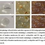 |
Figure 1: Morphology of Beads Before and After Exposure to SGI Using Optical Microscopy 30X. |
SEM images (Figure 2) illustrate surface morphology of biopolymer gel network of alginate beads before exposure to SGI. SEM observations revealed clearly the beads’ spherical shape (Figure 2a, 2c, 2e). Beads without bamboo mushroom showed high agglomeration among particles, the calcium-alginate network is uniform and continuous in structure (Figure 2a-2b). The addition of either fruit body or egg of bamboo mushroom caused more wrinkles and irregularities on the capsules surface due to the presence of insoluble bamboo mushroom in system (Figure 2c-2f). The association of calcium-alginate in beads without bamboo mushroom promoted a more cohesive structure, indicating better characteristic for protection of probiotic in capsules. This result was in agreement with Sathybama et al.13 who found that the external surfaces of the beads prepared by co-encapsulation of probiotics and prebiotics such as sugar beet and chicory were rougher than those of the alginate beads without prebiotics.
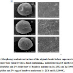 |
Figure 2: Morphology and Microstructure of the Alginate Beads Before Exposure to SGI. Click here to View figure |
As shown in Figure 3, after exposure to gastro-intestinal juice bead surfaces developed areas of roughness and cracks. However, wall microstructure of capsules containing only L. acidophilus (without bamboo mushroom) did not appeared clearly porous wall. On the other hand, pore size of the alginate network increased from 0.8 to 3% with the addition of fruit body or egg of bamboo mushroom. It is generally accepted that calcium-alginate gel forms a porous biopolymer network, which is well known as egg-box model.5,8,31,32 The gel is sensitive to extreme pH and influences the release and protection of core materials.26,33 Alginate beads ruptured when immersed in intestinal fluid. Under this condition, carboxyl groups of calcium-alginate hydrogel network are subjected to bile salt, and undergo ion exchange process. The electrostatic repulsion between negative charge COO– groups caused chains to relax and gel particles to rupture.6,34 Moreover, under co-encapsulation process, the crosslinking reaction between calcium-alginate networks may be disrupted from the presence of insoluble bamboo mushroom in system resulting in forming a less interconnected network and simple destroying by adverse condition. The result confirmed that co-encapsulation with bamboo mushroom affected the microstructure of hydrogel particles with inhomogeneous morphology and large porous biopolymer network. Similar results were reported by Huq et al.34, and Khorasani and Shojaosadati.35 The surface of alginate capsules was rough and crack due to severe damage from the bio-composite including low acid, HCL, bile salt and enzymes under gastrointestinal condition.
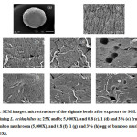 |
Figure 3: SEM Images, Microstructure of the Alginate Beads After Exposure to SGI. |
Cells Survival under Simulated Gastrointestinal Condition
For simulation the conduct of beads and determination their resistance during gastrointestinal tract was carried out. This study proposed to mimic the stomach pH, intestinal pH, bile salt and enzymes in gastrointestinal tract.26,36 Such determination shows the ability of the beads to protect the probiotic as well as investigate the role of bamboo mushroom on survivability of L. acidophilus. An in vitro viability of L. acidophilus demonstrates in Table 3.
The initial number of probiotic cells of L. acidophilus in all samples varied 9.40-9.93 log cfu/g, higher than recommended minimum probiotic count in probiotic food (>6 log cfu/g) by US FDA.37 Cells decreased after 4 hours of SG and SI phase, demonstrating the role of stomach pH, intestinal pH, bile salt and enzymes in the gastrointestinal tract to reduce the probiotics. In particular, the log reduction of free cells was 5.16 and 0.85 (log cfu/g) under SG and SI phase, respectively that corresponded to 6.46 log reduction or survival rate of 34% after SGI stage (Table 3). Free L. acidophilus cells were obviously more sensitive to environmental conditions in the stomach than those in the intestine. Accordingly, the study of de Farias et al.7 also indicated free L. casei was more resistant to intestinal conditions than those in the stomach. The low pH of HCL (pH 2) and pepsin in gastric condition can severely damage cell membranes, loss of enzymatic activity and function of probiotic cells.37,38,39 Furthermore, acid and base in gastrointestinal tract also destroy peptidoglycan, proteins and lipids in bacteria cell membranes resulting in the loss of balancing reactions in cells.37 This was consistent with the findings of Peredo et al.26 that cell retention of free cells of L. casei Shirota, and L. plantarum 33 and 17.2b under simulated gastrointestinal digestion was 24-48%. In addition, 6.9 and 6.7 log reduction of free L. casei was reported after passage through in vitro gastric and intestinal step, respectively.5 Huq et al.34 stated the reduction of free Lactobacillus rhamnosus of 7.81 log cfu/g under simulated gastric system. Interestingly, this result suggested that L. acidophilus TISTR 2365 is more tolerant to gastrointestinal conditions when compared to other microorganisms5,26,34, a favorable characteristic for probiotics in commercial application.37
The survival of encapsulated and co-encapsulated cells with bamboo mushroom in alginate capsules was significantly higher than those in free cells during SG and SI phases (p<0.05). The log reduction of both encapsulated and co-encapsulated cells under SG and SI stages varied within the range of 1.61-2.24 and 0.81-1.38 (log cfu/g), respectively that corresponded to 2.25-3.25 log reduction or survival rate of 63-68% during SGI stage (Table 3), indicating the efficient protection of probiotic cells by encapsulation. This suggests alginate forms a hydrogel barrier restraining the transmission of gastric and intestinal fluids into cells, protecting cells during passage through the gastrointestinal tract.26 In both encapsulation cases in this study, the high viability of probiotic, >6 log cfu/g, after transit through SGI would be expected to provide a beneficial effect on human health.37,40 Our results were consistent with many earlier researches, which found that the encapsulation process with alginate can not only protect but also and greatly improve the viability of probiotics in the gastrointestinal tract.5,31,41,42 According to Zaeim et al.43 showed that co-encapsulation of probiotic and prebiotic including inulin and resistant starch improved survival of probiotics as compared to free cells under simulated gastrointestinal condition.
Probiotic counts under encapsulation and co-encapsulation with either fruit body or egg of bamboo mushroom at different concentrations did not differ significantly after exposure to SG and SI stage (p≥0.05) (Table 3). These results implied the addition of bamboo mushroom in co-encapsulation did not enhance the stability of probiotic as compared to alginate beads without bamboo mushroom. However, survival rate of co-encapsulated probiotics in this study (63-68%) was within the reported survival rate of co-encapsulated probiotics in alginate beads containing different prebiotic materials (57-75%), specially potato starch, Plantago psyllium and inulin26, and Hi-maize plus chitosan (64%)33 under gastrointestinal digestion. Likewise, Silva et al.6 has also reported that the survival rate of L. acidophilus in alginate-gelatin microbeads was higher than that of alginate-gelatin-fructooligosaccharide microbeads in gastrointestinal tract in vitro, but both encapsulation cases provided greater viability of probiotic cells than that of free cells. Nevertheless, these results are contrary to those reported by Darjani et al.5 that the probiotics encapsulated in alginate beads containing oligofructose or inulin prebiotics showed higher viability relative to alginate beads without prebiotics. The study by Etchepare et al.30,33 showed also that the addition of Hi-maize, and Hi-maize plus chitosan in co-encapsulation improved survival of L. acidophilus during exposure to severe environment in the gastrointestinal stage as compared to alginate beads without prebiotic.
In the current study, even though the probiotic cells were co-encapsulated with bamboo mushroom, the porosity of gel particles (Figure 3c-3h), allowed the diffusion of SGI in beads, decreasing the viability of probiotic microorganisms. However, the reduction of cell counts in co-encapsulation was similar to that without bamboo mushroom (Table 3). This suggests the presence of prebiotic in matrix helped to protect the probiotic from contact with gastric-intestinal fluids. This result proved that the encapsulation with or without bamboo mushroom improved viability. Alginate beads were able to sustain part of their structure during transmission through the gastrointestinal stage. High amounts of L. acidophilus were maintained within the beads, preparing a satisfying environment for survival. Further, the incorporation of probiotic and prebiotic is known as a synbiotic.26,40 Thus, co-encapsulation of L. acidophilus and bamboo mushroom enhance the efficiency of probiotic food by employing synergic effect between probiotic and prebiotic components. When probiotics reach the large intestinal, prebiotics are potential energy source via probiotic catabolism, thereby supporting the growth and/or activity of probiotics.37,40 The combination of bamboo mushroom in beads also improved the health benefits resulting from their phenolic acids and antioxidant activity, supporting the concept of functional food.
Table 3: Survival of Free Cells, Encapsulated Cells and Co-encapsulated Cells with Bamboo Mushroom in Gastrointestinal Condition
| Formulations | Conc.(% w/v) | Number cells before digestion (log cfu/g) | SG | SI | Log reduction after SGI | Survival rate after SGI (%) | ||
| Viability of cells (log cfu/g) | Log reduction | Viability of cells (log cfu/g) | Log reduction | |||||
| Free cells | – | 9.93±0.50 | 4.32±0.28 b | 5.16 a | 3.47±0.28 b | 0.85 | 6.46 a | 34b |
| Control | – | 9.37±0.34 | 7.76±0.30 a | 1.61 b | 6.72±0.25 a | 1.04 | 2.65b | 67a |
| EB | 0.8 | 9.65±0.31 | 7.55±0.26 a | 2.10 b | 6.55±0.16 a | 1.00 | 3.10b | 65 a |
| 1 | 9.53±0.29 | 7.72±0.23 a | 1.81 b | 6.34±0.31 a | 1.38 | 3.16b | 63 a | |
| 3 | 9.46±0.28 | 7.61±0.41 a | 1.85 b | 6.37±0.14 a | 1.24 | 3.06b | 64 a | |
| FB | 0.8 | 9.60±0.26 | 7.36±0.25 a | 2.24 b | 6.36±0.31 a | 1.00 | 3.24b | 64 a |
| 1 | 9.40±0.34 | 7.60±0.30 a | 1.80 b | 6.76±0.12 a | 0.84 | 2.64b | 68a | |
| 3 | 9.61±0.38 | 7.45±0.23 a | 2.16 b | 6.64±0.10 a | 0.81 | 2.52b | 66a | |
Values are means ± SD
Values in same column with different superscripts indicate significant differenced (p<0.05)
Control (sample without bamboo mushroom), EB (egg of bamboo mushroom), FB (fruit body of bamboo mushroom), Conc (concentration of bamboo mushroom), SG (simulated gastric juice), SI (simulated intestinal juice), SGI (simulated gastrointestinal condition)
Conclusion
The addition of egg or fruit body of bamboo mushroom at different levels to alginate matrix resulted in the less interconnected network, causing larger pores when compared to alginate bead without bamboo mushroom. However, both beads with and without bamboo mushroom protected probiotic L. acidophilus, improving their survival in gastrointestinal condition as compared to free cells. Bamboo mushroom acted as prebiotic source and helped to protect probiotic when subjected to gastrointestinal condition. This study showed a successful co-encapsulation of bamboo mushroom and L. acidophilus in alginate beads with gratifying survival of probiotic cells, which should be further developed for functional foods.
Acknowledgments
The authors wish to thank Professor Dr. Frederick W.H. Beamish (Faculty of Science, Barapha University) for English proofreading, Department of Food Science, Program of Biological Science and Faculty of Science, Burapha University for equipment support.
Funding Source
The authors received no financial support for the research, authorship, and/or publication of this article.
Conflict of Interest
The authors declare no conflict of interest.
References
- FAO/WHO. Probiotics in Foods Health and Nutrition Properties and Guidelines for Evaluation. Rome: FAO/WHO; 2006.
- Panesar, P. S., Marwaha, S. S. Biotechnology in Agriculture and Food Processing Opportunities and Challenges. New York: Taylor and Francis Group; 2014.
CrossRef. - Ray, R.C., Montet, D. Microorganisms and Fermentation of Traditional Foods. New York: Taylor and Francis Group; 2015.
CrossRef. - Guarner, F., Khan, G. A., Garisch, J., Eliakim, R., Gangl, A., Thomson, A., Krabshuis, J., Lemair, T. Probiotics and Prebiotics. USA: World Gastroenterology Organisation; 2011.
- Darjani, P., Nezhad, M. H., Kadkhodaee, R., Milani, E. Influence of Prebiotic and Coating Materials on Morphology and Survival of Probiotic Strain of Lactobacillus casei Exposed to Simulated Gastrointestinal Conditions. LWT-Food Sci Technol. 2016;73:162-167.
CrossRef. - Silva, K., Cezarino., E. C., Michelon, M. Symbiotic Microencapsulation to enhance Lactobacillus acidophilus Survival. LWT-Food Sci Technol. 2018;89:503-509.
CrossRef. - de Farias, T., Ladislau, H., Stamford, T., Medeiros, J., Soares, B., Arnaud, T., Stamford, T. Viabilities of Lactobacillus rhamnosus ASCC 290 and Lactobacillus casei ATCC 334 (in Free form or Encapsulated with Calcium Alginate-chitosan) in Yellow Mombin Ice Cream. LWT-Food Sci Technol. 2019;100:391-396.
CrossRef. - Ravishankar, R. V. Advances in Food Biotechnology. Oxford: John Wiley and Sons; 2016.
- Iyer, C., Kailasapathy, K. Effect of Co-encapsulation of Probiotics with Prebiotics on Increasing the Viability of Encapsulated Bacteria under in Vitro Acidic and Bile Salt Conditions and in Yogurt. J Food Sci. 2005;70:99-104.
CrossRef. - Manjana, K. M., Pramod Kumer, T. M., Shivakumar, B. Calcium Alginate Cross-linked Polymeric Microbeads for Oral Sustained Drug Delivery in Arthritis. Drug Discov Ther. 2010;4(2):109-122.
- Krasaekoopt, W., Watcharapoka, S. Effect of Addition of Inulin and Galactooligosaccharide on the Survival of Microencapsulated Probiotics in Alginate Beads Coated with Chitosan in the Simulated Digestive System, Yogurt and Fruit Juice. LWT-Food Sci Technol. 2014;57:761-766.
CrossRef. - Jayachandran, M., Xiao, J., Xu, B. A Critical Review on Health Promoting Benefits of Edible Mushrooms though Gut Microbiota. Int J Mol Sci. 2017;18:1-12.
CrossRef. - Sathyabama, S., Kumar, M., Bruntha, P., Vijayabharathi, R. Co-encapsulation of Probiotics with Prebiotic on Alginate Matrix and Its Effect on Viability in Simulates Gastric Environment. Int Food Res J. 2014;57:419-425.
CrosRef. - Liao, W., Luo, Z., Liu, D., Ning, Z., Yang, J., Ren, J. Structure Characterization of a Novel Polysaccharide from Dictyophora indusiata and Its Macrophage Immunomodulatory Activities. J Agric Food Chem. 2015;63:535-544.
CrossRef. - Liu, X., Chen, Y., Wu, L., Wu, Huang, Y., Liu, B. Optimization of Polysaccharides Extraction from Dictyophora indusiata and the Determination of Its Antioxidant Activity. Int J Biol Macromol. 2017;103:175-181.
CrossRef. - Hua, Y., Yang, B., Tang, J., Ma, Z., Gao, Q., Zhao, M. Structural Analysis of Water-soluble Polysaccharides in the Fruiting Body of Dictyophora indusiata and They Are in Vivo Antioxidant Activities. Carbohydr Polym. 2012;87:343-347.
CrossRef. - Ishiyama, D., Fukushi, Y., Ohnishi-Kameyama, M., Nagata, T., Mori, H., Inakuma, T., Inakuma, T., Ishiguro, Y., Li, J., Kawagishi, H. Monoterpene-alcohols from a Mushroom Dictyophora indusiate. Pytcas. 1999;50:1053-1056.
CrossRef. - Lee, K., Yun, B. S., Han, G., Cho, D. H., Kim, Y. H., Yoo, I. D. Dictyoquinazols A, B, and C, New Neuroprotective Compounds from the Mushroom Dictyophora indusiata. J Nat Prod. 2002;65:1769-1772.
CrossRef. - Deng, C., Shang, J., Fu, H., Chen, J., Liu, H. Mechanism of the Immunostimulatory Activity by a Polysaccharide from Dictyophora indusiata. Int J Biol Macromol. 2016;91:752-759.
CrossRef. - Wang, J. H., Zhang, Y. K., Yao, Y. F., Lin, Y., Xu, J. L., Sun, H. J. Structure Identification and Antioxidant Activity of a Novel Triple Helical Polysaccharide Isolated from Dictyophora indusiata. J Chem Pharm Res. 2015;7(1):678-684.
- Zhang, J., Shi, R., Li, H., Xiang, Y., Xiao, L., Hu, M., Ma, F., Huang, Z. Antioxidant and Neuroprotective Effect of Dictyophora indusiata Polysaccharide in Caenorhabditis Elegans. J Ethnopharmacol. 2016;192:413-422.
CrossRef. - Association of Official Analytical Chemists. Official Methods of Analysis of Association of Official Analysis Chemistry International (17th edition). Washington DC: AOAC; 2000.
- Karagozler, A., Eedag, B., Emek, Y. C., Uygun, D. A. Antioxidant Activity and Proline Content of Leaf Extracts from Dorystoechas hastata. Food Chem. 2008;111:400-407.
CrossRef. - Mozzi, F., Raya, R., Vignolo, G. M. Biotechnology of Lactis Acid Bacteria Oxford: John Wiley and Sons; 2016.
CrossRef. - Gandomi, H., Abbaszadeh, S., Misaghi, A., Bokaie, S., Noori, N. Effect of Chitosan-alginate Encapsulation with Inulin on the Survival of Lactobacillus rhammnosus GG During Apple Juice Storage and under Simulated Gastrointestinal Conditions. LWT-Food Sci Technol. 2016;69:365-371.
CrossRef. - Peredo, A. G., Beristain, C. I., Pascual, L. A., Azura, E., Jimenez, M. The Effect of Prebiotics on the Viability of Encapsulated Probiotic Bacteria. LWT-Food Sci Technol. 2016;73:191-196.
CrossRef. - Ashwar, B. A., Gani, A., Shah, A., Masoodi, F. A. Production of RS4 from Rice Starch and Its Utilization as an Encapsulating Agent for Targeted Delivery of Probiotics. Food Chem. 2018;239:287-294.
CrossRef. - Habtemariam, S. The Chemistry, Pharmacology and Therapeutic Potential of the Edible Mushroom Dictyophora indusiate (Vent ex. Pera.) Fischer (Synn. Phallus indusiatus). J Biomed. 2019;7(98):1-21.
CrossRef. - Oyetayo, V. O., Dong, C. H., Yao, Y. J. Antioxidant and Antimicrobial Properties of Aqueous Extract from Dictyophora indusiata. Open Mycol J. 2009;3:20-26.
CrossRef. - Etchepare, M., Raddatz, G. C., Cichoski, A. J., Flores, É. M. M., Barin, J. S., Zepka, L. Q., Jacob-Lopes, E., Barin, J. S., Grosso, C. R. F., de Menezes, C. R. Effect of Resistant Starch (Hi-maize) on the Survival of Lactobacillus acidophilus Microencapsulated with Sodium Alginate. J Funct Foods. 2016;21:321-329.
CrossRef. - Bosnes, L. A., Moschakis, T., Nigam, P. S., Biliaderis, C. G. Growth Adaptation of Probiotics in Biopolymer-based Coacervate Structures to Enhance Cell Viability. LWT-Food Sci Technol. 2017;77:282-289.
CrossRef. - Pliszczak, D., Bourgeois, S., Bordes, C., Valour, J. P., Mazoyer, M. A., Orecchioni, A. M., Nakache, E., Lanteri, P. Improvement of an Encapsulation Process for Preparation of the Preparation of Pro- and Prebiotic-loaded Bioadhesive Microparticals by using Experimental Design. Eur J Pharm Sci. 2011;44:83-92.
CrossRef. - Etchepare, M., Raddatz, G. C., Cichoski, A. J., Flores, É. M. M., Barin, J. S., Zepka, L. Q., Jacob-Lopes, E., Barin, J. S., Grosso, C. R. F., de Menezes, C. R. Effect of Resistant Starch and Chitosan on the Survival of Lactobacillus acidophilus Microencapsulated with Sodium Alginate. LWT-Food Sci Technol. 2016;65:511-517.
CrossRef. - Huq, T., Fraschini, C., Khan, A., Riedi, B., Bouchard, J. Alginate Based Nanocomposite for Microencapsulation of Probiotic: Effect of Cellulose Nanocrystal (CNC) and Lecithin. Carbohydr Polym. 2017;168:61-69.
CrossRef. - Khorasani, A. C., Shojaosadati, S. A. Bacterial Nanocellulose-pectin Bionanocomposites as Prebiotics Against Drying and Gastrointestinal Condition. Int J Biol Macromol. 2016;83:9-18.
CrossRef. - Brinques, G. B., Ayub, M. A. Z. Effect of Microencapsulation on Survival of Lactobacillus plantarum in Simulated Gastrointestinal Conditions, Refrigeration, and Yogurt. J Food Eng. 2011;103:123-128.
CrossRef. - Tripathi, M. K., Giri, S. K. Probiotic Functional Food: Survival of Probiotics During Processing and Storage. J Funct Foods. 2014;9:225-241.
CrossRef. - Nunes, G. L., Etchepare, A., Cichoski, A. J., Zepka, L. Q., Lopes, E. J., Barin, J. S., Flores, M., Silva, C. B., Menezes, C. R. Inulin, Hi-maize, and Trehalose as Thermal Protectants for Increasing Viability of Lactobacillus acidophilus Encapsulated by Spray Drying. LWT-Food Sci Technol. 2018;89:128-133.
CrossRef. - Rodrigues, V. C. C., Silva, L. G. S., Simabuco, F. M., Venema, K., Antunes, A. E. C. Survival, Metabolic Status and Cellular Morphology of Probiotics in Dairy Products and Dietary Supplement After Simulated Digestion. J Funct Foods. 2019;55:126-134.
CrossRef. - Boye, J. I. Nutraceutical and Functional Food Processing Technology. Oxford: John Wiley and Sons; 2016.
- Moumita, S., Goderska, K., Johnson, E. M., Das, B., Indira, D., Yadav, R., Kumari, S., Jayabalan, R. Evaluation of the Viability of Free and Encapsulated Lactic Acid Bacteria using In-vitro Gastro Intestinal Model and Survivability Studies of Symbiotic Microcapsules in Dry Food Matrix During Storage. LWT-Food Sci Technol. 2017;77:460-467.
CrossRef. - Sohail, A., Turner, M. T., Coombes, A., Bostrom, T., Bhandari, B. Survivability of Probiotics Encapsulated in Alginate Gel Microbeads using a Novel Impinging Aerosols Method. Int J Food Microbiol. 2011;145:168-178.
CrossRef. - Zaeim, D., Sarabi-Jamab, M., Ghorani, H., Kadkhodaee, R. Double Layer Co-encapsulation of Probiotics and Prebiotics by Electro-hydrodynamic Atomization. LWT-Food Sci Technol. 2019;110:102-109.
CrossRef.

This work is licensed under a Creative Commons Attribution 4.0 International License.