
Extraction and Characterization of Nanopowder from Snakehead Fish (Channa striata) Bones
 , Agusriansyah Saputra, Indah Widiastuti, Sabri Sudirmanand Chalidazia
 Department of Fisheries Product Technology, Universitas Sriwijaya, Indralaya, Indonesia
Corresponding Author Email: herpandi@fp.unsri.ac.id
DOI : http://dx.doi.org/10.12944/CRNFSJ.14.2.11
Download this article as:

This research investigates the extraction and characterization of nanopowder from snakehead fish (Channa striata) bones using ultrasonic treatment and variations in calcination temperatures. The fish bones were calcined at 500℃, 750℃, and 1000℃ for 5 hours to eliminate organic components and improve the crystallinity of the nanopowder. Ultrasonic waves were also employed to create cavitation conditions, promoting the formation of nanoparticles with reduced size. Characterization was performed using Scanning Electron Microscopy-Energy Dispersive X-ray Analysis (SEM-EDX), X-ray Diffraction (XRD), and Fourier Transform Infrared Spectroscopy (FTIR). Five samples were analyzed: Sample A (untreated fish bone powder), Sample B (ultrasonic-treated), Sample X (ultrasonic-treated and calcined at 500℃), Sample Y (ultrasonic-treated and calcined at 750℃), and Sample Z (ultrasonic-treated and calcined at 1000℃). The results showed that increasing calcination temperature drove morphological transformation from heterogeneous platy fragments toward uniform spherical nanoparticles, while simultaneously enhancing calcium and phosphorus content and HAp crystallinity. Calcination at 1000°C yielded the optimal outcome, a mean particle size of 341 ± 197 nm, Ca and P concentrations of 31.02% and 18.25%, respectively, and crystallinity reaching 95%. These findings establish ultrasonic-assisted calcination at 1000°C as an effective and scalable route for producing high-crystallinity HAp nanopowder from an underutilized fishery by-product, with potential applications in bone tissue engineering and biomedical implants.
KEYWORDS:FTIR; Nanopowder; Snakehead fish bone; SEM-EDX; XRD
Introduction
Snakehead fish (Channa striata) is a freshwater fish species native to Southeast Asia. It is widely recognized as a popular product and is commonly found in markets.1 Due to its high consumption, Channa striata is commonly utilized as a raw material in the production of various processed fish-based products. However, this practice generates significant by-products, including fish bones, which are often discarded.2 Fish bones hold high potential for further utilization due to their rich calcium content, as the primary components of fish bones are calcium, phosphorus, and carbonates.3 The organic components found in the bones have smaller proportions compared to inorganic elements. This is because organic components primarily function as the matrix that forms the bone or shell, while inorganic elements act as fillers that provide mechanical properties.4
Calcium is an essential mineral that plays a crucial role in the skeletal, cardiovascular, endocrine, and nervous systems. Around 99% of the body’s calcium is stored in the bones, where it provides structure and rigidity to the skeletal system. It also acts as a calcium reservoir. The remaining calcium is involved in key metabolic processes, including blood vessel and muscle contraction, nerve transmission, cellular membrane transport, enzyme activation, and hormone regulation.5 The calcium content of fish bone powder varies with the species of fish used and the processing methods applied.6 Several studies have explored the utilization of fish bone waste as a calcium-rich powder. One such study involved the extraction of nano calcium from tilapia bones using acid and alkaline solutions,7,8 while another extracted calcium from tuna bones through protein hydrolysis.9 In addition to employing different methods, researchers have also processed various fish species to produce calcium-rich bone powder using alkaline treatment on bones from six fish species, including tilapia, catfish, grouper, snapper, tuna, and mackerel.10 Although these studies have generally succeeded in utilizing calcium from fish bone waste, several limitations remain underexplored. Most studies have focused on producing conventional bone powder without reducing particle size to the nanoscale. At the same time, research that systematically combines sonication with variations in calcination temperature remains very limited, and no study has specifically characterized nanopowder derived from the bone waste of snakehead fish (Channa striata).
The primary focus of this study is to utilize waste from snakehead fish (Channa striata) by exploring the mineral potential in its bones to extract calcium. By applying nanotechnology methods, this research aims to produce snakehead fish bone powder in nano form. Nanoparticles are particles with nanometer-scale dimensions, typically ranging from 1 to 1000 nm.11 The application of nanoparticles in pharmaceuticals and traditional medicine has shown significant potential in enhancing pharmacological activity, bioavailability, and drug dissolution. Due to their extremely small size, nanoparticles can penetrate biological membranes more efficiently, including the blood-brain barrier.12 Sonication is a technique that utilizes sound energy to agitate particles within a sample for various purposes. This method employs sound waves to move particles, accelerating the dissolution process by breaking intermolecular bonds, thereby producing nanoparticles.13
Sonication involves applying ultrasonic treatment to a material under specific conditions, resulting in chemical reactions induced by the treatment.14 Ultrasonic waves are applied to fish bones immersed in a liquid solution, creating cavitation conditions. Due to cavitation and heating, fish bones undergo structural changes, leading to the formation of nanoparticles.15 The most effective method for reducing organic material in fish bone powder is by boiling it in a NaOH solvent.16 Additionally, the optimal sonication duration for synthesizing fish bone into nanoparticles is 45 minutes.17 Sonication can disrupt biological materials using high-frequency sound waves. However, the energy produced by sonication is often insufficient to break down hard templates, which are better removed through alternative methods like calcination and dissolution.18
Calcination is the process of heating materials at high temperatures in a controlled environment to remove volatile components and organic matter, while also inducing chemical and physical changes, crystallizing, and densifying the product.19 Organic material was completely removed from tuna bones at temperatures above 600°C.20 In another study, calcination of fish bone powder at temperatures above 900°C for 5 hours successfully removed organic matter and proteins.21 The main factors influencing the properties of the final product from calcination include calcination temperature, duration, extraction method, and bone type. Higher temperatures tend to enhance crystallinity and the purity of the calcium phosphate phase.22 Based on this gap, this study investigates the effect of calcination temperature variation on the physicochemical characteristics of snakehead fish (Channa striata) bone nanopowder processed via sonication, including surface morphology, particle size, mineral composition, functional groups, and crystallinity. The findings are expected to address the lack of data on the characterization of snakehead fish bone nanopowder and provide a broader reference for optimizing process parameters in the production of nano-calcium derived from fish bone waste.
Materials and Methods
Sample Preparation
Snakehead fish (Channa striata) bones were obtained from 26 Ilir Market, Palembang, Indonesia, and then washed thoroughly using running water. The fish bones were then boiled at 80°C for 30 minutes and washed again until no dirt remained. After that, the fish bones were boiled in a pressure cooker (Ramesia, Indonesia) for 3 hours, with lime juice added as a catalyst. In the next stage, the fish bones were dried in an oven (Memmert, Germany) at 80°C for 12 hours. The dried bones were then ground in a dry food grinder (Ossel, China) and sieved through a 100-mesh sieve (KZM, Indonesia) to obtain a uniform particle-size powder.23
Extraction and Calcination Process
10 grams of snakehead fish (Channa striata) bone powder were weighed and then extracted using 30 mL of 1 N NaOH solution at 40 kHz and 50°C for 45 minutes in an ultrasonic bath. After the extraction process was complete, the solution was cooled and filtered (Whatman No. 42), and then the precipitate obtained was washed with distilled water until it reached a neutral pH. Next, the precipitate was dried in an oven at 105°C for 3 hours.24 The calcination process was followed by a modification method. A 2-gram sample was placed in a crucible and heated using a furnace (Thermo Scientific FB1410M-33, USA). Three temperature levels were tested: 500°C, 750°C, and 1000°C, with a heating time of 5 hours for each treatment. These conditions are used to obtain pure HAp.25
Nanopowder Characterizations
Morphological characterization and particle size distribution of the nanopowder were analyzed using a scanning electron microscope (SEM) to observe the surface structure, while particle size distribution was determined using ImageJ. Elemental composition was analyzed using energy-dispersive X-ray spectroscopy (EDX) integrated with SEM (JEOL JSM-6510LA, Japan).26 The functional group of the nanopowder derived from snakehead fish bone was analyzed using a Fourier transform infrared spectrometer (FTIR, Brucker Tensor 27) according to a previously reported method.27 The crystalline structure of the samples was characterized using X-ray diffraction (XRD, Rigaku Miniflex 600).28 The data are presented as the mean of two replicates and were analyzed descriptively.
Results
SEM-EDX Images Analysis
Morphological characterization is an important stage in the analysis of calcium phosphate-based materials (Figure 1), particularly hydroxyapatite (HAp) derived from biological sources.2,14 SEM results on Sample A (untreated fish bone powder) showed highly heterogeneous particle morphology, dominated by flat (platy) plates and irregular angular fragments with a very wide size distribution. Sample B produced significant morphological changes compared to Sample A. Sample B exhibited a flatter and more homogeneous matrix surface. There was a noticeable reduction in the number of large agglomerates, although some relatively large single particles remained on the matrix. Sample X, which underwent ultrasonic treatment followed by calcination at 500°C for 5 hours, showed characteristic morphological changes that differed from the two previous samples. Sample Y, calcined at 750°C for 5 hours, exhibited a highly porous structure with a morphology resembling cauliflower formations or interconnected foam aggregates, forming a network of submicrometer-sized particles with significant interparticle space. Sample Z, calcined at the highest temperature (1000°C for 5 hours), displayed the most distinct and regular morphology compared to the other samples. SEM images clearly showed spherical to semi-oval particles distributed relatively uniformly with more homogeneous sizes, forming a dense and compact mass. The elemental (mineral) composition analysis conducted using Scanning Electron Microscopy-Energy Dispersive X-ray Spectroscopy (SEM-EDX) revealed an interesting trend among the five samples (A, B, X, Y, Z), as shown in Table 1.
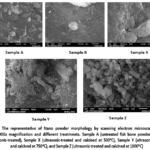 |
Figure 1: The representative of nanopowder morphology by scanning electron microscopy (SEM) with 15000x magnification and different treatments. |
Table 1: Mineral Compositions of nanopowder from Snakehead Fish Bones
|
Mineral Fish Bone Element Content |
Sample (%) | ||||
|
A |
B | X | Y |
Z |
|
| C | 32.70 | 19.33 | 12.07 | 7.59 |
– |
|
N |
9.69 | – | – | – | – |
| O | 31.32 | 51.33 | 48.41 | 46.91 |
49.39 |
|
Mg |
0.32 | 0.52 | 0.44 | 0.50 | 0.49 |
| Al | 0,07 | – | – | – |
– |
|
P |
9.8 | 11.62 | 15 | 17.09 | 18.25 |
| Ca | 15.63 | 16.84 | 23.15 | 27.61 |
31.02 |
|
Zn |
0.46 | – | – | – | – |
| Na | – | 0.35 | 0.42 | 0.29 |
– |
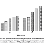 |
Figure 2: Calcium and Phosphorus Content from EDX Elemental Analysis with different treatments. |
FTIR Analysis
Fourier transform infrared (FTIR) analysis was conducted to identify the functional groups and changes in the chemical composition of snakehead fish (Channa striata) bone nanopowders subjected to different treatments. The FTIR spectra of the five samples are presented in Figure 3, revealing differences in the characteristic absorption bands with increasing calcination temperature. Overall, all samples exhibited absorption patterns typical of hydroxyapatite (HAp, Ca₁₀(PO₄)₆(OH)₂), the primary mineral component of bone.
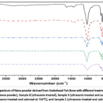 |
Figure 3: FTIR spectrum of nanopowder derived from Snakehead Fish Bone with different treatments. |
X-Ray Diffraction (XRD) Analysis
X-ray diffraction (XRD) analysis was used to determine the crystalline phases formed, the degree of crystallinity, and the lattice parameters of hydroxyapatite (HAp) nanopowder extracted from the bones of the snakehead fish (Channa striata). Hydroxyapatite (HAp) is a crystalline form of calcium phosphate with a chemical composition closely resembling the primary mineral found in human bones and teeth, having the chemical formula Ca₁₀(PO₄) ₆(OH)₂. Crystallinity refers to the percentage of material arranged in an orderly crystalline structure compared to the amorphous or disordered regions, as shown in Figure 4.
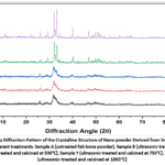 |
Figure 4: X-ray Diffraction Pattern of the Crystalline Structure of nanopowder Derived from Snakehead Fish Bone with different treatments. |
Discussion
SEM-EDX Images Analysis
Sample A (untreated fish bone powder) exhibited irregular particle morphology with a rough and heterogeneous surface.29 Organic substances such as proteins, collagen, and lipids adhering to the bone matrix caused particles to aggregate and form larger clusters.30 This is consistent with the fact that fresh snakehead fish bones contain significant organic components, including proteins and fats, resulting in non-uniform particle surfaces prior to chemical treatment.14 Sample A exhibited an average particle size of 946 ± 348 nm. Ultrasonication during the synthesis and processing of biological nanoparticles has been shown to improve particle dispersion, reduce average particle size, and enhance size distribution homogeneity.31
Sample B exhibited an average particle size of 466 ± 433 nm. After ultrasonic treatment in 1 N NaOH solution at 40 kHz and 50°C for 45 minutes (Sample B), the particles appeared more fragmented and smaller compared to Sample A. However, since Sample B did not undergo calcination, the organic components remained intact, and the true morphology of the HAp crystals was not fully revealed. The ultrasonication process generates acoustic cavitation, the rapid formation and collapse of microbubbles, which produces significant mechanical forces at the microscopic scale. This cavitation effect disrupts particle aggregates while facilitating NaOH solution penetration into the bone matrix to dissolve organic components. Ultrasonication significantly disrupts fish bone particle surfaces through cavitation, triggering the release of hydroxyapatite crystals from the mineralized collagen matrix and increasing the effective surface area of the particles.32
In samples subjected to ultrasonic treatment followed by calcination at 500°C (Sample X), 750°C (Sample Y), and 1000°C (Sample Z), progressively more dramatic morphological changes were observed. Samples X and Y exhibited average particle sizes of 863 ± 1207 nm and 769 ± 515 nm, respectively. Calcination at 500°C removed residual organic components that remained in Sample B, resulting in a cleaner particle surface and increased porosity.33 However, 500°C remains below the threshold for complete HAp recrystallization, which typically occurs above 600°C; consequently, these particles retain irregular, angular morphology with low porosity.34 Research on Catla fish bones demonstrated that within the temperature range of 650–750°C, most organic matter undergoes thermal degradation, resulting in more uniform and compact morphology.35 In agreement with this, that increased calcination temperature promotes HAp particle grain growth and partial sintering, characterized by progressively denser morphology and enlarged grain size due to thermal recrystallization.36,37 Large, elongated particle shapes with small surface protrusions were also observed, characteristic of the HAp recrystallization transition stage at elevated temperatures.36
The very significant morphological transformation in Sample Y is directly related to two main processes: (1) the complete thermal decomposition of all organic components, which releases volatile gases (CO₂, H₂O, NOₓ) on a massive scale, leaving macro- and mesopores in the particle structure, and (2) accelerated recrystallization and growth of HAp crystals at 750°C, resulting in larger crystal domains with more defined crystal boundaries.38 Sample Z exhibits an average particle size of 341 ± 197 nm. In Sample Z (1000°C), the particles exhibit a more compact structure with increased agglomeration. This phenomenon occurs because at elevated temperatures, nanoparticles undergo intense surface diffusion, causing sintering and coalescence of crystal grains.39 Previous study reported that calcination at temperatures ≥900°C of various fish bone sources produces a more regular hexagonal morphology, consistent with the formation of a purer and more crystalline HAp phase.40 Overall, the SEM results indicate that the combination of ultrasonic treatment and calcination successfully transformed the morphology of cork fish bone particles from irregular shapes with high organic content into nanoscale particles with homogeneous and pure structures.
The carbon (C) content decreased significantly from Sample A (untreated fish bone powder) to Sample Z (calcined at 1000°C), indicating that the removal of organic materials became more effective at each stage of synthesis. Sample A, with untreated fish bone powder, had the highest carbon content at 32.70%, whereas in Sample Z, carbon was not detected at all. Oxygen (O) content remained relatively high across all samples, ranging from 31.32% to 51.33%. In Sample A, phosphorus and calcium were present at 9.80% and 15.63%, respectively, while in Sample Z, these elements reached 18.25% and 31.02%. This increase indicates a higher purity in Sample Z, suggesting that the synthesis process at 1000°C was more optimal, as shown in Figure 2. Other elements (Table 1), such as magnesium (Mg), were present in small but consistent amounts across all samples, indicating that magnesium content remained relatively stable throughout the synthesis process. Sodium (Na) was detected in small amounts only in Samples B (ultrasonic-treated), X (ultrasonic-treated and calcined at 500°C), and Y (ultrasonic-treated and calcined at 750°C), likely due to the use of ultrasonic waves in the presence of a NaOH solution during synthesis.
FTIR Analysis
Sample A (untreated fish bone powder) exhibits absorption bands at approximately 1630–1650 cm⁻¹ and 1540 cm⁻¹, corresponding to C=O stretching (Amide I) and N-H bending (Amide II), respectively, which are characteristic markers of collagen in the bone matrix. Absorption bands at 2850–2960 cm⁻¹ reflect C–H stretching from lipids and aliphatic compounds, while the broad band at 3200–3600 cm⁻¹ indicates O–H stretching from water molecules and organic hydroxyl groups.41 Phosphate (PO₄³⁻) bands at approximately 900–1150 cm⁻¹ (P-O stretching) and 560–600 cm⁻¹ (O-P-O bending) confirm the presence of HAp mineral, albeit embedded within the organic matrix. Carbonate (CO₃²⁻) bands at 1410–1450 cm⁻¹ and 873 cm⁻¹ indicate carbonated hydroxyapatite (B-type substitution), which is typical of biogenic HAp from animal sources.14
After ultrasonic treatment with NaOH (Sample B), the intensity of the Amide I and Amide II bands decreased dramatically, confirming successful deproteinization. A noticeable decrease in the C–H stretching bands was also observed, indicating partial elimination of lipids. The relative intensity of the phosphate bands at ~1030 cm⁻¹ (ν₃ PO₄³⁻) and 560–600 cm⁻¹ (ν₄ PO₄³⁻) increased markedly, consistent with purification of HAp minerals from the organic matrix.14 NaOH treatment effectively removed protein components from fish bones, as evidenced by the disappearance of amide bands in the FTIR spectra of the treated samples.42
Sample X (500°C), the Amide and C–H stretching bands have almost entirely disappeared, indicating nearly complete thermal decomposition of organic components. The PO₄³⁻ bands at ~1030 cm⁻¹ and ~960 cm⁻¹ (ν₁ PO₄³⁻, symmetric stretching) have become sharper and more intense. Carbonate bands remain detectable in this sample. The previous study reported that calcination at 650–750°C produces type-B carbonated hydroxyapatite in HAp from Catla fish bones, with carbonate bands persisting at those temperatures.35 Calcination at 950°C completely eliminates all organic components in HAp from carp bones, resulting in highly resolved characteristic phosphate (632 cm⁻¹) and hydroxyl (3572 cm⁻¹) bands.21
Samples Y (750°C) and Z (1000°C), the FTIR spectra show increasingly sharp and well-defined PO₄³⁻ bands at 1090 cm⁻¹, 1046 cm⁻¹, 962 cm⁻¹ (ν₃ and ν₁), as well as 602 cm⁻¹ and 571 cm⁻¹ (ν₄). The OH⁻ bands at ~3572 cm⁻¹ and ~630 cm⁻¹ progressively intensified with increasing temperature, confirming elevated hydroxyl group content characteristic of stoichiometrically pure HAp. Carbonate bands (1410–1450 cm⁻¹ and 873 cm⁻¹) weakened with increasing calcination temperature, indicating decomposition of substituted carbonates. Previous research reported that HAp from black tilapia bones confirmed that higher calcination temperatures produce sharper PO₄³⁻ bands, with the molar Ca/P ratio approaching 1.67 at optimal calcination temperature.37 Overall, the FTIR analysis confirms that the combination of ultrasonic-NaOH treatment and calcination successfully produced biological HAp with a functional group profile progressively approaching that of stoichiometric pure HAp as the calcination temperature increased.
X-Ray Diffraction (XRD) Analysis
HAp is composed of calcium, phosphate, and hydroxyl (OH⁻) ions in a specific ratio, making it highly similar to the mineral components of human bone.43 The diffraction peaks were indexed to JCPDS standard No. 09-0432, confirming the presence of HAp. The degree of crystallinity was calculated by comparing the crystalline area fraction with the sum of the crystalline and amorphous area fractions.44 Based on the analysis results, Sample A (untreated fish bone powder) exhibited a diffraction pattern with broad and weak HAp peaks, indicating a largely amorphous structure with very low crystallinity. This composition is characteristic of fresh biological bone, where the collagen matrix predominates. Research by Fatmawati on biogenic HAp from various animal bone sources confirmed that raw bone powder exhibits broad XRD diffraction peaks due to very small crystallite sizes and high amorphous phase content.45
Sample B (ultrasonic-treated) showed slight improvement in the clarity of diffraction peaks compared to Sample A, though the resulting structure remained relatively amorphous. The identified characteristic HAp peaks include the crystallographic planes (002), (211), (112), (300), (202), (310), (222), (213), and (004), located at their characteristic 2θ angles. This pattern is consistent with the hexagonal crystal system of HAp (JCPDS Card No. 09-0432) with lattice parameters a = b = 9.418 Å and c = 6.884 Å. The slight increase in peak intensity in Sample B relative to Sample A can be attributed to the ultrasonication effect, which removed the protein matrix previously covering the HAp crystal surface, thereby exposing the underlying crystalline structure. Sample B has a crystallinity of 75%. The decrease in crystallinity in Sample B is not an anomaly but rather evidence that ultrasonication without subsequent calcination can damage the crystalline integrity of the HAp naturally present in fish bone. This phenomenon has important implications for the fabrication process. Ultrasonication should be treated as a stage for particle preparation and homogenization, not as an independent method for increasing crystallinity.46
The most dramatic changes occurred in the calcined samples. In Sample X (500°C), the HAp diffraction peaks became sharper, and their intensities increased significantly. The main diffraction peaks at 2θ ≈ 25.9° (002), 31.8° (211), 32.2° (112), and 32.9° (300) became increasingly distinct. This sharpening and intensification reflect the onset of thermal recrystallization, which transforms the amorphous phase into a crystalline one. The study of HAp from horse bone reported that at 500°C calcination, apatite crystallization had already increased significantly, as evidenced by a more structured diffractogram; however, some transitional phases remained. Sample X exhibited a crystallinity of 85%.40
In Sample Y (750°C), the XRD pattern shows sharper and more intense HAp peaks with a smaller Full Width at Half Maximum (FWHM) compared to Sample X. This reduction in FWHM correlates directly with an increase in crystallite size according to the Scherrer equation: D = Kλ/(β·cosθ), where D is crystallite size, K is the Scherrer constant (≈0.9), λ is the X-ray wavelength (0.1542 nm for Cu-Kα), β is the FWHM in radians, and θ is the diffraction angle. Higher calcination temperatures promote greater crystal growth, as reported by a previous study for HAp derived from tilapia bones, where both crystallinity and crystal size increased monotonically with calcination temperature.37 Sample Y exhibited a crystallinity of 93%. This indicates that nearly all the material in these samples is arranged in a highly ordered crystalline structure. Crystalline regions consist of tightly packed molecular chains arranged in an orderly manner, endowing them with higher tensile strength than amorphous regions.47 Amorphous regions have a disordered arrangement of molecular chains.
Sample Z (1000°C) exhibited the sharpest and most intense XRD pattern among all samples, with very narrow peaks at 2θ ≈ 25.9°, 31.8°, 32.2°, 32.9°, 39.9°, 46.7°, 49.5°, 53.2°, and 56.5°, all consistent with the pure hexagonal HAp phase (P63/m). The absence of β-tricalcium phosphate (β-TCP) peaks, which typically appear at temperatures >1000°C, indicates that the formed HAp remains stable at 1000°C. Previous study reported that at 900°C, the apatite structure is highly crystalline with very narrow and sharp diffraction peaks, indicating a high crystal quality of 95%.40 The well-formed hexagonal crystal structure of HAp in Sample Z holds great biomedical potential; it has minimal amorphous content, thereby providing superior physical properties, including good stability and mechanical strength.
XRD analysis confirmed the progressive transformation of the amorphous structure in fish bone powder (Sample A) into highly crystalline HAp (Sample Z) through a series of ultrasonic treatments and increasing calcination temperatures. Thermal treatment, specifically calcination, consistently produces more crystalline HAp compared with alkali hydrolysis or physical treatment.48 This transformation is supported by an increase in the Ca/P ratio, which approaches the theoretical stoichiometry of HAp in Sample Z (1.7), consistent with the EDX data. Thus, this study successfully demonstrates that corkfish bones are a promising source of biological HAp, with XRD characterization confirming the formation of a high-quality crystalline phase under optimized treatment conditions.
Conclusion
The calcination temperature for synthesizing nanopowder from snakehead fish (Channa striata) bones was determined to be 1000°C. SEM analysis revealed a more symmetrical and uniformly distributed particle morphology, with a mean particle size of 341 ± 197 nm, indicating smaller, more homogeneous particles than those produced at other calcination temperatures. The phosphorus and calcium contents were 18.25% and 31.02%, respectively. Furthermore, calcination at 1000°C yielded hydroxyapatite with a crystallinity of 95%. Future research should explore incorporating the optimized HAp nanopowder into bio ceramic- or polymer-based composite scaffolds to evaluate its biocompatibility, mechanical performance, and osteogenic potential for bone tissue engineering applications.
Acknowledgement
The authors would like to express their gratitude to Universitas Sriwijaya.
Funding Sources
The research or publication of this article was funded by DIPA of Public Service Agency of Universitas Sriwijaya 2024, Number: SP DIPA-023.17.2.677515/2024, on November 24th, 2023. In accordance with the Rector’s Decree Number: 0012/UN9/LP2M.PT/2024, On May 20th, 2024.
Conflict of Interest
The authors do not have any conflicts of interest.
Data Availability Statement
This statement does not apply to this article.
Ethics Statement
This research did not involve human participants, animal subjects, or any material that requires ethical approval.
Informed Consent Statement
This study did not involve human participants, and therefore, informed consent was not required.
Clinical Trial Registration
This research does not involve any clinical trials.
Permission to reproduce material from other sources
Not applicable.
Author Contributions
- Herpandi: Conceptualization, Writing-original draft, Supervision, Review & Editing.
- Agusriansyah Saputra: Writing-original draft, Visualization, Review & Editing.
- Indah Widiastuti: Methodology, Writing-original draft, Supervision, Review & Editing.
- Sabri Sudirman: Methodology, Writing-original draft, Supervision, Review & Editing.
- Chalidazia: Data Collection, Analysis, Visualization, Writing-original draft
References
- Gustiano R, Kurniawan K, Kusmini II. Bioresources and diversity of snakehead, Channa striata (Bloch 1793): a proposed model for optimal and sustainable utilization of freshwater fish. IOP Conf Ser Earth Environ Sci. 2021; 762:1-13. doi:10.1088/1755-1315/762/1/012012
CrossRef - Herpandi H, Manurung AS, Sudirman S, et al. Characteristics of nanocalcium derived from snakehead fish (Channa striata) bone meal using ultrasound-assisted extraction. J Food Chem Nanotechnol. 2025;11(2):97-103. doi:10.17756/jfcn.2025-207
CrossRef - Antu MAR, Ali MS, Ferdous MJ, et al. Recovery and characterization of calcium-rich mineral powders obtained from fish and shrimp waste: a smart valorization of waste to treasure. Sustainability. 2024;16(14):1-18. doi:10.3390/su16146045
CrossRef - Hou Y, Shavandi A, Carne A, et al. Marine shells: potential opportunities for extraction of functional and health-promoting materials. Crit Rev Environ Sci Technol. 2016;46(11-12):1047-1116. doi:10.1080/10643389.2016.1202669
CrossRef - Shlisky J, Mandlik R, Askari S, et al. Calcium deficiency worldwide: prevalence of inadequate intakes and associated health outcomes. Ann N Y Acad Sci. 2022;1512(1):10-28. doi:10.1111/nyas.14758
CrossRef - Savlak N, Çağındı Ö, Erk G, et al. Treatment method affects color, chemical, and mineral composition of seabream (Sparus aurata) fish bone powder from by-products of fish fillet. J Aquat Food Prod Technol. 2020;29(6):592-602. doi:10.1080/10498850.2020.1775742
CrossRef - Senphan T, Mungmueang N, Choommongkol V, et al. Improving calcium bioavailability from fish bone waste: the role of sodium hydroxide in bio-calcium extraction from tilapia (Oreochromis niloticus) bones. Food Chem X. 2025; 31:1-12. doi: 10.1016/j.fochx.2025.103126
CrossRef - Lekahena V, Faridah DN, Syarief R, et al. Physicochemical characterization of nano calcium from tilapia bone extracted by alkaline and acid solution. J Teknol dan Ind Pangan. 2014;25(1):57-64. doi:10.6066/jtip.2014.25.1.57
CrossRef - Nemati M, Huda N, Ariffin F. Development of calcium supplement from fish bone wastes of yellowfin tuna (Thunnus albacares) and characterization of nutritional quality. Int Food Res J. 2017;24(6):2419-2426
- Kusumawati P, Triwitono P, Anggrahini S, et al. Nano-calcium powder properties from six commercial fish bone waste in Indonesia. Squalen Bull Mar Fish Postharvest Biotechnol. 2022;17(1):1-12. doi:10.15578/squalen.601
CrossRef - Saputra A, Rahmawati L, Herpandi H, et al. Preparation and characterization of chitosan and κ-carrageenan-based nanocomposite coatings utilizing different technologies for food packaging applications. Mater Plast. 2025;62(1):64-77. doi: 10.37358/Mat.Plast.1964
CrossRef - Gunasekaran T, Haile T, Nigusse T, et al. Nanotechnology: an effective tool for enhancing bioavailability and bioactivity of phytomedicine. Asian Pac J Trop Biomed. 2014;4(Suppl 1):S1-S7. doi:10.12980/APJTB.4.2014C980
CrossRef - Kadam SU, O’Donnell CP, Rai DK, et al. Laminarin from Irish brown seaweeds Ascophyllum nodosum and Laminaria hyperborea: ultrasound assisted extraction, characterization and bioactivity. Mar Drugs. 2015;13(7):4270-4280. doi:10.3390/md1307427
CrossRef - Herpandi, Hanif I, Widiastuti I, et al. Hydroxyapatite characteristics from snakehead fish (Channa striata) bone via alkali treatment followed by calcination method. Trop J Nat Prod Res. 2024;8(2):6147-6151. doi:10.26538/tjnpr/v8i2.12
CrossRef - Guo J, Zhu S, Chen H, et al. Ultrasound-assisted solubilization of calcium from scale ground fish bone particles. Food Sci Nutr. 2022;10(10):712-722. doi:10.1002/fsn3.2696
CrossRef - Talib A, Zailani K. Extraction and purification of yellowfin tuna fishbone flour as an ingredient of future traditional medicine. IOSR J Pharm. 2017;7(11):8-14.
- Tawali A, Wakiah N, Qanitah K, et al. The effect of sonication time on physicochemical profiles of the nanocalcium from snakehead fish bone (Channa striata). IOP Conf Ser Earth Environ Sci. 2019; 355:1-6. doi:10.1088/1755-1315/355/1/012091
CrossRef - Han Z, Mu Z, Yin W, et al. Biomimetic multifunctional surfaces inspired from animals. Adv Colloid Interface Sci. 2016; 234:27-50. doi: 10.1016/j.cis.2016.03.004
CrossRef - Hasan MR, Yasin NSM, Ghazali MSM. Proximate and morphological characteristics of nano hydroxyapatite extracted from fish bone. J Sustain Sci Manag. 2020;15(8):9-21. doi:10.46754/jssm.2020.12.002
CrossRef - Venkatesan J, Qian ZJ, Ryu B, et al. A comparative study of thermal calcination and an alkaline hydrolysis method in the isolation of hydroxyapatite from Thunnus obesus Biomed Mater. 2011;6(3):1-12. doi:10.1088/1748-6041/6/3/035003
CrossRef - Hammood AS, Hassan SS, Alkhafagy MT, et al. Effect of calcination temperature on characterization of natural hydroxyapatite prepared from carp fish bones. SN Appl Sci. 2019;1(5):1-12. doi:10.1007/s42452-019-0396-5
CrossRef - Terzioğlu P, Öğüt H, Kalemtaş A. Natural calcium phosphates from fish bones and their potential biomedical applications. Mater Sci Eng C. 2018; 91:899-911. doi: 10.1016/j.msec.2018.06.010
CrossRef - Putranto HF, Asikin AN, Kusumaningrum I. Properties of belida (Chitala) fish bone powder as calcium source based on protein hydrolysis method. ZIRAA’AH. 2015;40(1):11-20. doi:10.31602/zmip. v41i1.315
- Malahayati N, Wardhani TW, Alsoyuna NS. The effect of extraction time on the physicochemical characteristics of nanocalcium powder from chicken and duck eggshells. Potravin Slovak J Food Sci. 2021; 15:712-722. doi:10.5219/1675
CrossRef - Venkatesan J, Kim SK. Effect of temperature on isolation and characterization of hydroxyapatite from tuna (Thunnus obesus) bone. Materials (Basel). 2010; 3:4761-4772. doi:10.3390/ma3104761
CrossRef - El-Deeb MM, Ads EN, Humaidi JR. Evaluation of the modified extracted lignin from wheat straw as corrosion inhibitors for aluminum in alkaline solution. Int J Electrochem Sci. 2018;13(5):4123-4138. doi:10.20964/2018.05.49
CrossRef - Huang YC, Hsiao PC, Chai HJ. Hydroxyapatite extracted from fish scale: effects on MG63 osteoblast-like cells. Ceram Int. 2011;37(6):1825-1831. doi: 10.1016/j.ceramint.2011.01.018
CrossRef - Olokoba SW, McIver FA. Synthesis and characterization of graphene oxide from agricultural waste: cocoa pod (Theobroma cacao). J Mater Environ Sci. 2024;15(9):1208-1218
- Abdulrahman I, Tijani HI, Mohammed BA, et al. From garbage to biomaterials: an overview on eggshell-based hydroxyapatite. Sci World J. 2014;2014(1):1-6. doi:10.1155/2014/802467
CrossRef - Hasanuddin DNA, Dewi SP, Fatmawati S, et al. Nano calcium from marine fish bones via high-energy ball milling: a potential natural supplement for bone health. J Bone Metab. 2025;32(4):285-296. doi:10.11005/jbm.25.875
CrossRef - Jin B, Bang H, Suslick KS. Applications of ultrasound to the synthesis of nanostructured materials. Adv Mater. 2010;22(10):1039-1059. doi:10.1002/adma.200904093
CrossRef - Guo J, Zhu S, Chen H, et al. Ultrasound-assisted solubilization of calcium from micrometer-scale ground fish bone particles. Food Sci Nutr. 2022;10(3):712-722. doi:10.1002/fsn3.2696
CrossRef - Figueiredo M, Fernando A, Martins G, et al. Effect of the calcination temperature on the composition and microstructure of hydroxyapatite derived from human and animal bone. Ceram Int. 2010;36(8):2383-2393. doi: 10.1016/j.ceramint.2010.07.016
CrossRef - Shi P, Liu M, Fan F, et al. Characterization of natural hydroxyapatite originated from fish bone and its biocompatibility with osteoblasts. Mater Sci Eng C. 2018; 90:706-712. doi: 10.1016/j.msec.2018.04.026
CrossRef - Bee SL, Noor SNFM, Ul-Hamid A, et al. Effect of calcination temperature on the physicochemical properties of natural hydroxyapatite derived from Catla fish bone. J Phys Conf Ser. 2024;2907(1):012010. doi:10.1088/1742-6596/2907/1/012010
CrossRef - Haberko K, Bu MM, Brzezi J, et al. Natural hydroxyapatite—its behaviour during heat treatment. J Eur Ceram Soc. 2006; 26:537-542. doi: 10.1016/j.jeurceramsoc.2005.07.033
CrossRef - Dermawan SK, Ismail ZMM, Jaffri MZ, et al. Effect of the calcination temperature on the properties of hydroxyapatite from black tilapia fish bone. J Phys Conf Ser. 2022;2169(1):012034. doi:10.1088/1742-6596/2169/1/012034
CrossRef - Guo X, Yan H, Zhao S, et al. Effect of calcining temperature on particle size of hydroxyapatite synthesized by solid-state reaction at room temperature. Adv Powder Technol. 2013;24(6):1034-1038. doi: 10.1016/j.apt.2013.03.002
CrossRef - Kokubo T, Kim HM, Kawashita M. Novel bioactive materials with different mechanical properties. Biomaterials. 2003; 24:2161-2175. doi:10.1016/S0142-9612(03)00044-9
CrossRef - Touareb D, Latifi S, Saoiabi S, et al. Influence of calcination temperature on equine bone hydroxyapatite structure and lead adsorption efficiency. Sci Rep. 2025; 15:33990. doi:10.1038/s41598-025-11961-4
CrossRef - Charlena, Bikharudin A, Wahyudi ST, et al. Synthesis and characterization of hydroxyapatite-collagen-chitosan (HA/Col/Chi) composite by using ex-situ wet precipitation method. Rasayan J Chem. 2017;10(3):766-770. doi:10.7324/RJC.2017.1031768
CrossRef - Muñoz F, Haidar ZS, Puigdollers A, et al. Efficient hydroxyapatite extraction from salmon bone waste: an improved lab-scaled physico-chemico-biological process. Molecules. 2024;29(17):4002. doi:10.3390/molecules29174002
CrossRef - Ismail NF, Hamzah S, Razali NA, et al. Preparation and characterisation of hydroxyapatite extracted from fish scale waste for the removal of gallic acid as inhibitor in biofuel production. Malaysian J Anal Sci. 2019;23(6):938-949. doi:10.17576/mjas-2019-2306-03
- Salem KS, Kasera K, Rahman A, et al. Comparison and assessment of methods for cellulose crystallinity determination. Chem Soc Rev. 2023; 52:6417-6446. doi:10.1039/d2cs00569g
CrossRef - Fatmawati S, Istiqomah SM, Hasanah N, et al. Physico-chemical characterization of natural nano calcium extracted from different fish bones in catfish (Clarias gariepinus) and snakehead fish (Channa striata). Case Stud Chem Environ Eng. 2025; 11:101080. doi: 10.1016/j.cscee.2024.101080.
CrossRef - Guo J, Zhu S, Chen H, Zheng Z, Pang J. Ultrasound-assisted solubilization of calcium from micrometer-scale ground fish bone particles. Food Sci Nutr. 2022; 10:712–722. doi:10.1002/fsn3.2696
CrossRef - Shi CY, Qin WY, Qu DH. Semi-crystalline polymers with supramolecular synergistic interactions: from mechanical toughening to dynamic smart materials. Chem Soc Rev. 2024; 15:8295-8310. doi:10.1039/d4sc02089h
CrossRef - Granito RN, Muniz Renno AC, Yamamura H, de Almeida MC, Menin Ruiz PL, Ribeiro DA. Hydroxyapatite from fish for bone tissue engineering: a promising approach. Int J Mol Cell Med. 2018;7(2):80–90. doi: 10.22088/IJMCM.BUMS.7.2.80
Abbreviations
EDX Energy Dispersive X-ray
FTIR Fourier Transform Infrared Spectroscopy
HAp Hydroxyapatite
SEM Scanning Electron Microscopy
XRD X-ray Diffraction
 PlumX:
 Views: 1868
  PDF Downloads:
257
PDF Downloads:
257 
Accepted on: 29 Jun 2026
Second Review by: Sudhanya Nath
Final Approval by: Dr. Rajesh Jeewon





Web of Science Coverage
Emerging Sources Citation Index (ESCI)
2024 Journal Impact Factor: 1.1
Scopus Journal Metrics
CiteScore 2025: 2.6
CiteScore Details
Sustainable Nutrition: Food Systems, Nutrient Retention, and Public Health Impact

This journal is a member of, and subscribes to the principles of, the Committee on Publication Ethics (COPE)