
Introduction
Pathophysiology of Diabetes mellitus
The escalating prevalence of diabetes mellitus (DM) remains a challenge in the medical field. The World Health Organization (WHO) report on the ascending resistance in microorganisms and the Global Antimicrobial Resistance Surveillance System (GLASS) report predict a critical future for humankind1. Similarly, the International Diabetes Federation Atlas (IDFA) previously estimated a population of greater than 425 million with DM including 4 million deaths annually2 while the report of 2022 and 2023 highlight further complications of DM leading non healing foot wounds among others. The American Diabetes Association (ADA) in 2024 estimated 643 people to have diabetes by 2030 if left untreated3. If left untreated, DM may not only have devastating effects but represent consequent economic burden universally4. Several microorganisms namely, Acinetobacter species, Bacillus species (Bacillus sp.), candida albicans, Enterococcus sp., Escherichia coli (E. coli), Klebsiella sp., Morganella morganii (M. Morganii) Pseudomonas aeruginosa (P. aeruginosa), Proteus sp., Staphylococcus aureus (S. aureus), among others have been detected as invasive microbiota of foot ulcers5,6,7,8. It should be noted that microvascular complications of DM lead to retinopathy9 and neuropathy diseases while macrovascular complications lead to coronary diseases and cerebrovascular diseases10. The limited volume of epidermal growth factors in non-healing foot wounds (NHFW) contribute to chronic foot ulcers (25%), which blend in treatment failures, lower limb amputation notably 85% of all amputations, poor quality of life and shortly premature death5.
Treatment; currently used antiseptics
The disordered physiological process that causes DM is yet to considered as formerly distinguished medicines like insulin-sensitizing biguanide, also known as metformin and thiazolidinedione (rosiglitazone and pioglitazone) aim at hindering single molecules, such as adenosine monophosphate-dependent kinase (AMPK)-6-2 enzyme and peroxisome proliferator activated receptor gamma (PPARˠ) 96-311 also known as the reverse insulin receptor. However, due to health complications such as lactic acidosis resulting from derivatives of the above, phenformin and buformin are no longer used in various countries12. Topical antiseptics are praiseworthy as they act on bacteria, fungi, viruses, with minimal side effects unlike several drugs that have restricted targets13. They also allow movement of keratinocyte into wound site to start the re-epithelisation, and eventually wound healing, Timolol has been currently the choice of topical in the treatment of foot ulcers14. Sponges, hydrogels, films, hydrocolloids and hydrofiber mats have been simultaneously used as innovative dressings due to their ability to provide safe and suitable environment while distributing active ingredients to wound site to ease the healing process14. Other commonly used antiseptic compounds include alcohol, iodine, biguanides, halophenols, bisphenols, silver, gold and zinc nanoparticles15 hydrogen peroxide. However, owing to the distinctive biological and the complex system of tissue repair, new effective and targeted cures are constantly required14. A more recent technique to better understand the microbiota of organisms colonising NHFW is the 16s-ribosomal DNA sequencing, which helps limit inflammatory process thus aid in the healing process16. Hence, this review paper highlights the potential therapeutics for chronic wounds infections in terms of antimicrobials from the marine environment.
Traditional herbs and freshwater algae as antimicrobials
In addition to antimicrobial properties, plants contain anti-inflammatory substances due to their phytochemical constituents that lighten human pain by reducing swelling17 while traditional herbs are often recognized to mimic drugs, known as phytopharmaceuticals18 which eventually help in recovery of wounds. Moreover, there are about 2.7-4 million species of endophytes residing in plants19 which rather than harming their internal tissues, develops similar bioactive substances as the host plants and endophyte richness is especially observed among tropical plants. The discovery of novel antibiotics in terms of fungal endophytes is a promising substitute to overcome drug resistance by pathogens ever since Alexander Fleming was credited with the discovery of penicillin in 1928 and may be supported by their antibacterial, antiparasitic and antifungal properties and their accessibility20. Ficus religiosa, from the family of Moraceae, has been reported to be effective against DM as its constituents have similar effect as enzymes α-glucosidase and α-amylase, that degrade polysaccharide into glucose21. Phytochemicals in green algae exhibit antimicrobial properties22. For instance, chlorellin derived from freshwater green microalgae Chlorella, inhibits human pathogens isolated from NHFW22. Similarly, Dunaliella salina and freshwater Pseudokirchneriella subcapitata microalgae have bactericidal properties against S. aureus, P. aeruginosa, E. coli, and Klebsiella sp.23, which are the most common pathogens isolated from NHFW.
Aspects of common antibiotics and antimicrobial resistance
Men and microbes have been forever co-existing and co-evolved. Antibiotics have been widely used for healing purposes1 and for increasing human life expectancy as infections with resistant organisms lead to several diseases and death in men. Antibiotics should have selective toxicity, either be bactericidal, that is they completely kill bacteria or bacteriostatic, that is they inhibit the growth of bacteria24. Satisfactory antimicrobials should also be able the hinder the growth of organisms regardless of the affected area with minimal side effects on the host; should reach the target at effective concentration; have tolerance residue and should have a defined Minimum Inhibitory Concentration (MIC). The spectrum activity, economic aspect and availability are key elements that should be considered on selection of antibiotics25. The overuse, inappropriate prescribing, incorrect dosages and duration of treatment has progressively led to antimicrobial resistance (AMR)26; one of the major drawbacks in the treatment of long-term complications of DM. AMR arises when bacterial pathogens evolve to mutate; that is acquire new genes in a way to decrease or annihilate the potency of antibiotics27. In this way, the antibiotics is less effective, which permit bacteria to thrive28. The comprehension of the biochemical and genetic basis of AMR is of paramount importance as antimicrobial selection may differ according to the two resistance mechanism25. In the intrinsic resistance mechanisn, there is inhibition of cell wall synthesis and permeability through it as well as inhibition of protein synthesis while in the acquired resistance mechanism, the function of nucleic acids is hampered by mutations25.
Resistance according to classes of antibiotics and multidrug resistance
Multidrug resistance (MDR) is the resistance of microorganisms to the administered antimicrobials despite previous sensitivity to them. MDR organisms have emerged from both the hospital environment and community settings28 by bacterial adaptation. A report published by WHO stated that infections are generated by resistant pathogens such as E. Coli against broad spectrum antibiotics namely cephalosporin and fluoroquinolones, Klebsiella pneumoniae (K. pnemoniae) against cephalosporin and carbapenems, S. aureus against narrow spectrum methicillin and Streptococcus pneumoniae (S. pneumoniae) against penicillin25. Organisms secrete several enzymes including beta-lactamase that often break down drugs resulting in beta lactamase antibiotic resistance29. Tetracyclines group of antibiotics, widely used in several treatment prevent the attachment of aminoacyl-tRNA to the ribosomal acceptor and operate by inhibiting protein synthesis. The three main modes of action of resistance to tetracyclines are: 1) efflux of the antibiotics; 2) ribosome protection and 3) modification of the binding sites30. Aminoglycosides, another group of antibiotics exert their activity by irreversible binding of ribosomes and inhibiting protein synthesis30. The mechanisms of aminoglycoside resistance include: 1) Decreased permeability or reduced uptake by cell membrane modification; 2) alterations at ribosomal binding sites through mutations and 3) production of aminoglycosides modifying enzymes30,31. Fourth group of antibiotics, Fluoroquinolones antimicrobial activity is due to interference with deoxyrobonucleic acid (DNA) replication and transcription in bacteria through inhibition of certain bacterial topoisomerase enzymes30. The two mechanisms of resistance to fluroquinolones include 1) modification in drug target enzymes; 2) changes that decrease penetratiom of the drug to the site30. Macrolides, a fifth group of antibiotics meddles with amino acid during protein synthesis. The mechanisms of resistance to macrolides are 1) post-transcriptional modification; 2) the presence of efflux proteins and 3) enzymatic inactivation32. Glycopeptides, the sixth group, achieve bacteriostatic effect by interfering with D-alanyl-D-alanine side chains. Resistance mechanism to glycopeptide is by the production of innovative genes which promote regeneration of the peptidoglycan side chain33. Sulfonamides, the seventh group of antibiotics proceed by competitively blocking the p-amino-benzoic acid (PABA) which is a chief compound in bacterial folic acid synthesis. Also, constantly evolving genes render organisms resistant to Sulfonamides, as the latter fail to bind and attack the new structure of microbials34. Eighth, the oxazolidones interfere with the 80S initiation complex at the P site and prevent protein biosynthesis. Resistance to oxazolidone drugs is generally due to mutations in the six domains V of 23S, nucleotide long component ribosomal ribonucleic acid (rRNA) of the organisms. Fortunately, owing to the presence of multiple copies of redundant rRNA genes in bacterial genomes, resistance to oxazolidones hardly occurs and develops steadily25. The search for new antibiotics remains vital for a better management of public health35.
Benefits of the marine environment
The marine environment is associated with wide range of thermal pressure, has vast surfaces which receive sunlight as well as aphotic zones, with a wide range of nutrients and low oxygen zones which are about 300m to 700m deep down36. The ocean therefore comprises of several living organisms including plants and algae, among others, that produce extensive secondary metabolites to safeguard themselves against other species such as marine herbivores, for reproduction purposes as well as to keep pace with competitors for survival37. By evidence, marine organisms constantly evolve to adapt themselves in harsh environmental conditions37. As contrast to terrestrial organisms, various organic products are derived from marine organisms due to the elaborated living diversity of species and hence have more powerful biodiversities38. Sponges, molluscs, tunicates and macroalgae are the common marine organisms39. Algae are neither plants nor animals but rather belong to a group of living things called protists. Seaweed comprises of species of macroscopic, multicellular algae sticking to rocks and found along the seashore in abundance in every ocean40.
New sources of therapeutics in the marine environment
There have been several controversies surrounding the responsiveness of existing drugs. Despite vigorous attempts from the whole world to enhance knowledge and tackle AMR, since July 2017, only eleven new drugs, most associated with pre-existing classes with restricted clinical benefits have been approved41. The marine organisms produce several residues by break down of their natural components that could be used to fuel the futured discovery pipeline of drugs which are currently less explored42. During the period 1998-2008, initially the global marine pharmaceutical analysis included 806 chemicals which expressed antibacterial, antifungal, among others, with actions on the cardiac, hormonal and sense organs43. By 2022, there has been a huge increase in seawater pharmaceuticals as around 37, 542 novel natural products have been recorded and authorized for official use in the market as pharmaceuticals44. Eminent marine drugs are derived from marine species of bacteria, virus, algae, fungi and sponge37, which have been modified to fight against competitive organisms by showing defence mechanisms against those that swallow them, plants that grow on them for support and organisms that pollute the surrounding with excrement45. The possible rationale for the enhancement of antibiotic production in microorganisms in terms of bacteria is due to their fierce competition for space, nutrient and potential to kill competitor bacteria in their environment38. One of the major side effects of NHFW is of inflammation and amphichopyrones A, a marine fungus derived component has been shown to reduce inflammatory effects by inhibiting nitric oxide production44. Phomoxanthone A, another marine fungus derived natural product, was seen to have cytotoxic effects by aiming at the inner mitochondrial membrane of cancer cells, hence having a specific target44. Also, advanced technology like CRISPER (Clustered regularly interspaced short palindromic repeat) gene editing technique can be used to alter the host plant genomes to achieve the goal46. The above could hence be implemented in genome mining of coastal greens to hamper the regulatory hurdles in diabetic foot treatment and enhance existing therapies. The various species of microorganisms indigenous to the sea that portray antimicrobial is noticeable, hence isolation and identification of specific antibiotics have been attempted, but few of them are in preclinical and clinical trials47. Exploring the marine microbials for their coaction with existing drugs is therefore worthwhile45.
Mangroves and algae
Mangroves are shrubs or trees that grow mainly in coastal areas and brackish water in tropical regions, which also adapt in harsh costal conditions48 during climate change. Mangroves are distributed over an area of 152,000 km2 globally49. 84 mangrove species are known worldwide, while 80 of them considered true mangroves and the other 14 are semi-mangrove50. The inhabitants of the coastal regions have acknowledged the benefits of mangroves as the latter have been used as alternatives to drugs for a plethora of diseases51. Flavonoids, saponins, alkaloids and phenolics are the bioactive constituents of Excoecaria agallocha, whose methanolic extracts displayed antibacterial activity52. Other mangroves used in the medical field to treat hypoglycemic activities include Acanthus ilicifolius, Avicennia marina, Rhizophora apiculate, Bruguiera gymnorhiza and Rhizophora mucronatae, Xylocarpus granatum, Xylocarpus moluccensis, Xylocarpus granatum, Sonneratia species52.
Seaweeds (macroalgae) possess fat, water soluble vitamins, essential minerals and other bioactive agents to combat attributes of the ecosystem such as UV photo damage, high sodium chloride, oxygen toxicity, and stress caused by bacterial colonization53. Previous studies demonstrated that methanolic extract of Halimeda species, part of the green macroalgae family inhibited the growth of Bacillus subtilis (B. subtilis), Bacillus cereus (B. cereus) and S. aureus54 while methanolic extract of Sargassum polycystum, a brown macroalgae inhibited E. coli, Proteus vulgaris, K. pneumoniae among others55. Literature review revealed that green algae have higher potential to inhibit gram positive bacteria than gram negative bacteria54. Prorocentrum isolates, which forms part of the microalgae family, have been previously identified to have desirable antagonistic effects against Methicillin-resistant S. aureus (MRSA), Enterococcus faecalis (E. faecalis), among others56. Marine structures are grouped into different chemical classes that is alkaloids, polyketides, peptides, shikimates, sugars and terpenes57. Chemical substances from biologically active seaweeds have been assessed in tissue repair by increasing the feasibility of collagen fibrils or by preventing cell degeneration, or by enhancing DNA synthesis while the brown algae were described as anti inflammatory agent58.
Impact of coastal greens on public health and economy
Our public health policy is to elevate the health status of our citizens and at the same time guarantee that health goals are met. Thus, the merit of use of marine herbs in routine use, is to mitigate antimicrobial resistance with the intention to provide medications to patients in a brief delay as they are easily and freely available in several countries. Using coastal greens as drugs are presumed to be sustainable as they will be constantly available and reproducible, with a minimum cost to pharmaceutical companies. Some countries found using traditional herbs for the treatment of DM highly decrease the burden of the cost of conventional medicine as they are easily available and reproducible despite climate change as native plants adapt to their surroundings59. WHO predicted 10 million of deaths on a yearly basis by 2050 due to AMR60. Since AMR is a global issue, several pharmaceutical companies may have an increased turnover by business interaction with foreign companies owing to production of drugs with their unique biodiversity of marine herbs. On the other hand, harvesting marine herbs may be laborious in bad weather conditions and their extensive exploitation may lead to their depletion60,61 and may hence cause an imbalance of the natural habitat of the oceans leading to deaths of marine organisms. Restoring the damage and caused to the ecosystem might take considerable time and be costly.
Review methodology
Relevant literature and representative case reports were collected by scrutinising scientific electronic database namely EBSCO, PubMed and Science Direct from the year 2015 to 2024. Keywords marine drugs, traditional drugs, antihyperglycemic, pharmaceutical properties and pharmacological activities were used. Article assemblage was accomplished based on a search strategy. Primarily, a generalised search was executed in the above-mentioned databases, following a screening of the papers which led to their inclusion in this study. Inclusion criteria were based on diversity of marine organisms, their sustainability and availability, their antimicrobial properties and their use in the future to relieve the burden of AMR. Also, genome mining and other novel technologies using bioinformatic analysis for identifying the structure of marine organisms were searched for reproducibility. Data was then gathered and compiled as appropriate as shown in the flow diagram below.
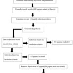 |
Figure 1: A flowchart showing the methodology of the review paper |
Discussion
DM forms part of the major universal health pathophysiology’s of the current era. There is an undeviating link between family history, increased mechanization, sedentary lifestyle, weight gain, education level, dietary pattern and prevalence of DM2. Diabetic foot ulcer (DFU) is defined as the presence of diabetic foot pathologies such as neuropathy and ischaemia, which NHFW. Neutrophils and macrophages are inflammatory cells that play a part in cleanup the wound of pathogens, cell debris and damaged extracellular matrix, and are therefore favourable and vital for restoring damaged skin62. Excessive inflammation rate hampers the typical formation of new cells that migrate and multiply in infected sites, which eventually leads to compromised wound healing63.
Evolving ailments have overburdened the existing drug resources and led to AMR and as the pursue for novel drugs proceeds, confrontation to known drugs continue to rise47. Accumulated evidence from another study of suggest that the highly impermeable cell wall of gram-negative bacteria may significantly contribute to AMR64. For instance, penicillin was primarily used to treat S. aureus infections, followed by methicillin, which has now changed to vancomycin 65. These researchers also enumerate that resistance to macrolide classes of antibiotics is due to mutations in the ribosomal proteins and enzymatic modification of ribosomal target during drug binding. Hence, treatments to reach specific targets need to be updated in different ecological niches.
About 500,000 natural compounds have been describe globally, among which, 80,000 are bacterial and fungal-derived compounds; 29% of which is derived from actinomycetes66. Roughly 80% of the antibiotics used from 1950 to 1980 were isolated from actinomycetes from the genus Streptomyces, by 1980, at most eleven actinomycetes genera were disclosed, subsequently one hundred genera by 2005 and two hundred and twenty genera by 201066. From the same study, it was shown that out of thirty new antibiotics launched worldwide since 2000, two were natural products (NP), twelve were NP-based and sixteen were synthetic66. A category of organisms adjusts exceptionally in salty environments and gravitational pressure exerted by the ocean due to anthropogenic activities, hence the assortment of microorganisms in marine habitats is quite remarkable67. Reactive oxygen species (ROS) are end products of biochemical pathways of metabolism, which upon excess accumulation prevents the body to clean these free radicals, thus causing aerobic damage, which leads to further diabetic complications53. Chemical compounds from oceanic sources inherit altered shapes in the
foundation or side chain in contrast to human origin, which make them the design of choice for drugs. The peptides taken from ascidians, sponges, as well as mollusks also provide huge great stability from enzymatic degradation as well as thermal conditions44. A wide selection of bioactive peptides are the end products of the association that links microorganisms and the marine creatures68.
Moreover, cephalosporins, usual drugs were refined from the marine fungus Cephalosporium acremonium37. Other antibiotics from saltwater fungi include Peniciadametizine A and B from Penicillium adametzioides in addition to other Penicillium spp. from sponges associated, mangroves associated, and marine algae associated69. Since then, extended investigation has been conducted to reveal the bioactivity of marine microbes such as bacteria, fungi, actinomycetes and microalgae-cyanobacteria and the results have been fruitful as antibiotics. The active components derived from several marine organisms and their corresponding antimicrobial effect on common pathogens isolated from NHFW are listed in Table 1. The mechanisms of action of each strain or bioactive compound have not been documented and the marine organisms have been collected from several coastline globally.
Table 1: Some bioactive compounds isolated from marine organisms.
|
Organisms isolated from |
Marine organisms |
Bioactive compound |
Coastal region |
Antimicrobial activity against |
Reference |
|
Bacteria |
Alteromonas spp. |
strain 1, strain 6 | Rameswaram and Pudhumadam | S. aureus | [70] |
| Strain 20 | S. aureus, E. coli |
[70] |
|||
|
Pseudomonas spp. |
Strain 4, strain 5, strain 13, strain 24, strain 25 | S. aureus | [70] | ||
| Pseudomonas spp. | Strain 18 | S. aureus. E. coli |
[70] |
||
|
Marinobacter spp. |
Strain 8 | S. aureus | [70] | ||
| Marinobacter spp. | Strain 14 | E. coli |
[70] |
||
|
Bacillus spp. |
Strain 16 | S. aureus, E. coli | [70] | ||
| Bacillus spp. | Bogorol | MRSA, vancomycin resistant Enterococcus (VRE) |
[71] |
||
|
Pseudomonas bromoutilis |
2,3,4-tribromo-5(1′hydroxy,2′,4′- dibromophenyl) | Puerto Rico | S. aureus | [71] | |
| Pseudomonas spp.* | Pyrone-I | MRSA, VRE |
[71] |
||
|
Hahella spp. |
prodigiosin | Nagasaki Prefecture, Japan | S. aureus | [72] | |
| Halobacillus litoralis YS3106 strain | halolitoralins A | C. albicans |
[72] |
||
|
Pseudomonas UJ-6 |
1-acetyl-beta-corboline | S. aureus | [72] | ||
| S. arenicola | Salinaphthoquinones | S. aureus, E. faecalis |
[72] |
||
|
Bacillus spp. |
Bacicyclin | mussel Mytilus edulis | S. aureus, E. faecalis | [72] | |
| Pseudoalteromonas s. | Heat tolerant cell-free culture supernatant | Coral (Platygyra sp.) | B. cereus, S. aureus |
[72] |
|
|
S. sampsonii SCSIO 054 |
Julichrome Monomers | Mollusk Batillaria zonalis | S. aureus | [72] | |
| B. flexus EED 15 and S. lienomycini EED 16 | Seagrasses (Cymodocea sp., Enhalus acoroides, Syringodium sp., and Thalassia hemprichii) | E. coli, S. aureus |
[72] |
||
|
Actinomyces |
Nocardiopsis dassonville | kahakamides A | Hawaii | B. subtilis | [71] |
| Nocardiopsis dassonville | N-(2-hydroxyphenyl)- 2-phenazinamine | Hawaii | Candida albicans |
[71] |
|
|
Micromonospora spp. |
Tetroarcin | Toyama Bay, Japan | B. subtilis, S. aureus | [72] | |
| Streptomyces | Streptomyces** | RM80 strain | S. aureus, C. albicans |
[71] |
|
|
Streptomyces spp. |
1492 Strain | Diu Island, Arabian Sea | S. aureus, A. baumanii | [72] | |
| Streptomyces MUSC 135T | bacitracin A | Mangrove forest, Peninsular Malaysia, east coast | S. aureus |
[72] |
|
|
Streptomyces CTF9 |
Indole-3-lactic acid,phenylacetic acid | C. albicans | [72] | ||
| Streptomyces B8080 | chalcomycin | Mangrove sediment in Pohoiki, Hawaii | S. aureus, B. subtilis, E. coli |
[72] |
|
|
Streptomyces spp. |
Aborycin | Deep-sea sediments of the South China Sea | S. aureus | [72] | |
| Streptomyces SMS806 | albonoursin | deep-sea sediments in the South China Sea | S. aureus |
[72] |
|
| Cyanobacterium | Hormoscilla spp. | anaephenes A-C | S. aureus | [72] | |
| sponges | Holothuria tubulosa | peptide fractions | Surface adherent Staphylococci, P. aeruginosa | [71] | |
| Xestospongia testudinaria | S. aureus, E. coli, K. pneumoniae, P. aeruginosa | [71] | |||
| Xestospongia exigua | diethyl ether fraction | S. aureus, E. coli | [71] | ||
| Agelas clathrodes | ethanoic extract | S. aureus | [71] | ||
| Phyllospongia lamellose t | Phyllospongin D, phyllospongin E, | Red sea | |||
| Phyllospongia lamellose t | 12α-acetoxy-13β,18βcyclobutan-20,24-dimethyl-24oxoscalar-16-en-25β-ol | B. subtilis, S. aureus | [71] | ||
| Hertios erectus | B.subtilis | [71] | |||
| Suberea mollis | Subereamollines A, Subereamollines B, Subereaphenols B, and C | S. aureus, P.aeruginosa, K. pneumoniae | [71] | ||
| Negombata magnifica | Latrunculin T | Red sea | S. aureus, B. cereus, C. albicans | [71] | |
| Amorphinopsis spp. | Eutypella species, Lasiodiplodia theobromae, Fusarium solani | Indonesia mangrove | ESBL E. coli | [71] | |
| Aplysina aerophoba | cyclic lipopeptides from Bacillus species | Mediterranean Sea | C. albicans, S. aureus, E. coli | [71] | |
| Fungus | Penicillium minioluteum | Purpurides E and F | Red sea | MRSA, E. coli, C. albicans | [71] |
| Soft Coral | Gorgosten-5(E)-3β-ol, Sarcoaldosterol A. | B. subtilis, S. aureus, E. coli | [71] |
*The mechanism of action of the Pyrone-I targets the bacterial cell membrane
** RM80 strain acts on the N-acetylglucosamine
Since the exploration of species not formerly cultivated, could relieve the pressing demand to resistance against currently used antibiotics66, ethnomedicines have been recognized by WHO as successful therapeutic agents with application to aquaculture due to the loss of antimicrobial efficiency and the struggle of pathogens to antibiotics50. In line with the above, seaweeds are classified according to their pigments content, structure and physiological attributes in a trio namely red algae (Rhodophycea), brown algae (Phaeophyceae) and green algae (Chlorophyceae)53. The bactericidal activity of the seaweed extracts seems to be restrained to associates of Phaeophyceae and partly among the species of Rhodophyceae and Chlorophyceae73.
The methanolic extract of red algae Grateloupia lithophila Boergesen (G. Lithophila) showed biocidal effect against E. coli, P. aeruginosa and S. aureus73. Compounds found in Sphaerococcus coronopifolius has been shown to have antibacterial activity against S. aureus74.In vitro analysis showed that the bromophenol 3,4-dibromo-5-(2-bromo-3,4-dihydroxy-6-ethoxymethyl benzyl) benzene-1,2-diol, derived from the red algae Rhodomela confervoides along with its synthetic analog 3,4-Dibromo-5-(2-bromo-3,4-dihydroxy-6-isopropoxymethyl benzyl) benzene-1-2,diol (HPN), have convincing Protein tyrosine phosphatase 1B (PTP1B); a significant protein target in the treatment of T2DM75. PBDE (Polybrominated diphenyl ethers) compounds comprising of bromophenol was isolated as well from the Indonesian aquatic sponge Lamellodysidea herbacea which is known to exhibit antibacterial effect against K. Pneumoniae and E. coli76. Though extensive research on antibacterial activity has been done on Symphyocladia latiuscula, a phenolic compound isolated from another red algae Symphyocladia linearis inhibited S. aureus and had an MIC of 280 μg/mL alongside B. Cereus, with an MIC of 1,024 μg /mL extract77.The mass of the brown algae breed is exposed to extensive study owing to their potentials as active ingredients in pharmaceutical leads and nutraceuticals78. Phlorotannins have shown in vitro to act differently by blocking the enzyme site by prohibiting the enzymes such as α-amylase, α-glucosidase, angiotensin-converting enzymes (ACE), aldose reductase, dipeptidyl peptidase-4, and PTP 1B enzyme78. Ecklonia Stolonifera Okamura forms part of the algal family Lessoniaceae, and its methanolic extract is rich in the bioactive compound phlorotannins which is familiar with strong inhibition effect on enzymes present in tissues that develop diabetic conditions both in rat lenses and human79. Phlorotannins from Ecklonia kurome Okamura simultaneously had capacity to inhibit α-glucosidase and α-amylase activities in vitro and decreased blood glucose levels after meals in vivo. Corresponding activity was also shown by dichloromethane fraction squeezed from Saccharina japonica and fucoxanthin removed from Eisenia bicyclis and Undaria pinnatifda80. The methanolic, ether, aquatic and phenolic extracts of Colpomenia sinuosa seaweeds, have been identified as antimicrobials against gram positive organisms, namely B. subtilis, E. coli, K. Pneumoniae, Ps. Aeruginosa and S. aureus81.
Likewise, there is scope for green algae to screen for antidiabetic compounds since there is at present a terpene (Dysidine), derived from the sponge Dysidea villosa that forms part of experiments in search of the cure of diabetes75. Another study outlined that Spirulina had antihyperglycemic effect in clinical trials which could maintain euglycemia82. The green microalgae Chlorella species and diatom Nitzschia laevis showed the maximum obstructive action against the development of total advanced glycation end-products (AGEs), especially pentosidine and Ne-Carboxymethyllysine75. AGEs are the principal process whereby toxic compounds are made when protein or fat amalgamate with sugar in the blood circulation. In the same study, they enumerated that Cladophora rupestris have been investigated to suppress α-glucosidase and α-amylase in vitro while Derbesia marina inhibits PTP1B in vitro75.
Alongside, phytochemical screening showed that mangroves such as Avicennia marina, Rhizophora apiculate and Sonneratia alba exhibit antibacterial effect against the human pathogen S. aureus83. Also, Xylocarpus granatum contain the alkaloids xylogranatinin, granatoin, acanthicifoline and trigonelline, which has glucose lowering properties by reinfocing the hormones from the islets of Langerhans or by increasing the flow of blood glucose level to the peripheral tissue52. The same researchers also mentioned that Avicennia marina (A. marina), Xylocarpus granatum (X. granatumand) and Bruguiera sexangula (B. sexangula) are known to possess phytochemicals namely quercetin, kaempferol, catechin, epicatechin and rutin, which aid in stopping β-cell necrosis, encouraging β-cell propagation and insulin flow, thus amplifying insulin activity52. Triterpenoid, referred to saponin, is a glucose lowering activity and found in A. marina and Barringtonia Racemose (B. rasmose) and may have phenolic compounds that might induce hypoglycemia by promoting insulin levels, raising the responsiveness of tissues to glycaemic action, encouraging glucose exploitation and restraining the action of α-amylase52. Rhizophora mucronata (R. mucronata) and A. marina have likely been referred to as insulin inhibitors50. Studies showed that mangrove plant extracts inhibited Staphylococcus sp., E. coli and Pseudomonas sp.84. Similarly, petroleum ether, chloroform, ethyl acetate, ethanol and sterilized water extracts was used to test for the antimicrobial activity of A. marina, Exoecaria agallocha (E. agallocha) and B. sexangula against S. aureus, which showed considerable results. Ethanolic extract of Acalypha indica (100mg/ml) and Datura metel (100 mg/ml) had highest antimicrobial activity against P. aeruginosa, E. coli and B. subtilis and least bactericidal activity against S. aureus, while the ethyl acetate extract of the plants showed similar effects against E. coli. It should also be noted that the above plants restrained the growth of fungal pathogens84. Phyllanthus amarus, also inhibited the growth of S. aureus, E. coli, Enterobacter spp.; E. faecalis, K. pneumoniae, P. mirabilis, P. aeruginosa, among others. Mangifera indica hampered the growth of K. pneumoniae, S. aureus, P. mirabilis, P. aeruginosa and E. coli84. One hundred and sixty-eight endophytic fungi were isolated from the greenery of Acanthus ilicifolius var. Xiamenensis, out of which, twenty-eight fungal strains showed bacteriostatic activity against E. coli, B. subtilis, S. aureus, C. albicans in one of the various studies conducted in this line51. From the above it can be concluded that screening for antimicrobials in the marine environment has been in the limelight. The combination of the extracts of several plants as antimicrobials in topicals can be taken into consideration for future investigations.
It should be noted that new technologies with genome mining could be implemented for more specific target of marine organisms instead of only conventional ethanolic or methanolic extracts in drug design. Also, the structure of marine organisms can be defined using nuclear magnetic resonance spectroscopy and single X-ray diffraction76.
Challenges in using marine derivative organisms as microbials.
Attempts have been previously made to validate the use of marine sources using in vitro and in vivo models, however under reporting lead to underestimation of their scientific importance. The microbes in the marine basement are assumed to be up to ten percent of the total living biomass carbon in the biosphere, however, the microorganisms use these compounds for their own defence mechanisms and hence not many of them have been explored for their pharmaceutical potential47. Unlike terrestrial actinomyces, some unusual marine actinomyces demand unique growth conditions, hence it has been observed that many bacterial cells in environments are viable but not culturable (VBNC), as approximately only 1% of bacterial cells can grow on isolation media by traditional methods has been mentioned in a study66. Antibiotic production is altered both qualitatively and quantitatively by nature of culture medium, low nutrient media may be used to imitate the low nutrient content of deep-sea environments. Sampling difficulties are the main challenges in understanding marine metabolites as most organisms inhabiting these sites are obligate piezophiles, sensitive to shifts in atmospheric.
The fact that some secondary metabolites found in Rhodophytae (Red algae) are poisonous is a major drawback for their use. Manauealide and polycavernoside found in Pacific members of Gracilariaceae do have antiviral effects but at the same time are known to have tumour promoting activities52. Depending upon their dissolvability and polarity, solvents show a range of antimicrobial activity, so it is vital to optimize each antibacterial activity by selecting the best solvent system53. However, low marketing value, non culturability, laborious isolation, large scale production and purification procedures, long term clinical trials, high toxicity of the active compounds, environmental consequence on natural territory and inadequate investment by pharmaceutical companies39 are among the cause of underutilization of red algae, despite their potent benefits.
Most studies of mangroves are limited to muddy regions as only few species like Pemphis acidula are growing only in coral sand substrates84. Each mangroves species may vary in their salinity tolerance and adaptative mechanisms may be vital to maintain proper growth and competitive dominance84. Moreover, due to growing human population, land conversion, overharvesting and pollution, the equilibrium of mangroves is under pressure49.
Though the mentioned bioactive compounds from marine organisms have not yet been totally linked as antibiotics for the treatment of pathogens isolated in NHFW, they have been identified to inhibit pathogens like those found in NHFW. Their synergy with commonly used antibiotics can be used at uncovering antimicrobials with favourable outcomes like Chevalone E, derived from the fungus Aspergillus milianensis exhibited favourable synergism with the drug oxacillin against MRSA43. Similar models could be in trial in vivo as S. aureus is one of the commonly used pathogens isolated in NHFW. Howeover, animal disease models are used during pre-clinical testing, and though these models are known to copy human conditions, finding the best animal model remains a challenge85. The equilibrium of drug exposure in disease targeted tissues as compared to healthy tissue is overseen which mislead the selection of drugs in terms of toxicity and dose efficacity further leading to failed trials85.
Conclusion
There is a desperate need to develop new risk-free drugs to treat the pathophysiology of DM. Owing to their rich biodiversity, low toxicity and precious natural wealth the marine environment is a vast pool of unique species in terms of chemical structure, biological activity, eco-friendly, bio-friendly, cost effective with relatively untapped sources for new bioactive therapeutics. The progress in biological therapy and the substantial improvements in modern multitarget drug design manufacturing has made discovery of marine drugs a feasible goal. Recent advances in genome mining and biosynthetic pathways of natural components of oceanic fungi are an important source of pharmaceuticals.
Future perspectives
Other technologies such as bioinformatic analysis, gene knockout and heterologous expression should be implemented in clinical practice to elucidate the genome of aquatic plants and conduct in vivo studies to validate the efficacity of their identified bioactive compounds. The merit of technologies includes harvesting specific organisms in a small quantity to keep a balance in the ecological system. This paper provides supplementary scientific data for selection, essential insights and offers support for the sustained research of new feasible drugs for human therapeutics from cultured marine species.
Acknowledgement
The University of Mauritius is thanked for research and administrative support to Ms B Rakhal during her studentship. Rajesh Jeewon would also like to thank the DSFP programme of King Saud University, Riyadh, Saudi Arabia. RajeshKumar KC also thankS the Director, MACS Agharkar Research Institute, Pune, for motivation in our work.
Funding Sources
The authors received no financial support for the research, authorship and publication of this review paper.
Conflict of Interest
The authors declare no conflicts of interest.
Data Availability Statement
This statement does not apply to this review paper.
Ethics Statement
This review paper did not involve human participants, animal subjects, or any material that requires ethical approval.
Informed Consent Statement
This review paper did not involve human participants, and therefore, informed consent was not required.
Permission to reproduce material from other sources
This review paper does not contain any figures or tables that have been published.
Clinical Trial Registration
This review paper does not involve any clinical trials.
Author Contributions
- Bhavna Rakhal: Methodology, Writing, Data Collection, Original Draft
- Abdulwahed Alrefaei: Supervision and Project Administration
- Nadeem Nazurally: Correction of initial and final Drafts
- Kunhiraman C. Rajeshkumar: Writing, and editing different versions of manuscript
- Rajesh Jeewon: Supervision, Project Administration Original Draft, Review, Editing and submission.
References
- Santos J. D, Vitorino I., Reyes F., Vicente F., Lage O. M. From Ocean to Medicine: Pharmaceutical Applications of Metabolites from Marine Bacteria. Antibiotics. 2020;9(8):1-30. doi:https://doi.org/10.3390/antibiotics9080455
CrossRef - Seifu Y., Tsegaw D., Haji Y., Ejeso A. Prevalence and Associated Factors of Diabetes Mellitus Among Adult Population in Hawassa Zuria Woreda, Sidama Region, Ethiopia. Diabetes Metab. Syndr. Obes. 2020; 13:4571-4579. doi:https://doi.org/10.2147/dmso.s280230
CrossRef - Tiwari D., Aw T.C. The 2024 American Diabetes Association Guidelines on Standards of Medical Care in Diabetes: Key Takeaways for laboratory. Explor Endocr Metab Dis. 2024;1:158–66. https://doi.org/10.37349/eemd.2024. 00013
CrossRef - Lee J. W., Wang J. H. Teo S. S. Antidiabetic Effect of Kappaphycusalvarezii Extracts on Streptozotocin-Induced Type II Diabetic ICR Mice. OPEN ACCESS. 2017;1-5. Accessed January 26, 2025. https://www.remedypublications.com/open-access/antidiabetic-effect-of-kappaphycusalvarezii-extracts-on-streptozotocin-induced-type-ii-diabetic-icr-mice-1108.pdf
- Gangawane A. K., Bhatt B. Skin Infections in Diabetes: A Review. Diabetes Metab. 2016;07(02): 1-4. doi:https://doi.org/10.4180/2155-8080.1000804
CrossRef - Morsli M., Salipante F., Magnan C., Dunyach-Remy C., Sotto A., Lavigne J.P. Direct Metagenomics Investigation of Non-Surgical Hard-to-Heal Wounds: A review. ANN CLIN MICROB ANTI. 2024; 2-17. https://doi.org/10.1186/s12941-024-00698-z
CrossRef - Gunasekaran P. Assessment of major microorganisms involved in diabetic foot infection which delays wound healing. IAIM. 2017; 4(8): 87-90
- Palani H. View of Non-Anthrax Bacillus Species Association In Non Healing Diabetic Foot Ulcers And Their Drug Susceptibility. org.2023; 8; 5883-5890. Accessed January 26, 2025. https://www.nveo.org/index.php/journal/article/view/5000/4001
- Ansari, P., Tabasumma, N., Snigdha, N., Siam, N., Panduru, R., Azam, S., Hannan, JMA., & Abdel-Wahab, Y. Diabetic Retinopathy: An Overview on Mechanisms, Pathophysiology and Pharmacotherapy. Diabetology. 2022; 3(1), 159-175. https://doi.org/10.3390/diabetology3010011
CrossRef - Mezil S. A, Ahmed B. Complication of Diabetes Mellitus. Ann Rom Soc Cell Biol. 2021;25(3):1546-1580. https://www.researchgate.net/publication/ 351438082_Complication_of_Diabetes_Mellitus.
- Vieira I, Barros L.m, Baptista F, Rodrigues D.M, Paiva I.M. Recommendations for Practical Use of Metformin, A Central Pharmacological Therapy in Type 2 Diabetes. Clin Diabetes. 2022;40(1):97–107 https://doi.org/10.2337/cd21-0043
CrossRef - Bailey C.J. Metformin: Therapeutic Profile in The Treatment of type 2 Diabetes. Diabetes Obes Metab. 2024;26(3):3–19
CrossRef - Hoang T.P. N, Ghori M. U, Conway B.R. Topical Antiseptic Formulations for Skin and Soft Tissue Infections. Pharmaceutics. 2021; 13, 558. https://doi.org/10.3390
CrossRef - Menezes J.V.F., Senthamizhan S. Utility of Topical Timolol in the Management of Chronic Non-healing Foot Ulcers. Surg. J. 2023;10(4):806-807. doi:https://doi.org/10.18203/2349-2902.isj20230980
CrossRef - Negut I., Grumezescu V., Grumezescu A. Treatment Strategies for Infected Wounds. Molecules. 2018;23(9):2392. doi:https://doi.org/10.3390/ molecules23092392
CrossRef - Baig M.S, Banu A., Zehravi M. An Overview of Diabetic Foot Ulcers and Associated Problems with Special Emphasis on Treatments with Antimicrobials. Life. 2022;12(7):1054. doi:https://doi.org/10.3390/life12080054
CrossRef - Krishna Freddy I.K, Yerragopu A.k, Chamarthi S.K. A Review on Plants Having Anti Inflammatory Activity. Pharm. Res. 2020; 4(4):1-5. DOI: 10.23880/oajpr-16000217
CrossRef - Alves A.S.S, Nascimento C.C.H.C, Barreto A.S, Stephens P.R.S, Saranraj P, Dire G.F. The Role of Medicinal Plants on Drug Development: A Biotechnological Overview. IAJMR. 2020; 6(4):2098-2102. DOI: 10.22192/iajmr.2020.6.4.5
- Sikarwar V, Ranawat M.S. Review Article. Chem. J.10(1):21-32. Available online www.tpcj.org
- Muhammad N, Liaqat I. Endophytes: Potential Source of Antimicrobial and Antibiofilm Agents. Nova Publishers. 2021; 17-60. DOI:52305/OETH8715
CrossRef - Tiwari P., Mahalingam G. Antidiabetic Activity of Endophytic Fungi Isolated from Ficus Religosa. Asian J Pharm Clin Res. 2017;10(4):80. doi:https://doi.org/10.22180/ajpcr.2017.v10i4.14808
CrossRef - Little S.M., Senhorinho G.N,A., Saleh M., Basiliko N., Scott J.A. Antibacterial Compounds in Green Microalgae from Extreme Environments: A Review. ALGAE. 2021;36(1):80-80. doi:https: //doi.org/10.4490/algae.2021.36.3.6
CrossRef - Otolorin I., Osuala O.J., Chinyere C., Ezemba A.S, Etikudike V. Antimicrobial Activities of Green Algae on Microbial Isolates. J. Stat. 2020;1:37-43. Published online January 1, 2023. doi:https: //doi.org/10.2139/ssrn.4435239
CrossRef - E., Arikekpar I., Antibiotics: Classification and Mechanisms of Action with Emphasis on Molecular Perspectives. ResearchGate. 2016 (4):90-101. https://www.researchgate.net/ publication/319881509_Antibiotics_ Classification_and_mechanisms_of_action_with_ emphasis_on_molecular_perspectives
- Fomnya, Ngulde J., Amshi S.I., Bilbonga G. Antibiotics: Classifications and Mechanism of Resistance. Appl. Microbiol. Biotechnol. 2021, 9, 38-50.
- Giacomini E., Perrone V., Alessandrini D., Paoli D, Nappi C,, Degli Esposti L. Evidence of Antibiotic Resistance from Population-Based Studies: A Narrative Review. Drug Resist. 2021; 14:849-880. doi:https ://doi.org/10.2147/idr.s289801
CrossRef - Hanrahan D. Combatting Resistence: Federal Efforts to Reduce Overuse, Curb Misuse, and Incentivize the Development of Antibiotic Drugs. Law Commons. 2017: (26):67-91. Accessed January 26, 2025. https://lawecommons.luc.edu/annals/vol26/iss1/5/
- Munita J. M., Arias C.A. Mechanisms of Antibiotic Resistance. 2016: (4): 1-25.https: //www.researchgate.net/publication/ 303808080_Mechanisms_ of_Antibiotic_Resistance
CrossRef - Ahmed D.A., Beta-Lactamases Enzymes: Mechanism and classification. Biochem Cell. Arch. 2021;21(1):1903-1916. https://www.researchgate.net/publication/ 354208804_ Beta-Lactamases Enzymes: Mechanism and classification
- Dowling A. Antibiotics: Mode of Action and Mechanisms of Resistance. 2017:1-694. https://www.researchgate.net/publication/318081480_Antibiotics_ Mode_of_action_and_mechanisms_of_resistance
- Garneau-Tsodikova S., Labby K.J., Mechanisms of Resistance to Aminoglycoside antibiotics: Overview and Perspectives. Med Chem Comm. 2016;7(1):11-27. doi:https: //doi.org/10.1039/c5md00344j
CrossRef - Miklasińska-Majdanik M. Mechanisms of Resistance to Macrolide Antibiotics among Staphylococcus aureus. Antibiotics. 2021;10(11):1406. doi:https: //doi.org/10.3390/antibiotics10111406
CrossRef - Kang H.K., Park Y. Glycopeptide Antibiotics: Structure and Mechanisms of Action. J Bacteriol Virol. 2015;45(2):80. doi:https: //doi.org/10.4180/jbv.2015.45.2.80
CrossRef - Venkatesan M., Fruci M., Verellen L. A. Molecular Mechanism of Plasmid-borne Resistance to Sulfonamide Antibiotics. Commun. 2023;14(1). doi:https: //doi.org/10.1038/s41480-023-39808-7
CrossRef - Stonik V.A., Makarieva T.N., Shubina L.K. Antibiotics from Marine Bacteria. Biochemistry (Moscow). 2020;85(11):1380-1380. doi:https: //doi.org/10.1134/s0008098020110080
CrossRef - Stramma L, Schmidtko S. Oxygen and Nutrient Trends in The Tropical Oceans. Ocean Sci. J. 2021;17(3):1-25. https://doi.org/10.5194/os-17-833-2021
CrossRef - Navgire T. D. Study Of Marine Drugs. IJCRT.ORG 2021: (9): 729-736. https://ijcrt.org/papers/IJCRT2109294.pdf
- Pimpliskar M., Pagare P., Jadhav R. Antagonistic Activity of Marine Bacterial Isolates on Common Human Pathogenic Bacteria. Biomed. eng. 2017;1(2). doi:https: //doi.org/10.29280/pbb2017.v1i2.18
CrossRef - Jaspars M., De Pascale D., Andersen J.H., Reyes F, Crawford A.D., Ianora A. The Marine Biodiscovery Pipeline and Ocean Medicines of Tomorrow. JMBA. 2016;96(1):151-180. doi:https: //doi.org/10.1017/s0025315415002106
CrossRef - Ismail G.A., Gheda S.F., Abo-Shady A.M. In vitro Potential Activity of Some Seaweeds as Antioxidants and Inhibitors of Diabetic Enzymes. Sci. Technol. 2020;40(3):801-801. doi:https: //doi.org/10.1800/fst.18019
CrossRef - Fleury Y. Marine Antibiotics 2020. Drugs. 2021;19(6):351. doi:https: //doi.org/10.3390/md19080351
CrossRef - Tortorella E., Tedesco P., Palma Esposito F. Antibiotics from Deep-Sea Microorganisms: Current Discoveries and Perspectives. Drugs. 2018;16(10). doi:https: //doi.org/10.3390/md18000355
CrossRef - Liu Y. Renaissance of Marine Natural Product Drug Discovery and Development. ICES J. Mar. Sci. 2012;02(02). doi:https ://doi.org/10.4180/2155-9910.1000e106
CrossRef - Han C., Song A., He Y., Yang L., Chen L., Dai W., Wu Q., Yuan S. Genome Mining and Biosynthetic Pathways of marine-Derived Fungal Bioactive Natural products. 2024 Microbiol. 15:1520446. doi: 10.3389/fmicb.2024.1520446
CrossRef - Pérez M., Falqué E., Domínguez H. Antimicrobial Action of Compounds from Marine Seaweed. Drugs. 2016;14(3):52. doi:https: //doi.org/10.3390/md14030052
CrossRef - Tang J. Research and Prospect of CRISPER System in Marine Biological System. Highlight Sci Eng Technol. 2023;36;1-7.
CrossRef - Nweze J.A., Mbaoji F.N., Huang G. Antibiotics Development and the Potentials of Marine-Derived Compounds to Stem the Tide of Multidrug-Resistant Pathogenic Bacteria, Fungi, and Protozoa. Drugs. 2020;18(3):145. doi:https://doi.org/10.3390/md18030145
CrossRef - Biswas P.L., Biswas S.R. Mangrove Forests: Ecology, Management, and Threats. Encyclopedia of the UN sustainable development goals. Published online January 1, 2019:1-14. doi:https://doi.org/10.1007/980-3-319-80080-5_26-1
CrossRef - Palacios O.A., Adame-Gallegos J.R., Rivera-Chavira B.E., Nevarez-Moorillon GV. Antibiotics, Multidrug-Resistant Bacteria, and Antibiotic Resistance Genes: Indicators of Contamination in Mangroves? Antibiotics. 2021;10(9):1103. doi:https://doi.org/10.3390/antibiotics10091103
CrossRef - Morales-Covarrubias M.S., García-Aguilar N., Puello-Cruz A.C. Biopotentials of mangroves. IJAEB. 2019;04(05):280-281. doi:https://doi.org/10.35410/ijaeb.2019.4445
CrossRef - Sadeer N.B., Gokhan Z., Jeewon R., Mahomoodally M.F. Fungal Endophytes Associated with Mangroves – Chemistry and Biopharmaceutical Potential. J. Bot.. 2029: 134: 1-26. doi:https://doi.org/10.1016/j.sajb.2019.12.016
CrossRef - Das S.K., Samantaray D., Patra J.K., Samanta L., Thatoi H. Antidiabetic Potential of Mangrove Plants: A Review. Life Sci. 2015;9(1):80-88. doi:https://doi.org/10.1080/21553807.2015.1091386
CrossRef - Emer S., Abu-Ghannam N. Seaweeds as Nutraceuticals for Health and Nutrition. Phycologia. 2019;80(5):803-807. doi:https://doi.org/10.1080/00318884.2019.1800533
- Wan R., Ross R., Farinna N., Rahim A., Khairul Adzfa. Antimicrobial activity of Marine Green Algae Extract Against Microbial Pathogens. J. Biochem. Mol. Biol. 2018;2(2):42-46. https://www.researchgate.net/publication/328073921_Antimicrobial_activity_of_marine_green_ algae_extract_against_ microbial_pathogens
- Kousalya M., Rao G.M.N. Antimicrobial Activity of Marine Algae. 2015;6(1):80-87. https://www.researchgate.net/publication/ 280518024_Antimicrobial_ activity_of_Marine_algae
- Mussai P., Larsen J., Abdulwahed F.A, Jeewon R. Ribosomal DNA Sequence-Based Taxonomy and Antimicrobial Activity of Prorocentrum spp. (Dinophyceae) from Mauritius Coastal Waters, South-West Indian Ocean. drugs. 2023;21(4):216-216. doi:https: //doi.org/10.3390/md21040216
CrossRef - Mayer A, Rodríguez A., Taglialatela-Scafati O., Fusetani N. Marine Pharmacology in 2009–2011: Marine Compounds with Antibacterial, Antidiabetic, Antifungal, Anti-Inflammatory, Antiprotozoal, Antituberculosis, and Antiviral Activities; Affecting the Immune and Nervous Systems, and other Miscellaneous Mechanisms of Action. Drugs.2013;11(7):2510-2803. doi:https: //doi.org/10.3390/md11080510
CrossRef - Premarathna A.D., Wijesekera S.K., Jayasooriya A.P. In Vitro and in Vivo Evaluation of the wound Healing Properties and Safety Assessment of Two Seaweeds (Sargassum ilicifolium and Ulva lactuca). Biophys. Rep. 2021; 26:100986. doi:https: //doi.org/10.1016/j.bbrep.2021.100986
CrossRef - Salehi B, Ata A, Sharopov F, et al. Antidiabetic Potential of Medicinal Plants and Their Active Components. Biomolecules. 2019;9(10):551. doi:https: //doi.org/10.3390/biom9100551
CrossRef - Bharathi D., Lee J., Recent Advances in Marine-derived Compounds as Potent Antibacterial and Antifungal Agents: A Comprehensive Review. Drugs.2024, 22(8), 348; https://doi.org/10.3390/md22080348
CrossRef - Domanski A., Laverty A.L. Ecosystem-Service Scaling Techniques to Evaluate the Benefits of marine Debris Removal. 2022; https;//doi.org/10.1007/s00267-022-01636-5
CrossRef - Rohl J., Zaharia A., Rudolph M., Murray R.Z. The role of inflammation in cutaneous repair. IHBI. 2015; 23(8),1-15.
- Lin TK, Zhong L, Santiago J. Anti-Inflammatory and Skin Barrier Repair Effects of Topical Application of Some Plant Oils. Int J. Mol. Sci. 2017;19(1):80. doi:https://doi.org/10.3390/ijms19010080
CrossRef - Udhav S.B., Sivagurunathan M.S. Antibiotic susceptibility testing: a review on current practices. J. Pharm. 2016;6(21):11-17
- Gherardi G. Staphylococcus aureus Infection: Pathogenesis and Antimicrobial Resistance. 2023;24(9):8182. doi: https: 3//doi.org/10.3390/ijms24098182
CrossRef - Subramani R., Sipkema D. Marine Rare Actinomycetes: A Promising Source of Structurally Diverse and Unique Novel Natural Products. Drugs. 2019;17(5):249. doi:https: //doi.org/10.3390/md18050249
CrossRef - Isaac A. An Essay on The Special Adaptations of Organisms to The Estuarine Environment. Researchgate. 2021; 1-6.DOI:13140/RG.2.2.32276.83843
- Youssef F.S., Ashour M.L., Singab A.N.B., Wink M. A Comprehensive Review of Bioactive Peptides from Marine Fungi and Their Biological Significance. Drugs. 2019;17(10):580. doi:https: //doi.org/10.3390/md18000580
CrossRef - Wang C., Tang S., Cao S. Antimicrobial compounds from marine fungi. Phytochemistry Reviews. 2020;20(1):85-117. doi:https: //doi.org/10.1007/s11101-020-09805-5
CrossRef - Jeganathan P., Rajasekaran K.M., Karuppusamy S. Antimicrobial activity and Characterization of Marine bacteria. 2013;1(04):38-44. doi:https: //doi.org/10.30800/ijpbr.1.4.8
CrossRef - Alahmari A.N., Hassoubah S., Alaidaroos B.A. Sponges-associated Marine Bacteria as Sources of Antimicrobial Compounds. Res. Microbiol. J. 2022;6(5):1802-1807. doi:https: //doi.org/10.21808/nrmj.2022.280424
CrossRef - Srinivasan R., Kannappan A., Shi C., Lin X. Marine Bacterial Secondary Metabolites: A Treasure House for Structurally Unique and Effective Antimicrobial Compounds. Drugs. 2021;19(10):530. doi:https: //doi.org/10.3390/md19100530
CrossRef - Ponmudi P., Shanthi, N., Kotteswari, Subbiah, M. Antibacterial Activity of Marine Red Alga Grateloupia Lithophila Boergesen. j. Pharm. Biol. Sci. 2018, 8, 1125-1129.
- Rodrigues D., Alves C., Horta A. Antitumor and Antimicrobial Potential of Bromoditerpenes Isolated from the Red Alga, Sphaerococcus coronopifolius. Drugs. 2015;13(2):803-806. doi:https: //doi.org/10.3390/md13020803
CrossRef - Lauritano C, Ianora A. Marine Organisms with Anti-Diabetes Properties. Drugs. 2016;14(12):220. doi:https://doi.org/10.3390/md14120220
CrossRef - Hanif N, Ardan M.S, Tohir D, Setiawan A, Voogd N. J, Farid M, Murni A, Tanaka J. Polybrominated Diphenyl Ethers with Broad Spectrum Antibacterial Activity from The Indonesian Marine Sponge Lamellodysidea herbacea. J. Appl. Pharm. Sci. 2019; 9(12): 001-006. DOI: 10.7324/JAPS.2019.91201
CrossRef - Lee J.M., Yim M., Kim H.S. Antioxidant, Anti-inflammatory and Antibacterial Activities of a 80% Ethanol-Symphyocladia Linearis Fish Aquatic Sci. 2022;25(11):809-806. doi:https://doi.org/10.48053/fas.2022.e52
CrossRef - Gunathilaka T.L., Samarakoon K., Ranasinghe P., Peiris LDC. Antidiabetic Potential of Marine Brown Algae—a Mini Review. Diabetes Res. 2020;2020:1-13. doi:https://doi.org/10.1155/2020/1230218
CrossRef - Kumari A., Bharadvaja B.N. A comprehensive Review on Algal Nutraceuticals as Prospective Therapeutic Agent for Different Diseases. 3 Biotech. 2023;13(2). doi:https://doi.org/10.1007/s13205-022-03454-2
CrossRef - Remya R.R., Samrot A.V., Kumar S.S. Bioactive Potential of Brown Algae. Sci. Technol. 2022;2022:e9104835. doi:https://doi.org/10.1155/2022/9104835
CrossRef - Omar F. M, Godeh M. M, Said A.A. Study The Effectiveness of Different Seasons of extracts Colpomenia sinuosa (Mertens Ex Roth) Derbes et Soler Against Some Types of Humans Pathogenic Bacteria. IJAS. 2019;5(4):15-19. DOI: https://doi.org/10.53555/eijas.v5i4.109
- Metwally N.S., Maghraby A.S., Farrag E.K, El A., Rawi S.M. Efficiency of the Algae Spirulina Platensis as Antidiabetic Agent. WJPR. 2015;4(11):18 54. https://www.researchgate.net/ publication/283480217_Efficiency_of_the_ Algae_Spirulina_Platensis_as_Antidiabetic_Agent
- Baldevia J. H, Acanto R. B, Conlu M. T, Langcoy M. G, Lirazan S, Mario J. Phytochemical Screening and Antibacterial Activity of the Selected Species of mangroves. Researchgate. 2022; 49: 65-80. DOI: 10.7719/jpair.v49i1.640
CrossRef - Saranraj J.P. Mangrove Medicinal Plants: A Review. 2015;7(3):146-180. https://www.researchgate.net/publication/280385348_Mangrove_ Medicinal_ Plants_A_Review
- Sun D., Gao W., Hu H., Zhou. S. Why 90% of Clinical Drug Development Fails and How to Improve it?. APSB. 2022;12(7):3049-3062.
CrossRef
Abbreviations
ACE angiotensin-converting enzymes
A. marina- Avicennia marina
AMPK-6-2- Adenosine monophosphate-dependent kinase
AMR Antimicrobial resistance
B. cereus Bacillus cereus
B. subtilis Bacillus subtilis
B. rasmose Barringtonia aacemose
B. sexangular Bruguiera sexangula
CRISPER Clustered regularly interspaced short palindromic repeats
DFU Diabetic foot ulcer
DM Diabetes mellitus
DNA Deoxyribonucleic acid
E. coli Escherichia coli
E. faecalis Enterococcus faecalis
E. agallocha Exoecaria agallocha
GLASS Global Antimicrobial Resistance Surveillance System
HPN benzene-1-2,diol
IDFA International Diabetes Federation Atlas
K. pneumoniae Klebsiella pneumoniae
MIC Minimum Inhibitory Concentration
M. morganii Morganella morganii
MDR multidrug resistance
MRSA Methicillin resistant Staphylococcus aureus
NP Natural products
NHFW Non healing foot wounds
P. aeruginosa Pseudomonas aeruginosa
PABA p-amino-benzoic acid
PBDE Polybrominated diphenyl ethers
PPARˠ Peroxisome proliferator activated receptor gamma
PTP1B Protein tyrosine phosphatase 1B
R. mucronate Rhizophora mucronata
ROS Reactive oxygen species
SAR Structure activity relationship
S. aureus Staphylococcus aureus
Spp. Species
S. pnemoniae Streptococcus pneumoniae
STAR Structure tissue activity relationship
WHO World Health Organization
X. granatum Xylocarpus granatum

This work is licensed under a Creative Commons Attribution 4.0 International License.


