Low 25 Hydroxyvitamin D and High Leptin Level as Risk Factors of Metabolic Syndrome in Obese Women
Luh Putu Ratna Sundari1*
, I Ketut Tunas2 and I Wayan Weta3 1Department of Physiology, Faculty of Medicine, Udayana University, Bali, Indonesia.
2Department of Health Information Management, Faculty of Health Sciences, Bali International University, Bali. Indonesia.
3Department of Clinical Nutrition Science, Faculty of Medicine, Udayana University, Bali.
Corresponding Author Email: luhputu_ratnafk@unud.ac.id
DOI : http://dx.doi.org/10.12944/CRNFSJ.10.3.29
Download this article as:
![]()
Obesity is associated with low vitamin D levels and high leptin levels. The aim of this study is to see if low vitamin D levels and high leptin levels are risk factors for metabolic syndrome in obese women. This study use case control study to observe the difference of leptin and vitamin D plasma level as risk factor of metabolic syndrome on obesity women. There are 34 obese women as the sample of study. Subject is divided randomly into 2 groups, where each of groups consists of 17 obese with syndrome metabolic case and 17 obese women as control. Each group was tested for its vitamin D and leptin level. The relationship between vitamin D and leptin levels and metabolic syndrome was then investigated using the Chi Square test, and logistic regression was taken to discover odds ratio (OR). The result is p=0.031, and OR=5.25; CI 95% which means the group with low vitamin D has risk factors for metabolic syndrome by 5 times than the control group high vitamin D. It was obtained p=0.001, and OR= 256; CI 95% means that the case group with high leptin is the risk factors of the occurred metabolic syndrome by 256 times than low level of leptin in control group. It is concluded that in this study there is a link between low vitamin D levels and high leptin levels as risk factors of metabolic syndrome found in obesity.
KEYWORDS:Leptin; Metabolic Syndrome; Obesity; Vitamin D; Women
Introduction
Obesity caused various health problems in women, such as insulin resistance, reproduction disorders, infertility, increased obstetric risk, diabetes, cardiovascular disease, and osteoporosis. Women have high risk to obesity because of several factors, namely hormonal factor, diet, lifestyle, and several reproduction stages, such as, pregnancy, giving birth, breastfeeding, parenting, and menopause which are closely related with weight gain and fat accumulation in the women’s body. Obesity is an originator condition of other kinds of disease which can increase the death rate. Several research have proven that obesity is the risk factors of death in cardiovascular disease. Overweight or obesity is an important issue of women’s health. Nowadays, obesity prevalence of women in all over the world is increasing in developing and developed countries. Obese people have a variety of metabolic and biochemical abnormalities (atherogenic dyslipidemia, insulin resistance and hyperinsulinemia, endothelial dysfunction, chronic inflammatory and prothrombotic conditions) have caused obesity as an important role in metabolic syndrome.
The metabolic syndrome (MS) is a collection of metabolic disorders, including insulin resistance, impaired glucose tolerance, lipid abnormalities and hemostasis, endothelial dysfunction and hypertension, and obesity, all of which are risk factors for atherosclerosis, leading to the development of coronary heart disease and/or stroke. Until these days, there are 3 definitions of MS are submitted, namely World Health Organization (WHO), NCEP ATP-III and International Diabetes Federation (IDF). Those three definitions have a similar main component with different determination of criteria. In 1998, Alberti and Zimmet on behalf of WHO state the definitions of MS with other components, those are impaired glucose regulation or diabetes; insulin resistance; hypertension; dyslipidemia (plasma triglycerides > 150 mg/dL and/or high-density lipoprotein cholesterol (HDL-C) 35 mg/dL for men; 39 mg/dL for women); central obesity (men: waist-to-hip ratio > 0,90; women: waist-to-hip ratio > 0,85) and/or body mass index (BMI)>30 kg/m2; and microalbuminuria (Urea Albumin Excretion Rate >20 mg/min or albumin/creatinine ratio > 30 mg/g).1
Metabolic syndrome can be occurred if one of the first two criteria and two of last four criteria shown in a person. It means that the criteria from WHO in 1999 emphasize on the occurrence of impaired glucose tolerance or diabetes mellitus, and/or insulin resistance with at least the other 2 factors, such as hypertension, dyslipidemia, central obesity, and microalbuminuri.2 The people with hypovitaminosis D are significantly have higher risk to suffer from metabolic syndrome compared to people who did not have hypovitaminosis D.3 Previous research indicates that vitamin D plays a role in insulin and glucose metabolism. Obese people are more likely to have hypovitaminosis D and are more likely to develop metabolic syndrome disorders due to a lack of vitamin D.
Obesity causes hyperleptinemia, which is caused by an increase in leptin production in adipose tissue. High number of leptin in the circulation of obesity cannot carry out its physiology function due to leptin resistance. Leptin is a peptide hormone produced by adipose cells that regulates body fat reserves. It circulates and is associated with adipose tissue mass in energy balance homeostasis via central activity, particularly in fat accumulation and metabolism disorders. The elevated leptin level is most likely caused by hyperinsulinemia or decreased insulin sensitivity. It is particularly occurred in obesity as a symptom to be watch out that there has been an imbalance energy and poor diet which is closely related with cardiovascular disease and type 2 diabetes mellitus.4 The peripheral effect of leptin is to reduce lipogenesis in order to prevent excessive weight gain, either acutely or chronically. In line with that, the decreased activity of leptin in periphery can initiate increased lipogenesis and fat accumulation not only in fat tissue but also in other tissues, which is known as lipotoxicity phenomenon.5 It is proven that lipotoxicity has a function in the development of several components in metabolic syndrome, such as hepatic steatosis, insulin resistance, dyslipidemia, pancreatic B cell failure and type 2 diabetes mellitus.6 The increased of leptin, especially in obese person, must be considered as an alert that there is an imbalanced energy, poor diet, hyperinsulinemia and insulin resistance.4
Based on the condition of obesity described above, this study aims to prove whether the decreased of 25-OH vitamin D and the increased of leptin level are risk factors of metabolic syndrome in obesity, especially in obese women.
Material and Method
The design of this study is case control study to observe the difference of leptin level and vitamin D plasma as risk factors of metabolic syndrome in obese pre menopause women. The sample of study is obese women who live in Denpasar and were chosen by stratified random sampling from accessible population after fulfilling inclusive and exclusive criteria and after willing to be the subject of study and signing informed consent.
Criteria of Case Control
Case : Obese women live in Denpasar and have experienced metabolic syndrome
Control : Obese women live in Denpasar and have not experienced metabolic syndrome
Inclusive case: Obese woman with BMI>25 who are 19 – 35 years old, have experienced metabolic syndrome (2 or more than 4 criteria, except obesity) and show willingness to be involved in the study
Inclusive control: Obese women with BMI>25 who are 19 – 35 years old, have not experienced metabolic syndrome (0 out of 4 criteria, except obesity) and show willingness to be involved in the study
Exclusive criteria: Subjects who have 1 out of 4 criteria in metabolic syndrome, except obesity, have history of primary hyperlipidemia, diabetes and secondary obesity, undergoing treatment that affects the metabolic profile such as beta blockers, steroids and diuretics.
Sample
Sample was chosen by consecutive sampling from obese women who are 30 – 45 years old and have not menopause yet and have experienced metabolic syndrome in several cases, have not experienced metabolic syndrome as control, thus the ration of amount of sample is 1:1. The amount of sample was calculated with formula: 7
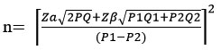
Based on the formula above, the amount of sample in each group is 15 people. In order to anticipate incomplete data, sample was added 10% so that the amount of sample is rounded up to 17 people in each group.
Tool and Material
The materials which needed were blood samples of obese women in case and control group. The variable which was checked, such as the level of vitamin D and leptin as independent variable. The parameter of metabolic syndrome is lipid profile, such as total cholesterol, triglycerides, LDL, and HDL. Fasting blood sugar levels and blood pressure are as dependent variable. Examination of vitamin D and leptin levels using the Elisa method with Kit 25 hydroxyvitamin D and leptin from Diagnostic Biochem Canada Inc. The examination of lipid profile by using Elisa method using Triglycerides, HDL dan Total Cholesterol from DiaSys Diagnostic Systems GmbH, Germany. Blood sugar level was checked with sphygmomanometer from Riester. The result in the form of quantitative value was analyzed with cross table and Chi Square test to examine the relation between the level of vitamin D and leptin with metabolic syndrome, which significance value is p<0,05. Then, odd ratio value was also calculated with IK 95%. This study has got Ethical Clearance (EC) No: 2185/UN14.2.2.VII.14/LT/2021.
Procedure
Obese women which were fulfilled by inclusive and exclusive criteria were recruited and signed informed consent. It was followed by blood pressure measurement and blood test in order to find out blood sugar level and blood cholesterol level to indicate diagnostic to metabolic syndrome. The obese women who have experienced metabolic syndrome are categorized as Case Group and those who have not experienced yet as Control Group. Then, blood tests were performed to determine the levels of leptin and vitamin D in each group.
Results And Discussion
From the table 1, it is obtained that the data of subject, such as age, weight, height, body mass index, level of vitamin D, leptin, TG, total cholesterol, HDL and LDL are normally distributed. The average of age is 24.9. This age is when the woman is productive in both hormonal and activity, so that by choosing the sample with those age range, it is expected that the subject has not experience hormonal changes which tend to indicate aging process. During this process, the function of cells is decreasing which can trigger the emergence of other comorbidities that can be confounding factors that influence vitamin D levels and leptin levels, other than obesity factors.
Table 1: Subject Characteristic and Level of Cholesterol, Blood Sugar, Vitamin D and Leptin
| Subject Characteristics | N | Average | SB | Minimum | Maximum | p |
| Age (th) | 34 | 24.9 | 5.3 | 20.0 | 37.0 | 0.142 |
| Weight (kg) | 34 | 75.9 | 12.1 | 54.9 | 101.0 | 0.829 |
| Height (cm) | 34 | 157.9 | 6.3 | 138.0 | 169.0 | 0.754 |
| BMI (kg/m2) | 34 | 30.3 | 3.9 | 25.2 | 38.5 | 0.731 |
| Level of Vitamin D (ng/ml) | 34 | 18.9 | 4.4 | 12.5 | 54.6 | 0.571 |
| Level of Leptin (ng/ml) | 34 | 26.9 | 11.5 | 3.4 | 57.9 | 0.862 |
| Level of TG | 34 | 350.5 | 69.8 | 218.15 | 579.5 | 0.661 |
| Level of LDL | 34 | 144.3 | 22.6 | 109.39 | 149.7 | 0.505 |
| Level of HDL | 34 | 54.6 | 9.7 | 47.77 | 66.5 | 0.215 |
| Total Cholesterol | 34 | 162.9 | 34.3 | 94.82 | 222.3 | 0.984 |
| Glucose | 34 | 105.3 | 34.9 | 43.88 | 181.8 | 0.897 |
| Systolic blood pressure | 34 | 128.9 | 2.0 | 104.0 | 162.0 | 0.948 |
| Diastolic blood pressure | 34 | 82.7 | 3.0 | 67.0 | 106.0 | 0.863 |
Normality value p (p>0.05)
The average of body mass index in the sample is 30.3 kg/m2. It shows the value is categorized as obese based on criteria from Asia Pacific (BMI>25kg/m2) and WHO (BMI>30kg/m2). Women have high risk of obesity due to several factors, namely hormonal factors, diet, lifestyle and several reproduction stages (pregnancy, giving birth, breastfeeding, parenting, and menopause) which are closely related with weight gain and fat accumulation in the women’s body. Based on many changes of metabolism and biochemical occurred in obese people (atherogenic dyslipidemia, insulin resistance and hyperinsulinemia, endothelial dysfunction, chronic inflammatory and prothrombotic conditions), it affects to get high risk to macrovascular disease.8
The average of vitamin D in sample group is 18.9 ng/ml, which it belongs to vitamin D deficiency (<25 ng/dl).9 Scientists are still debating the role of vitamin D in the pathophysiology of obesity. Despite the fact that numerous studies have found a negative relationship between obesity and vitamin D serum levels. Furthermore, the cause and effect are still unknown. 10
One study has shown a visible relation between the increased of BMI with low concentration of 25 hydroxyvitamin D serum. There are several mechanisms that have been studied and have resulted in low levels of 25 hydroxyvitamin D in obese people, such as decreased vitamin D synthesis in the skin, decreased absorption in the gut, and impaired vitamin D metabolism.11,12 There is no agreement yet about the condition of decreased vitamin D level in obese people. The common cause is that adipose tissue absorbs a lot of vitamin D which dissolve in fats.13 The previous study has shown that vitamin D serum has strong inverse relationship with fat level in the body and has weak inverse relationship with body mass index.14 The other hypothesis reveals that the low level of vitamin D is caused by the fact that obese people have a sedentary lifestyle so that they are rarely exposed to sunlight which certainly will inhibit the synthesis of vitamin D .15
The average of leptin level is 26.9 ng/ml. It indicates hyperleptinemia conditions, since the value of normal leptin is below 5 ng/ml. Obesity causes hyperleptinemia, which is caused by increased leptin production in adipose tissue. However, high leptin in the circulation of obese people cannot operate its function because of leptin resistance. Commonly, leptin resistance is described as the decreased of sensitivity towards anorexia effect as a respond of leptin given from outside. There are several factors affect the conditions, such as (1) Failure of leptin in the circulation to reach its target in the brain, (2) Decreased expression of the leptin receptor, and (3) There is an obstacle in the process of sending signals to certain neurons in certain areas of the brain.16
In addition, leptin level in brain fluid of obese people is less, even though the level of leptin in the serum is high. It is supported the notion that there is a failure of leptin transport inside the brain. Triglycerides are also considered as the cause of leptin resistance through impaired transport across the blood-brain barrier.17 The increase of leptin, especially in obese person, must be considered as an alert that there is an imbalanced energy, poor diet, hyperinsulinemia and insulin resistance. 4
In order to discover metabolic syndrome in the low level of vitamin D, Chi Square test is used based on cross-tabulation 2 x 2. The analysis is presented in Table 2 below.
Table 2: Low Vitamin D Levels as a Risk Factor for Metabolic Syndrome
| Variable | Group | OR | IK 95% | p | ||
| Case | Control | |||||
| Level of Vit D | Low | 14 | 8 | 5.25 | 1.09 – 25.21 | 0.031 |
| High | 3 | 9 | ||||
(p significancy <0.05)
In the Table 2 above, it shows that the level of vitamin D in case group is low and it is the risk factor of metabolic syndrome for 5 times (OR = 5.25; IK 95 % = 1.09 – 25.21; p = 0.031) more than high level of vitamin D in control group.
According to one study, a low level of 25 OH Vitamin D serum is associated with an increased risk of metabolic syndrome.18 It also reveals a negative relationship between 25 OH vitamin D levels and each component of metabolic syndrome, such as thick abdominal fat, hypertriglyceridemia and hyperglycemia. The prevalence of metabolic syndrome is lower in people who get enough vitamin D and calcium than in people who don’t get enough. The prevalence of metabolic syndrome is lower in people who get enough vitamin D and calcium than in people who don’t get enough. 19
The results show that vitamin D interacts with several components of the metabolic syndrome. In rats, a lack of vitamin D reduces glucose clearance and insulin secretion in response to a glucose tolerance test. Vitamin D supplementation supports the relationship between vitamin D and insulin metabolism, including a positive effect of vitamin D on blood sugar in diabetic animals. 20 Another study discovered that people with impaired glucose tolerance or diabetes have lower levels of 25 OH vitamin D than people with normal blood sugar levels. Another study discovered that people with impaired glucose tolerance or diabetes have lower levels of 25 OH vitamin D than people with normal blood sugar levels..21
Other mechanisms which can be used to describe the role of vitamin D in the immune system, where vitamin D can inhibit the production of interleukin 2 and interferon, which stimulate the effect of lymphocyte T helper type 2, is the link between vitamin D and metabolic syndrome. It initiates reduction of metalloproteinases matrix and inhibits the progression of atherosclerotic plaque.22 Inflammation is an important element in metabolic syndrome, and vitamin D is also as antiinflammation.23 This has explained the relationship of metabolic syndrome with vitamin D deficiency.24 Vitamin D deficiency is also related with high blood pressure through the lack of pressure from renin-angiotensin system.25,26 In the group with low vitamin D concentrations > 40 ng/ml, the prevalence of hypertriglyceridemia and HDL levels is significantly lower. In the group with low vitamin D concentrations > 40 ng/ml, the prevalence of hypertriglyceridemia and HDL levels is significantly lower. In the group with low vitamin D concentrations > 40 ng/ml, the prevalence of hypertriglyceridemia and HDL levels is significantly lower. 27, and most studies show that the level of vitamin D serum is related to the level of HDL-C and negatively to the level of cholesterol. 28 In the recent study, it is obtained that calcitriol inhibits development of triglyceride and reduces accumulation of liver fat, thus, further study is highly expected to be done to prove the functions of vitamin D in lipid metabolism.29
Chi Square test is used to find out metabolic syndrome in high level of leptin based on cross-tabulation 2 x 2. The result of analysis is presented in Table 3.
Table 3: The High Level of Leptin as The Risk Factor of Metabolic Syndrome
| Variable | Group | OR | IK 95% | p | ||
| Case | Control | |||||
| Level of Leptin | High | 16 | 1 | 256.0 | 14.70 – 4457.27 | 0.001 |
| Low | 1 | 16 | ||||
(p significancy <0.05)
In Table 3, It demonstrates that a high level of leptin in the case group is a risk factor for metabolic syndrome. it shows that the high level of leptin in case group is the risk factor of metabolic syndrome, that is 256 times (OR = 256; IK 95 % = 14.70 – 4457.27; p = 0.001) compared to low level of leptin in control group.
Martins, et al. (2012) claims that the increase of leptin, especially in obese person. It must be considered as a warning that there are imbalanced energy, poor diet, hyperinsulinemia and insulin resistance.4 Leptin receptor is inside of heart cells, leptin also has a role as modulator of insulin activity inside the cells. Leptin operates in opposite with insulin signal by reducing insulin triggered by tyrosine phosphorylation of insulin receptor substrates. This will increase the reaction of phosphoenolpyruvate carboxinase and decrease the glucokinase expression, which initiates increased gluconeogenesis and decreased glycolysis. The effect in heart occurred due to high level of leptin can cause insulin resistance. Recent study shows the direct effect of leptin in insulin gene transcription that reduces mRNA of preproinsulin up to 50%. In the other side, the failure of insulin sensitivity will increase the level of insulin and leptin through decreasing the regulation of leptin receptors in the hypothalamus or decreased sensitivity of leptin.30 It is consistent with a previous study that found leptin serum to be associated with metabolic syndrome and the risk of cardiovascular disease in Taiwanese adults.31 In a cohort study (Framingham Study), the concentration of leptin is related with metabolic syndrome.32 More importantly, the decreased level in obese children shows reduction of body fat and improvement of lipid levels and insulin sensitivity.33 Leptin affects toward appetite, energy consumption, adipose synthesis, and insulin function. As a hormone, leptin has biology function with wide spectrum. The main function is as regulator of adipose tissue and body mass.23,34 First, the inhibition of appetite and energy consumption is reached through the reduction of neuropeptide Y secretion (NPY) and melanocyte secretion as the result of combination between leptin and its receptor in hypothalamus. Second, the increase of energy consumption is reached thought release of heat in massive reserved energy. That process is caused by the increase of activity of sympathetic nerves inducted by leptin and activation of epinephrine receptor in membrane cells of adipose. Third, synthesis and resorption of adipose tissue is directly affected by leptin. One of study also estimates that leptin triggers the maturation of adipose cell.16 Leptin also affects other hormones, such as insulin which fasten leptin secretion, in the other hand, leptin has negative feedback towards synthesis and insulin secretion.
Conclusion
The conclusions of this study are as follow:
Obese women with low vitamin D levels and high leptin levels are at risk for metabolic syndrome.
Acknowledgment
We express high appreciation to LPPM of Udayana University which provide research grant funding which is delayed due to pandemic Covid-19, in accordance with SP DIPA: 023.17.2.677526/2021 on 23rd November 2020, so that this study can be well-conducted and finished.
Conflict of Interest
All authors declare no conflict of interest.
Funding Sources
The authors received financial support for the research from LPPM of Udayana University by research grant.
References
- Alberti, K.G and Zimmet, P.Z. Definition, Diagnosis and Classification of Diabetes Mellitus and Its Complications. Part 1: Diagnosis and Classification of Diabetes Mellitus Provisional Report of a WHO Consultation. Diabetic Medicine. 1998 ;15: 539-553.
CrossRef - Adriansjah, H and Adam, J. Sindrom Metabolik (Pengertian, Epidemiologi, dan Kriteria Diagnosis). Informasi Laboratorium. 2006; No. 4/2006. ISSN 0854-7165.
- Chiu, C.K., Chu Go, W.V.L., Saad, M.F. Hypovitaminosis D is Associated with Insulin Resistance and β Cell Dysfunction. Am J Clin Nutr. 2004; 79:820-5.
CrossRef - Martins, M., Faleiro, L., Fonseca, A. Relationship between leptin and body mass and metabolic syndrome in an adult population. Revista Portuguesa de Cardiologia (English Edition). 2012; 31(11): 711-719.
CrossRef - Muoio, D.M., Dohm, G.L., Tapscott, E.B., Coleman, R.A. Leptin opposes insulin’s effects on fatty acid partitioning in muscles isolated from obese ob/ob mice. Am J Physiol. 1999; 276(5): E913-21.
CrossRef - Unger, R.H. Lipid overload and overflow: metabolic trauma and the metabolic syndrome. Trends Endocrinol Metab. 2003;14(9):398-403.
CrossRef - Kasiulevicius, V., Sapoka, V. and Filipaviciūte, R. Sample Size Calculation in Epidemiological Studies. 2006. Vilnius University, Institute of Experimental and Clinical Medicine at Vilnius University.
- Carbone, S., Canada, J.M., Billingsley, H.E., Siddiqui, M.S., Elagizi, A., Lavie, C.J. Obesity paradox in cardiovascular disease: where do we stand? Vasc Health Risk Manag. 2019 ; 15:89-100.
CrossRef - Kennel, K.A., Drake MT, Hurley DL. Vitamin D deficiency in adults: when to test and how to treat. Mayo Clin Proc. 2010;85(8):752-758.
CrossRef - Lamendola, C.A., Ariel, D., Feldman, D., Reaven, G.M. Relations between obesity, insulin resistance, and 25-hydroxyvitamin D. Am J Clin Nutr. 2012;95(5):1055-9.
CrossRef - Kull, M., Kallikorm, R., Lember, M. Body Mass Index Determines Sun Bathing Habits: Implication on Vitamin D Levels. Med J. 2009; 39: 256-258.
CrossRef - Hewitt, S., Søvik, T., Aasheim, E.T., Kristinsson, J., Jahnsen, J., Birketvedt, G.S., Bøhmer,T., Eriksen, E.F., Mala, T. Secondary hyperparathyroidism, vitamin D sufficiency, and serum calcium 5 years after gastric bypass and duodenal switch. Obes Surg. 2013; 23(3):384-90.
CrossRef - Wortsman, J., Matsuoka, L.Y., Chen, T.C., Lu, Z., Holick, M.F. Decreased Bioavailability of vitamin D in obesity. Am J Clin Nutr. 2000; 72: 690–3.
CrossRef - Vimaleswaran, K.S., Berry, D.J., Lu, C., Tikkanen, E., Pilz, S., Hiraki, L.T., et al. Causal relationship between obesity and vitamin D status: bi-directional Mendelian randomization analysis of multiple cohorts. PLoS Med. 2013;10: e1001383.
CrossRef - Florez, H., Martinez, R., Chacra, W., Strickman-Stein, N., Levis, S. Outdoor exercise reduces the risk of hypovitaminosis D in the obese. J Steroid Biochem Mol Biol. 2007;10:679–81.
CrossRef - Myers, M.G., Cowley, M.A., Münzberg, H. Mechanisms of leptin action and leptin resistance. Annu Rev Physiol. 2008; 70:537-56.
CrossRef - Banks, W.A., Coon, A.B., Robinson, S.M., Moinuddin, A., Shultz, J.M., Nakaoke, R., Morley, J.E. Triglycerides induce leptin resistance at the blood-brain barrier. Diabetes. 2004; 53(5): 1253–1260.
CrossRef - Ford, E.S. Prevalence of the metabolic syndrome defined by the International Diabetes Federation among adults in the U.S. Diabetes care. 2005; 28(11), 2745–2749.
CrossRef - Liu, S., Song, Y., Ford, E.S., Manson, J.E., Buring, J.E., Ridker, P.M. Dietary Calcium, Vitamin D, and the Prevalence of Metabolic Syndrome in Middle-Aged and Older U.S. Women. Diabetes Care. 2005; 28(12): 2926-2932.
CrossRef - De Souza, W.N and Martini, L.A. The Role of Vitamin D in obesity and inflammation at adipose tissue. J Obes Metab Res. 2015; 2: 161-6
CrossRef - Scragg, R., Sowers, M., Bell, C. Serum 25-hydroxyvitamin D, diabetes, and ethnicity in the Third National Health and Nutrition Examination Survey. Diabetes Care. 2004; 27(12):2813-8.
CrossRef - Andress, D.L. Vitamin D in chronic kidney disease: a systemic role for selective vitamin D receptor activation. Kidney Int. 2006; 69 (1): 33–43.
CrossRef - Sundari, L.P.R., Bakta, I.M., Astawa, I.N.M., Adiatmika, I.P.G., Arijana, I.G.K.N., Tunas. I.K. The Effect of Vitamin D Administration on Leptin, Adiponectin and mRNA MCP-1 Levels in Adipose Tissue of Obese Female Wistar Rats. Current Research in Nutrition and Food Science. 2020;8 (2): 541-549.
CrossRef - Sharma, P Inflammation and the metabolic syndrome. Indian J. Clin. Biochem.2011; 26 (4): 317–318.
CrossRef - Li, Y.C., Qiao, G., Uskokovic, M., Xiang, W., Zheng, W., Kong, J. Vitamin D: a negative endocrine regulator of the renin-angiotensin system and blood pressure. J Steroid Biochem Mol Biol. 2004; 89-90 (1-5): 387-392.
CrossRef - Zhou, C., Lu, F., Cao, K., Xu, D., Goltzman, D., Miao, D. Calcium-independent and 1,25(OH)2D3-dependent regulation of the renin-angiotensin system in 1alpha-hydroxylase knockout mice. Kidney Int. 2008; 74(2): 170-179.
CrossRef - Wang, C.M., Chang, C.S., Chang, Y.F., Wu, S.J., Chiu, C.J., Hou, M.T., et.al. Inverse Relationship between Metabolic Syndrome and 25-Hydroxyvitamin D Concentration in Elderly People without Vitamin D deficiency. Sci Rep. 2018; 19;8(1):17052.
CrossRef - Jorde, R and Grimnes, G. Vitamin D and metabolic health with special reference to the effect of vitamin D on serum lipids. Lipid Res. 2011; 50(4): 303–312.
CrossRef - Cheng, S., So, W.Y., Zhang, D., Cheng, Q., Boucher, B.J., Leung, P.S. Calcitriol Reduces Hepatic Triglyceride Accumulation and Glucose Output Through Ca2+/CaMKKβ/AMPK Activation Under Insulin-Resistant Conditions in Type 2 Diabetes Mellitus. Curr Mol Med. 2016; 16(8):747-758.
CrossRef - Paz-Filho, G., Mastronardi, C., Wong, M.L., Licinio, J. Leptin therapy, insulin sensitivity, and glucose homeostasis. Indian J Endocrinol Metab. 2012;16(3): S549-S555.
CrossRef - Li, Wen-Cheng & Hsiao., Kuang-Yu & Chen., I-Chuan & Chang., Yu-Che & Wang., Shih-Hao & Wu, Kuan-Han. Serum leptin is associated with cardiometabolic risk and predicts metabolic syndrome in Taiwanese adults. Cardiovascular diabetology. 2011. 10. 36. 10.1186/1475-2840-10-36.
CrossRef - Ingelsson, E., Larson, M.G., Yin, X., Wang, T.J., Meigs, J.B., Lipinska, I., et.al Circulating ghrelin, leptin, and soluble leptin receptor concentrations and cardiometabolic risk factors in a community-based sample. J Clin Endocrinol Metab. 2008; 93(8): 3149-57.
CrossRef - Bocca, G., Corpeleijn, E., Stolk, R. Effect of obesity intervention programs on adipokines, insulin resistance, lipid profile, and low-grade inflammation in 3- to 5-y-old children. Pediatr Res. 2014 ;75: 352–357.
CrossRef - Houseknecht, K.L and Spurlock, M.E. Leptin regulation of lip homeostasis: dietary and metabolic implications. Nutrition research reviews.2003; 16(1): 83-96.
CrossRef
Accepted on: 16 Aug 2022
Second Review by: faramarz Mohtasham Iran
Final Approval by: Dr Jiwan S. Sidhu


Web of Science Coverage
Emerging Sources Citation Index (ESCI)
2024 Journal Impact Factor: 1.1
Scopus Journal Metrics
CiteScore 2025: 2.6
CiteScore Details
Sustainable Nutrition: Food Systems, Nutrient Retention, and Public Health Impact
![]()
This journal is a member of, and subscribes to the principles of, the Committee on Publication Ethics (COPE)


