Introduction
Nowadays more and more people are opening up to a healthier way of life and follow a diet that includes foods which promote healthy aging. Some of these foods are superfoods and superherbs that possessed functional health properties beyond its nutritive value. These foods containing exogenous nutrient and phytochemical antioxidants that could protect the human body from damage arising from chronic oxidative stress caused by excessive production of free radicals.
Phenolic compounds exhibit a considerable free-radical scavenging (antioxidant) activity, which is determined by their reactivity as hydrogen- or electron- donating agents, the stability of the resulting antioxidant-derived radicals, their reactivity with other antioxidants and their metal chelating properties.1
Extracts of herbs, fruits and other plant materials rich in phenolics are of increasing interest in the food industry because they retard oxidative degradation of lipids and therefrom improve the quality and nutritional value of food.2 Furthermore plant-derived polyphenols are of great importance because of their potential antioxidant, antifungal and antimicrobial properties. There are many reports concerning the antifungal and antimicrobial activity of plant extracts.
The main purpose of this work was the study of sixteen superfoods and superherbs produced in Greece and which consist the raw material for the production of innovative products. In this study, a variety of superfoods and superherbs cultivated in regions of Greece was investigated for the antioxidant activity. Moreover the potential of these superfoods/ superherbs to inhibit the growth of mycotoxigenic fungal was also studied.
Materials and Methods
Sampling and Treatment
Nine fruits-berries and seven herbs were investigated during this study: Cornus mas L. (cornelian cherries), Vaccinium corymbosum L. (blueberries), Rubus idaeus L. (raspberries), Morus alba L. (mulberries), Physalis peruviana L. (golden berries), Rosa canina L. (dog-rose), Photinia melanocarpa (Michx.) Elliott (black chokeberries), Hippophae rhamnoides L. berries (sea-buckthorn berries), Lycium barbarum L. (goji berries), Hypericum perforatum L. (St John’s wort), Echinacea purpurea (L.) Moench (purple coneflower), Crataegus monogyna Jacq. (common hawthorn), Cistus incanus L. (hairy rockrose), Hippophae rhamnoides L. leaves (sea-buckthorn leaves), Tribulus terrestris L. (puncturevine), Satureja montana L. (winter savory). Specimen of the cultivated plants were purchased from different growers while the native plants were collected from their natural habitats. All sixteen plant samples were originated from Greece. A representative and randomized quantity of each plant material was dried in a ventilated oven at 40°C. Every dried sample was homogenized before analysis.
Reagents
Folin–Ciocalteu’s phenol reagent were obtained from Merck KGaA (Germany). DPPH• (2,2-diphenyl-1-picrylhydrazyl) free radical and gallic acid was obtained from Alfa Aesar GmbH and Co KG (Germany) while L-ascorbic acid was obtained from Fisher Chemical, UK. Methanol (pro analysis) was from Merck (Darmstadt, Germany).
Apparatus
Ultrasound-assisted extraction was carried out using an ultrasonic bath device (Elmasonic S, Elma Schmidbauer GmbH, Germany) at a frequency of 37 kHz. For the evaporation of the extracts a rotary evaporator (Heidolph, Laborota 4000 efficient, WB eco) was used. For the spectrophotometric analyses a UV-vis spectrophotometer (Novaspek III visible spectrophotometer, Amersham Biosciences, USA) was used. A laminar flow (Telstar Bio II A, Madrid, Spain), an autoclave, Selecta-Autester-E Dry (PBI Milano, Italy), an incubator WTB Binder (Tuttlinger, Germany) and a centrifuge Sorvall RC-5B (HS-4) (Norwalk, USA) were utilized.
Culture Media
Aspergillus Flavus Parasiticus Agar (AFPA) was prepared by dissolving 2 g of yeast extract (Oxoid, Basingstoke, Hampshire, UK), 1 g of bacteriological peptone (Oxoid), 0.05 g of ferric ammonium citrate (Merck, Germany), 0.1 mL of Dichloran 0.2% in ethanol (Fluka Steinheim, The Netherlands), 0.01 g of chloramphenicol (Oxoid), and 1.5 g of agar (Oxoid) per 100 mL of distilled water, final pH 6.0–6.5. Potato Dextrose Agar (PDA) was prepared according to the label directions (BD Difco).
Extraction Procedure
For the preparation of the plants’ extracts, a combination of two extraction methodologies was performed. First a conventional extraction took place followed by an ultrasound-assisted extraction.3 Specifically, a ground particle sample (1 g) of each material was placed in separate beakers and mixed with 30 mL acidified with HCl methanol:water (80:20 (v/v) 0,1% HCl) for the extraction of the phenolic content by diffusion having samples shaken at regular intervals. After 24 h in the dark and at ambient temperature the beakers were sonicated for three times (with 10mL of solvent each time) in the ultrasonic device for 15 minutes. The extraction is completed, followed by filtration using Buchner funnel, to remove solids. The extracts were evaporated to dryness and there were rediluted to 5 ml of MeOH. Every sample was done in triplicate. The extracts were kept frozen at -20oC in sealed containers until analysis. 4,5
Determination of Total Phenolic Content
The same procedure was applied for all extracts to determine the total phenolic content using the same quantities of reagents. The total phenolic content (TPC) of each sample was determined by Folin–Ciocalteu’s colorimetric assay. 6,7
Twenty (20) μL of each extract, standard solutions or blank (methanol) were added to 1500 μL of water and 100 μL of the Folin–Ciocalteu reagent, mixed thoroughly and allowed to stand for 8 min. Then 300 μL of saturated sodium carbonate solution were added and mixed well. The cuvettes were incubated in a water bath at 40oC for 30 min. The absorbance of the resulting blue color was measured at 750 nm at room temperature with a UV-vis spectrophotometer. The experimental procedure and calculations were made in triplicate for each sample or standard solution, while different series of experiments were performed in the same day, but on different days as well. Final results are expressed in mg GAE g-1. 4,5,8
DPPH radical scavenging assay
Radical scavenging activity of each sample was evaluated using the stable 2,2-diphenyl-1-picryl-hydrazyl radical (DPPH•) according to a slightly modified method of Brand-Williams et al. (1995).9
Twenty (20) μL of extracts were placed in plastic cuvettes together with 1500 μL of DPPH 100 μM solution, left in the dark for 1 min and then the absorbance was measured at 516 nm, using a UV-vis spectrophotometer, every 10 min until the absorbance was stabilized in a minimum point at a plateau time. At the same time, the absorbance of DPPH solution used was measured so as to calculate the percentage of the halting at the plateau time. Also measurements of the blank were made to correct the error caused by the solvent. 10
L-ascorbic acid (AA), was used as standard compound to prepare the standard curve for quantification because ascorbic acid reacts rapidly and completely with DPPH radical The antiradical activity of samples was expressed as mg L-Ascorbic Acid Equivalents (AAE) per g, using a standard curve with 20-1800 μg AAE mL-1. The experimental procedure and calculations were made in triplicate for each sample or standard solution.4,5,8
Antifungal assay
The methanolic-aqueous extracts were tested for in vitro antifungal activity on Aspergillus parasiticus and Aspergillus carbonarius growth. The aflatoxigenic strain A.parasiticus speare (IMI 283883) utilized throughout this study was obtained from the International Mycological Institute (Engham Surrey, UK) and the ochratoxigenic strains A. carbonarius (ATHUM 2854) were obtained from the ATHUM Culture Collection of Fungi, Mycetotheca of the University of Athens.
For the preparation of spore inoculum of each fungus slants of stock cultures of PDA maintained at 25 °C were used. Fresh colonies of A.parasiticus and A. carbonarius were obtained on PDA after 7 days at 30 °C. Spore suspensions were prepared aseptically using 10 mL of sterile Tween 80 solution 0.01% v/v.11 Then the spore suspension was centrifuged and the supernatant fluid was rejected. A resuspension in 10 mL of sterile Tween 80 solution was followed. The procedure was repeated thrice. Dilutions (10-1, 10-2, 10-3, 10-4) from the initial spore suspension in sterile tubes containing 10 mL of Tween 80 0.05% v/v were made. Determination of the spore concentration was performed by the spread plate surface count technique using 0.1 mL of each dilution on AFPA plates (for A.parasiticus) and PDA plates (for A.carbonarius) after incubation at 30 °C for 48 h. AFPA and PDA plates with 10–100 colony-forming units (CFU) were selected and the preferred 102 spore quantity used in the present study was determined following the method described previously in details.12
For the antifungal assay, holes (1.5 cm in diameter) were punched at the center of Petri dishes (9 cm diameter), filled with AFPA (for Aspergillus parasiticus) or PDA (for Aspergillus carbonarius). For the study of the extracts’ effect on A.parasiticus growth, petri dishes were inoculated with 102 conidia of the fungi (inside the holes) and after that an addition of 100μL of each extract, was added around the agar-holes. Inoculated Petri dishes containing 100 μL MeOH:H2O (80:20 v/v) were used as control. The same procedure was applied for A.carbonarius as well. All Petri dishes were examined in 3 replicates. The cultures were incubated at 30 °C and the diametrical growth of fungi colonies was measured daily during 7 days. The percentage of inhibition was calculated based on growth of fungi in control Petri dishes.
Statistical analysis
All results presented are means of triplicates along with standard deviations. Correlations between antioxidant activity and phenolic content were determined using Pearson’s Correlation Coefficient Test.
Results and Discussion
Total Phenolics and Antioxidant Activity
The results for the determination of total phenolics and antioxidant activity varied widely and are shown in Table 1. Total phenolics content varied from 13.81±0.08 to 1231.74±4.10 mg GAE g-1 of dry sample. Among the sixteen (16) samples studied, low values were found in Morus alba L. (13.81±0.08 mg GAE g-1), Lycium barbarum L. (13.97±0.12mg GAE g-1), Physalis peruviana L. (16.40±0.10mg GAE g-1), Vaccinium corymbosum L. (20.21±0.19mg GAE g-1) while Crataegus monogyna Jacq., Hypericum perforatum L., Photinia melanocarpa (Michx.) Elliott, Satureja montana L., Cistus incanus L. and Hippophae rhamnoides L. leaves contained relatively high amounts of phenolics ranged from 588.44±2.80 mg GAE g-1 to 1238.74±4.10 mg GAE g-1. The highest level of phenolics was found in Hippophae rhamnoides L. leaves and it was 1238.74±4.10 mg GAE g-1.
Total antioxidant activity of the extracts, measured by the DPPH method, ranged from 47.92±0.02 to 116865.81±4.502021 mg AAE g-1 dry weight. Among the sixteen (16) studied plants, Photinia melanocarpa (Michx.) Elliott exhibited the highest antioxidant activity followed by Hippophae rhamnoides L. leaves and Cistus incanus L.
The results of the present study are in agreement with data from literature. Previously it was reported that Photinia melanocarpa (chokeberries) had significantly higher anthocyanin, phenolic content and antioxidant activity than Vaccinium corymbosum L. (blueberries) and other berries of Vaccinium family.13,14 Furthermore, many studies have demonstrated before the rich phenolic content and the antioxidants activity some of the studied extract. 4,15,16,17 Differences among the literature data concerning antioxidant profile of the plants, is probably due to the different origin, genetic background, climatic conditions etc.5
The antioxidant activity of samples might be influenced by several factors and could not be fully described by one single method. Most natural antioxidants are multifunctional and the antioxidant evaluation needs to take into account various mechanisms of antioxidant action.18
Table 1: Total phenolic content and DPPH• values of the evaluated berries and herbs
|
Samples |
||||
| Common name | Scientific names |
Total phenolics (mg GAE g-1) |
Antioxidant activity (mg AAE g-1) |
|
| Cornelian cherries | Cornus mas L. |
57.23 ± 0.40 |
359.21 ± 2.01 |
|
| Blueberries | Vaccinium corymbosum L. |
20.21 ± 0.19 |
65.07 ± 0.04 |
|
| Raspberries | Rubus idaeus L. |
57.12 ± 0.91 |
395.80 ± 1.12 |
|
| Mulberries | Morus alba L. |
13.81 ± 0.08 |
47.92 ± 0.02 |
|
| Golden berries | Physalis peruviana L. |
16.40 ± 0.10 |
53.23 ± 0.10 |
|
| Dog-rose | Rosa canina L. |
67.74 ± 1.02 |
458.34 ± 0.30 |
|
| Black chokeberries | Photinia melanocarpa (Michx.) Elliott |
721.20 ± 3.41 |
116865.81± 4.50 |
|
| Goji berries | Lycium barbarum L. |
13.97 ± 0.12 |
262.26 ± 2.13 |
|
| Sea-buckthorn berries | Hippophae rhamnoides L. berries |
104.54 ± 2.10 |
730.09 ± 3.24 |
|
| Sea-buckthorn leaves | Hippophae rhamnoides L. leaves |
1238.74 ± 4.10 |
9123.90 ± 2.50 |
|
| St John’s wort | Hypericum perforatum L. |
636.10 ± 3.21 |
3355.84 ± 1.03 |
|
| Purple coneflower | Echinacea purpurea (L.) Moench |
68.92 ± 0.90 |
345.20 ± 0.52 |
|
| Common hawthorn | Crataegus monogyna Jacq. |
588.44 ± 2.80 |
3362.82 ± 2.46 |
|
| Hairy rockrose | Cistus incanus L. |
839.11 ± 3.30 |
8387.24 ± 4.02 |
|
| Puncturevine | Tribulus terrestris L. |
111.41 ± 1.09 |
337.91 ± 2.10 |
|
| Winter savory | Satureja montana L. |
785.20 ± 3.01 |
3228.80 ± 4.03 |
|
| Data are expressed as means ± standard deviation (n=3) | ||||
The Pearson’s correlation coefficients between total antioxidant activity and the total phenolic content of the studied extract is shown in Figure 1. A positive but weak correlation (R2 = 0.1647) between the DPPH• value and total phenolic content showed that phenolic compounds may not be the main components that are responsible for the antioxidant activity. Nevertheless, if Photinia melanocarpa (Michx.) Elliott which possessed the strongest antioxidant activity were expelled, it would show a very strong positive correlation (R2 = 0.9076). Consequently it is possible the phenolic compounds of the other fifteen plants studied to be responsible for their antioxidant activity. Finally it must be mention that the contribution of individual phenolics to total antioxidant activity is generally dependent on their structure and content in plants.13
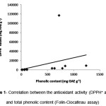 |
Figure 1: Correlation between the antioxidant activity (DPPH• assay) and total phenolic content (Folin-Ciocalteau assay)
|
Antifungal Activity
The effect of the sixteen studied extract against A.parasiticus and A.carbonarius was estimated by recording daily the diametrical mycelial growth of the fungi culture in AFPA and PDA medium respectively. The mycelial growth of the two fungi isolates has been influenced differently by the sixteen tested plant extracts (Figures 2, 3).
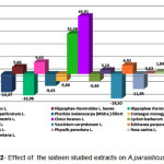 |
Figure 2: Effect of the sixteen studied extracts on A.parasiticus growth |
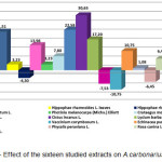 |
Figure 3: Effect of the sixteen studied extracts on A.carbonarius growth Click here to View figure |
Of the tested extracts obtained from the sixteen plant samples, three species (Morus alba L., Physalis peruviana L.,Satureja montana L.) showed antifungal activity only against A.parasiticus and five species (Lycium barbarum L., Crataegus monogyna Jacq., Photinia melanocarpa (Michx.) Elliott, Cornus mas L., Rubus idaeus L.) against A.carbonarius while six species (Hippophae rhamnoides L. leaves, Hypericum perforatum L., Tribulus terrestris L., Cistus incanus L., Echinacea purpurea (L.) Moench, Rosa canina L.) showed antifungal activity against both fungi. In all these cases mycelial growth inhibition values ranged from 45.91% to 1.08%.
Cistus incanus L., and Tribulus terrestris L. showed the strongest antifungal activity, inhibiting the A.parasiticus growth in a percentage of 45.91% and 32.08% respectively. Moreover, the highest efficiency against A.carbonarius was registered for Cistus incanus L. extract, followed by Tribulus terrestris L. extract (30.65% and 22.58% respectively).
Two of the tested extracts (Vaccinium corymbosum L., Hippophae rhamnoides L. berries) did not exhibit antifungal activity at all, on the contrary these extract stimulated the mycelium growth of both A.parasiticus and A.carbonarius (Figure 2,3). Furthermore the extracts of Rubus idaeus L., Cornus mas L., Photinia melanocarpa (Michx.) Elliott, Crataegus monogyna Jacq. and Lycium barbarum L. caused a stimulation of the A.parasiticus growth ranged from 5.66% to 14.45% while the extracts of Morus alba L., Physalis peruviana L. and Satureja montana L. stimulated the A.carbonarius growth at rate of 7.53%, 6.45% and 3.23% correspondingly.
From the above results, it is obvious that every extract have different modes.Τhe different plant species considerably varied in their antifungal potentials and the difference might increase from the variability in chemical constituents of the plants.19
Generally, the antifungal properties of plants extracts can be linked to their antioxidant activity and their phenolic content; however this is not always a pattern fact. The antifungal activity of plants might be due to the presence of diverse group of phytoconstituents.20 In this study the correlation between the antioxidant activity and the antifungal activity of the studied extracts seems to be weak. The extracts found to possess the highest antioxidant activity (Photinia melanocarpa (Michx.) Elliott, Hippophae rhamnoides L. leaves) showed low or no exhibited inhibitory effect on the growth of tested fungi. On the other hand Cistus incanus L. and Tribulus terrestris L. extracts exhibited the highest antifungal efficacy showed at the same time high antioxidant activity and phenolic content.
In conclusion, this study offers new information on the antioxidant function of these berries and herbs and revealed that they are an effective potential source of natural antioxidants. Moreover the use of these mixtures of natural active substances as non-pollutant and environmental friendly alternative antifungal agents is promising.
Acknowledgements
This work was supported in part by the University of Athens, Special Account for Research Grants (11400).
References
- Parsaeimehr A,Sargsyan E, Javidnia K. A Comparative Study of the Antibacterial, Antifungal and Antioxidant Activity and Total Content of Phenolic Compounds of Cell Cultures and Wild Plants of Three Endemic Species of Ephedra. Molecules;15:1668-1678: (2010).
CrossRef - Wojdyło A, Oszmiański J, Czemerys R. Antioxidant activity and phenolic compounds in 32 selected herbs. Food Chemistry;105: 940-949:(2007).
CrossRef - Toma M, Vinatoru M, Paniwnyk L, Mason T. Investigation of the effects of ultrasound on vegetal tissues during solvent extraction, Ultrasonics Sonochemistry; 8:137-142: (2001).
CrossRef - Roidaki A, Zoumpoulakis PG, Proestos C. Comparison of Extraction Methods for the Determination of Antioxidant Activity in Extracts of Hippophae Rhamnoides L. and Lippia Citriodora. The Effect of Seasonal Collection. Austin J Nutri Food Sci; 3(1): 1057: (2015).
- Kollia E, Markaki P, Zoumpoulakis P, Proestos C. Αntioxidant activity of Cynara scolymus L. and Cynara cardunculus L. extracts obtained by different extraction techniques. Natural Product Research; 1-5: (2016). Article in Press. DOI: 10.1080/14786419.2016.1219864
CrossRef - Singleton VL, Rossi JA. Colorimetry of total phenolics with phosphomolybdicphosphotungstic acid reagents. Am J Enol Vitic; 16:144-158: (1965).
- Craft BD, Kerrihard AL, Amarowicz R, Pegg RB. Phenol-Based Antioxidants and the In Vitro Methods Used for Their Assessment. Comprehensive Reviews in Food Science and Food Safety;11:148-173:(2012).
CrossRef - Lantzouraki DZ, Sinanoglou VJ, Zoumpoulakis PG, Glamoclija J, Ćirić A, Soković M, Heropoulos G, Proestos C. Antiradical–antimicrobial activity and phenolic profile of pomegranate (Punica granatum L.) juices from different cultivars: a comparative study. RSC Adv; 5:2602: (2015).
CrossRef - Brand-Williams W, Cuvelier ME, Berset C. Use of a free radical method to evaluate antioxidant activity. LWT – Food Sci Technol; 28:25–3: (1995).
- Nenadis N, Tsimidou M. Observations on the Estimation of Scavenging Activity of Phenolic Compounds Using Rapid 1,1-Diphenyl-2-picrylhydrazyl (DPPH•) Tests. Journal of the American Oil Chemists’ Society; 79(12):1191-1195: (2002).
CrossRef - Kollia E, Pyrri I, Kapsanaki-Gotsi E, Markaki P. Ochratoxin A production by Aspergillus section Nigri strains isolated from currants of Greek origin. International Journal of Environmental & Agriculture Research; 2(7): 110-120: (2016).
- Vergopoulou S, Galanopoulou D, Markaki P. Methyl jasmonate stimulates aflatoxin B1 biosynthesis by Aspergillus parasiticus. J Agric Food Chem; 7:3494–3498:(2001).
CrossRef - Zheng W, Wang SY. Oxygen radical absorbing capacity of phenolics in blueberries, cranberries, chokeberries, and lingonberries. J Agric Food Chem; 5(2):502-509: (2003).
CrossRef - Oszmianski J, Wojdylo A. Aronia melanocarpa phenolics and their antioxidant activity. Eur Food Res Technol; 221: 809–813:(2005).
CrossRef - Riehle P, Vollmer M, Rohnet S. Phenolic compounds in Cistus incanus herbal infusions — Antioxidant capacity and thermal stability during the brewing process. Food Research International;53:891–899:(2013).
CrossRef - Attaguile G, Russo A, Campisi A, Savoca F, Acquaviva R, Ragusa N, Vanella A. Antioxidant activity and protective effect on DNA cleavage of extracts from Cistus incanus L. and Cistus monspeliensis L. Cell Biology and Toxicology;16:83-90:(2000).
CrossRef - Zou Y, Lu Y, Wei D. Antioxidant activity of a flavonoid-rich extract of Hypericum perforatum L. in vitro. J Agric Food Chem; 52:(16):5032-5039: (2004).
CrossRef - Wong SP, Leong LP, Koh JHW. Antioxidant activities of aqueous extracts of selected plants. Food Chemistry; 99:775-783:(2006).
CrossRef - Ademe A, Ayalew A, Woldetsadik K. Evaluation of Antifungal Activity of Plant Extracts against Papaya Anthracnose (Colletotrichum gloeosporioides). J Plant Pathol Microb;4: 207:(2013).
CrossRef - Sethi S, Prakash O, Pant AK. Phytochemical analysis, antioxidant assay and antifungal activity of essential oil and various extracts of Alpinia malaccensis (Burm.f.) Roscoe leaves, Cogent Chemistry;2:1223781:(2016)
CrossRef

This work is licensed under a Creative Commons Attribution 4.0 International License.


