Review on Potential Vitamin D Mechanism with Type 2 Diabetes Mellitus Pathophysiology in Malaysia
Nurliyana Najwa bt Md Razip
Dept. of Biomedical Sciences, Faculty of Medicine and Health Sciences, Universiti Putra Malaysia, 43400 Serdang, Selangor, Malaysia.
Corresponding Author Email: huzwah@upm.edu.my
DOI : http://dx.doi.org/10.12944/CRNFSJ.6.1.01
Download this article as:
![]()
Evidences on vitamin D deficiency suggest there is increasing risk of diabetes. To date, some cohort, observation, cross-sectional studies on populations and randomized controlled trials in vitamin D supplements highlighting the potential of vitamin D are essentially in modifying Type 2 Diabetes Mellitus (T2DM) pathophysiology. Relevant literature sought in a various databases focus on the discovery of vitamin D studies in Malaysia, particularly in dietary, health status and disease study. However, recent data in Malaysia, the scope of the literature focuses on the deficient vitamin D mediated insulin impairment. The development of literary findings encompasses on the etiology of diabetes which highly correlates with decreased mechanism of action of vitamin D. It is important to understand diabetes etiology before explaining more about insulin resistance mechanisms which is strongly correlated with the involvement of c-Jun N-terminal kinase (JNK) pathways in insulin signalling. Furthermore, the vitamin D works synergistically with calcium homeostasis which is believed to have interaction with insulin. The purpose of this article is to illustrate the potential of vitamin D in modulating T2DM pathophysiology. Existing evidence showing the biochemical function of vitamin D is strongly involved in the pathogenesis of T2DM which requires considerable attention.
KEYWORDS:Calcium homeostasis; Vitamin D Type 2 diabetes mellitus; Insulin
Introduction
Historically, vitamin D is a “sunshine” antioxidant that effectively potentiates the maintenance of capacity on bone density. Despite of vitamin D adequate in Asian regions due to sufficiency sunlight exposure, yet 50% of population worldwide still in deficiency of vitamin D.1 This is attributed by the effect of weather, clothes, lifestyle, dietary intake, age, gender, predisposing of metabolic syndromes and genetic heredity which influence the bioavailability of vitamin D to function properly in the body system. In PubMed literature, vitamin D and diabetes research has been published more than 20,000 papers since and first literature on vitamin D which is written by Lusk entitled ‘Diet and Disease’. The literature is based on the diet properties which involved the classical understandings on metabolism of diet-doctrine dealt with proteins, fats, carbohydrates and the energy liberated within the body upon their oxidation with water and its by-product.57 Contrary, hypothetical evidence postulated the lack of nutrients at certain condition or environmental risk factors have influenced or permutated the metabolism at normal level. Extensive studies focus on T2DM as the pathological defect after the deficiency of vitamin D takes place were the dominant. However, substantive studies need to be explored more to validate the strong relationship of cause and effect between vitamin D and its predisposing risk factors. The literature review is focusing on T2DM pathophysiology regulates the defect of bioavailability of vitamin D and lead to the pathological effects. Many observational studies are linking between vitamin D and glucose homeostasis impairment that is predisposed to T2DM that has been received great attention and plays a significant role in alteration of insulin mechanism.2,4
Vitamin D Metabolism
Vitamin D can be obtained endogenous and exogenously by sun radiation or dietary intake before converted to cholecalciferol. The activated vitamin D is formed in two hydroxylation processes that takes place in the liver and kidney. Pre-vitamin D-3 is converted to vitamin D-3 via ultraviolet ray B (UVB) at 295-313 nm irradiation via 7-dehydrocholesterol under the skin. The research found that, every exposure of sunlight was about 10,000 IU (0.25 mg) of vitamin D naturally under the skin5 With the help of 25-hydroxylase (CYP2R1) enzyme, vitamin D-3 is converted to 25-hydroxyvitamin D-3 from the skin. It is an alternative source of vitamin D besides food from plant or animal origins. Vitamin D is stored in adipose tissue and utilized in the form of 25-hydroxyvitamin D-3 in the liver. Vitamin D-binding protein (DBP) is the carrier of 25-hydroxyvitamin D-3 in the bloodstream. Then, 25(OH)D-3-DBP complex will be excreted out into urine and reabsorbed through megalin, a scavenger receptor in the proximal tubules. 25-hydroxyvitamin D-3 is hydroxylated at the position of carbon-1 of the A ring, resulting in the conversion to 1,25-dihydroxyvitamin D-3, an active form of vitamin D which is responsible for biological activity either it is non-genomic or genomic activity.6 The key enzyme of the conversion of 1,25-dihydroxyvitamin D-3 from 25-hydroxyvitamin D-3 is 1-α-hydroxylase (CYP27B1) in the kidney.7 Importantly, the absence of this enzyme could result in vitamin D dependency rickets type 1 (VDDR1) that is caused by mutation. The previous study reported the knockout of gene Cyp27b1 in mice showed a mouse model of VDDR type.1 Parathyroid hormone (PTH) and calcitonin, act in concert to draw a negative feedback towards the calcium homeostasis.8 The reabsorption of calcium by the kidney depends on the level of calcium in plasma via the production of 1,25-dihydroxyvitamin D-3. Generally, 1,25-dihydroxyvitamin D-3 promotes calcium and phosphate absorption in the small intestines and regulates reabsorption of calcium in renal tubules. Low level of calcium affects the activation of CYP2B1 and causes the increase of 1,25-dihydroxyvitamin D-3 levels. In conjunction with this mechanism, it suggests the importance of maintaining the level of vitamin D by taking supplementation or diet in individuals needs to be taken into consideration. Recommended Nutrient Intake (RNI) of vitamin D supplementation has been identified among adult between 19-50 years old is 5 µg/day.
Vitamin D and Calcium
In calcium homeostasis, vitamin D has beneficial effects in controlling calcium plasma level. Vitamin D promotes the mineralization absorption efficiently for calcium by 30-40% and phosphorus by 80%.8,9 Historically, calcium is a mineral element that provide rigidity to the bone skeleton and it is believed to help in the insulin secretion.8 As calcium synergistically works with vitamin D, PTH is otherwise. Vitamin D deficiency results in calcium and phosphorus decrease while PTH increases antagonistically. Thus, it affects the endocrine system in the body. Decreased bone mineral density and porosity of bone could lead to osteoporosis. Calcium homeostasis are mainly regulated by parathyroid hormone (PTH), vitamin D and fibroblast growth factor (FGF)-23 which synergistically controlling several processes in different target tissue especially in the small intestine.10 The normal level of serum calcium is between 8.5-10.5 mg/dL and is tightly regulated by 1,25-dihydroxyvitamin D-3 also known as calcitriol in the kidney.8 If this level deviates slightly, parathyroid gland gives a signal to PTH to normalize the calcium level. Nevertheless, calcium depends on vitamin D which might affect PTH level.
However, in biochemistry perspective, the complex mechanism which are acquired to be explored in calcium homeostasis. Vitamin D is not only involved in non-genomic, but also, in genomic interaction with the nuclear receptor to modulate the expression of the targeted gene in insulin which occurs in nucleus.11 The activated 1,25-dihydroxyvitamin D-3 enters the plasma membrane with the help of vitamin D binding protein (DBP) and cause the activation of the VDR as shown in figure 2. Ligand binding to the VDR induces a conformational change in the receptor and subsequent heterodimerization with retinoid-X-receptor (RXR) in the nucleus.12
The VDR is known to recognize a specific DNA sequence or vitamin D response element (VDRE) comprised of two hexameric nucleotide half-sites separated by three base pairs.13 The RXR-VDR complex binds to the vitamin D response element (VDRE) which is located within the 5’ flanking region of target genes. Thereafter, co-repressor (CoR) proteins are released from the surface of the VDR, allowing interaction with co-activator (CoA) proteins.14 These molecules modulate chromatin structure and allow the interaction of the receptor with RNA polymerase II transcriptional complex, thus activating transcription of the target gene such as calbindin D28k, which is a calcium transporter and ATP transporter.14 Hence, the presence of vitamin D is significantly important for the interaction with the nuclear receptor. It becomes important from the role of vitamin D and calcium to gain an insight of diabetes study. With respect to its relevance in diabetes, the classical VDRE and other response sites are found within genes encoding insulin with important functions in pancreatic β-cells.
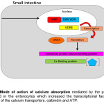 |
Figure 1: Mode of action of calcium absorption mediated by the presence of vitamin D-3 in the enterocytes which increased the transcriptional factor for the expression of the calcium transporters, calbindin and ATP Click here to View figure |
Prevalence of Vitamin D Deficiency in Malaysia
Vitamin D studies in Malaysia encompass the discovery of vitamin D status among Malaysian that has been associated with metabolic syndrome since 2011. Interestingly, research findings on the status of vitamin D and the interrelationships with T2DM pathophysiology were the major concern because its diabetic population is growing every year with the increment 7.5% (NHMS, 2008). In 2015, 9.2% was unaware of diagnose diabetes among Malaysian (NHMS, 2015). However, vitamin D deficiency has been speculated to have a functional role in many mechanisms of insulin signalling related to T2DM since a decade ago with tremendous research. Although this mechanism is not well understood, evidence suggests low vitamin D may increase the risk factor of diabetes through the disruption of pancreatic b-cell and increased the insulin resistance.
As Malaysia is located on the equator, sunlight sufficiency is more than enough. However, vitamin D deficiency in population is still increasing. About 30% -50% of people in the world have low levels of vitamin D and are recognized as global health problems worldwide despite the sufficiency of sun exposure. Previous diabetes intervention studies have been made to elucidate the relationship between vitamin D and the effects of reverse hyperglycemia revealing that vitamin D plays an important role in promoting β cell function and glucose homeostasis. Despite the alarming deficiency of vitamin D, previous studies revealed that vitamin D has a major role in promoting β-cell function and glucose homeostasis.2,33.,23 Many citations on vitamin D reported on deficiency of vitamin D and abnormal glucose tolerance involved in diabetes mellitus pathophysiology. In parallel, National Health and Nutrition Examination Survey (NHANES III) (1988-1994) also found that a higher serum vitamin D concentration leads to a lower risk of diabetes.38 Thus, by far, this occurrence was highlighted in this review. An animal study using diabetic mice fed on a vitamin D-depleted diet has an increased risk of developing glucose intolerance, develop at an earlier age, and more severe than control mice. The induction of vitamin D in obese Wistar rats with a diabetic condition indicates significantly decreased plasma glucose concentrations by 40%.37 In addition, human studies in adults have suggested that reduced vitamin D intake is associated with reduced insulin sensitivity and an increased risk of developing T2DM incidence. A cross-sectional survey of American adults at the age of 40-74 years old indicates an inverse correlation of vitamin D serum concentration to the T2DM incidence with the odd ratios for diabetes of 0.25 and the level of the cut-off point at more than 81 nmol/L.35 Regular consumption of dairy product which has a rich source of vitamin D is believed to be able to reduce glucose intolerance by 60%.40 This attribute may explain the presence of vitamin D ascertain the essential of having these hormones in modifying insulin mechanism of action.
The occurrence of vitamin D deficiency and diabetes-related highlights in Malaysia since 2011 when the first paper research by Moy and Bulgiba found the prevalence was 46% out of total population has been studied. The strong association of diabetes and deficiency of vitamin D was shown in this study.41 Given all the upsides of basking at least briefly evidence were reported on vitamin D deficiency in modulating insulin resistance, hyperglycemia and metabolic syndromes. A few cross-sectional has been done and many more will be followed through extensive research.
Recent update, vitamin D research’s directions in Malaysia led to cohort, retrospective and randomized controlled trial study which attributed to the significant of vitamin D in a clinical set up among Malaysians. As far, there were fourteen leading research papers linked with vitamin D in Malaysia which consulted the stratification of genders, ethnicity, level dosage of vitamin D, age and metabolic syndromes contributed to the deficiency of vitamin D in general. However, the preference of vitamin D deficiency commonly showed a risk factor amongst majority among women and postmenopausal women,42,43,41,21 increasing age,43 indoor athletes,44 using anti-epilepsy drugs,45 Malay Malaysian race,46 urban living with low income29 and work indoor.47 In summary, Malaysian’s population were the most having deficiencies of vitamin D which is enigmatic for the findings to the urgency of vitamin D supplementation dosage. However, the susceptible of vitamin D dosage is scarce according to the observational study in Malaysia.
Vitamin D revealed its insulinotropic effect due to the presence of vitamin D receptor (VDR) in the pancreas.22 Vitamin D acts indirectly on lipid impairment and diabetes pathogenesis. Vitamin D is has a potency to increase the secretion of osteocalcin, a hormone that improve the effectiveness of insulin secretion in pancreatic b-cells appears to improve glucose tolerance.5 In parallel, a Korean study showed the association of lipid impairment and vitamin D status.23 Triglycerides and vitamin D were demonstrated to have the strongest inverse correlation amongst adults who were above 50s. Higher body mass index leads to a low vitamin D concentration as it was sequestered in adipocytes. Generally, obese people indicated a deficiency of vitamin D.24 It was speculated that potential deficits in vitamin D concentration in an obese individual may have an association with metabolic syndromes development. The low concentration of vitamin D in blood manifested by sequestration of vitamin D in adipose tissue, insulinemia, visceral obesity and dyslipidemia.7,23,25 Furthermore, obesity is strongly associated with diabetes, which cannot be a negligible interference of vitamin D on insulin secretion and its action. Thus, obesity promotes the decreasing of vitamin D levels circulating in the bloodstream and strongly predisposed to diabetes.
Vitamin D Deficiency: Obesity
Obesity is the pre-disposing disease for T2DM and yet, the causality of insulin derangement due to the reduction of high density lipoprotein cholesterol, increasing of low density lipoprotein cholesterol and triglycerides.26 One of the reasons, the author proposed the JNK pathway as a primary indicator that could be explained by the effect of bioavailability vitamin D among obese people. A large amount of FFAs are being broken down to produce energy using Acetyl-Coa in Krebs cycle might defect the properties inside the cells.27 This finding suggests the sequestration of vitamin D in adipocytes.
In the Third National Health and Nutrition Examination Survey in the United States (NHANES III, 1988-1994), the prevalence of vitamin D deficiency is at less than 62.5 nmol/L was up to 57% of respondents during winter season.14 To date, clothes style, working indoor and avoiding sun exposure are the main pre-factors that contribute insufficiency of vitamin D. Observational study on Malay individuals of Malaysian population are the people who have deficiency of vitamin D because not only high body mass index and physical inactivity but also associated with diabetes.29
Obesity has a strong association with vitamin D plasma concentrations. Shi et.al (2001) revealed the adiposity reduction involving VDR and vitamin D promotes calcium influx in the intracellular mechanism whereby stimulates lipolysis and inhibits lipogenesis in aP2-agouti transgenic mice.30 Agouti gene is strongly linked with obesity which is believed caused the perturbation of calcium mechanism of action while vitamin D is indirectly mediated the calcium influx in the cells.31 From the point of view, the interaction of calcium and vitamin D indicates the imbalancing in the endocrine system which increased PTH, leads to obesity. We believe that body weight may implicate the vitamin D status because vitamin D is a fat soluble that is readily stored in subcutaneous fat, it may be sequestered in the larger body pool of fat of obese individuals. While body weight and vitamin D status have strong correlation with each other, obesity might be the implications of low plasma vitamin D7.32 This is strongly reported that vitamin D bioavailability has exhibited a decreased in obese individual.
Sunlight of UVB rays radiate to the skin and innately photolyzed 7-dehydrocholesterol to previtamin D-3.33 The capacity of the skin to produce vitamin D-3 by readily in the body contributes to the circulation of vitamin D in the bloodstream because it is transported throughout the cells. However, vitamin D production will be reduced due to increasing age. Older age is susceptibility to have vitamin D deficiency due to the reduction of vitamin D production beneath the skin.4 In elderly, the decline of vitamin D level is amongst women.34 It is suggested due to the insufficiency of vitamin D intake or renal dysfunction to produce the active form of 1,25-dihydroxyvitamin D-3 that constitute from the lack of substrate 25-hydroxyvitamin D-3.35 Cohort study on vitamin D of elder respondents was inversely associated with the incidents of diabetes especially in women by after 8 years follow-up.60 Furthermore, for darker skin people, longer exposure to sunlight is required because their pigmentation provides the protection from the sunlight. However, the period of exposure could be approximately 30 minutes without using sunscreen which can provide enough amount of vitamin D to the body.37
Vitamin D Deficiency: T2dm Pathophysiology
The hallmarks of T2DM include insulin resistance, pancreatic β-cells dysfunction and systemic inflammation. Over time the cycle of trafficking mishap reduce the sufficiency of vitamin D function properly. Nevertheless, hypothetical evidence claimed that vitamin D deficiency influences T2DM incidence4 since decades ago. To support this finding, prospective-cohort of the Nurses’ Health Study had a 33% lower risk of incident T2DM in dietary intake of calcium more than 1200 mg and vitamin D more than 800 IU.10 A large cohort study in Finland showed the strong association between vitamin D and the incidence of T2DM.14 Thus, there is mechanism of vitamin D has been proven by these two studies which suppressed the T2DM yet understanding of its pathophysiology is obscure.
Biologically understanding, vitamin D which has beneficial effects on insulin mechanism may act in two pathways either directly or indirectly.16 Direct action of vitamin D is enhancing insulin secretion and promoting the survival of β-cells. This feature could be explained by using diabetic mice which revealed that vitamin D deficiency causes diabetes mellitus incidence. The result suggests that the early stage of treatment vitamin D induction in three months reduced diabetes occurrence by 30%.17
Activated vitamin D may increase the secretion of insulin in pancreatic β-cells, thus improve glucose uptake in the cells by modulating signal molecules that are involved in the insulin signalling cascade and that reduce the notch effects of inflammatory cytokines.7 Postulations on the activated vitamin D and VDR trigger the orchestrated of modular enhancers on downstream signalling. Other than that, VDR acts as transcriptional regulator in pancreatic stellate cells and lead to the more inactive phenotype state. Moreover, vitamin D has the potency for immune modulation to prevent an autoimmune assault in insulin mechanism where it suppressed the cytokines that cause β-cells destruction.17 The idea on the cytokines suppression has been proven through the treatment of type 1 diabetic on supplementation of vitamin D and omega-3 fatty acids to preserve C-Peptide, the by-product of insulin.18
The indirect effect of vitamin D is also exerted by intracellular calcium and an influx of calcium in the cell membrane through the β-cells and peripheral insulin targeted tissues.37 In understandings of calcium homeostasis, vitamin D regulates the expression of calbindin, a calcium-binding protein found in the pancreas and acts as a modulator of depolarization, promote insulin stimulation via regulation of intracellular calcium.11 The idea reported that the depolarization of calcium can lead to insulin secretion via interaction of calcium to cells.20
In glucose homeostasis at the first phase, glucose converts to glucose-6 phosphate via glucokinase which is subsequently oxidized ATP as energy supply. This process inhibits the ATP-sensitive potassium (K+) channel to close its channel and not functionally after the events. The closure of ATP-K+ channel causes depolarization of the cell membrane that implicates the calcium channel to influx Ca2+ ions. This influx then stimulates fusion of the insulin vesicles to the cell membrane and secretion of insulin in beta cells which making enter to the bloodstream.19
Furthermore, the secretion of PTH depends on the level of vitamin D which indicates association with insulin synthesis and secretion.9 It is believed that pancreatic β-cells expressed VDR and vitamin D-3 which are responsible for promoting the insulin secretion. Vitamin D response element (VDRE), a transcription factor that partner with RXR that is bound to vitamin D regulates gene transcription in calcium homeostasis14 and also enhances the transcriptional activation of the insulin gene as well as insulin synthesis.14, 21
Insulin Resistance Implicated By Deficiency Vitamin D
Insulin, the dominant hormone driving metabolic processes in the fed state, acts in concert with growth hormone along with counter-regulatory hormones include glucagon, glucocorticoids and catecholamines during fasting states.48 Typically, insulin resistance is the pathogenesis of T2DM which tends to reduce the ability of the insulin receptor sensitivity to the cells. Other potential risk factors such as obesity, smoking, age, and metabolic syndrome are strongly associated with insulin resistance development over time.10,27,49 However, the causes of T2DM incidences are still scarce. Most of the cases, begin with risk factors, co-morbidities status and autoimmune which stimulates inflammatory mediators and leads to metabolic syndromes. Previous study investigating the relationship between inflammation and T2DM incident has unified enough data on the incidence of insulin resistance, particularly in the signal molecules involved in the insulin signal mechanism.50 The regulation of glucose in the cells has been confirmed by glucose transporters (GLUTs) types to serve its function in different tissue. GLUTs’ is a protein that facilitates glucose across the cells. Seven isoforms have been identified and its different functions GLUT-1, GLUT-2, GLUT-3, GLUT-4, GLUT-5, GLUT-6 and GLUT-7.51 Among those GLUTs, GLUT-4 is an insulin-dependent in muscle and brain.52 Possible mechanism in insulin resistance in most cases underlies at the cellular level via post-receptor defects in insulin signalling through down-regulation, deficiencies or genetic polymorphisms of tyrosine phosphorylation of the insulin receptor, insulin receptor substrate (IRS) protein or phosphatidylinositol 3-Kinase (PI3K) or abnormalities of the Glucose Transporter-4 (GLUT4) function.48 GLUT-4 is depending to the insulin when take up the glucose to the cells. Consequently, signalling molecules such as PI3K are obstructed by inflammation or any external mediators pathway that cause insulin to be less susceptible to the cells. PI3K promotes Akt phosphorylation, translocation of glucose via transporter proteins GLUT4, synthesis of glycogen, lipid and protein. These signalling molecules are involved in insulin mechanism of action that potentially leads to insulin resistance. For example, the elevation of free fatty acids (FFAs) is known to be potent in inhibiting phosphorylation of insulin substrate-1 (IRS-1) due to c-Jun component of the AP-1 transcript factor. This phenomenon is strongly associated with obesity that cause induced JNK, c-Jun N terminal kinase pathways by elevating FFAs (as mentioned in Figure 1 in adipocytes from activation of pro-inflammatory cytokines such as TNF-alpha, interleukin-6, interleukin-1β and resistin.53 Exclusively, the defect of IRS-1 is due to downregulation and upregulation of tyrosine and serine phosphorylation respectively. IRS-1 ser-307 phosphorylation is extensively studied revealing the downregulation of protein signalling. Its ability to associate with the insulin receptor and thereby inhibits downstream signaling of insulin action. The consequences of free radical from b oxidation (Kreb cycle) release FFAs chain breakdown in the cytosol and mitochondrial respiration for energy supply increased the accumulation of reactive oxygen species (ROS) due to increasing of free radical of oxygen. Based on our present literature findings, suggest the defect on insulin sensitizing on cells upon the vitamin D inadequately.
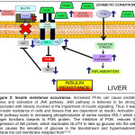 |
Figure 2: Insulin resistance occurrence. Increased FFAs can cause oxidative stress and activation of JNK pathway. JNK pathway is believed to be strongly associated with obesity involved in the impairment of insulin signalling. Click here to View figure |
The increased amounts of free fatty acids (FFAs) from the breakdown of lipid in adipocytes are released into the bloodstream. Elevated FFAs in turns caused a reduction of insulin sensitivity in the muscles by inhibiting-insulin mediated glucose uptake, thus caused the increasing the peripheral glucose.54 In addition, an increase in blood glucose concentration, and to some extent, circulating FFAs increase the insulin secretion, leading to hyperinsulinemia. The potential sources of lipid accumulation in the pancreas may be represented by circulating FFAs, de novo lipogenesis and dietary fat intakes. It suggested that the metabolic derangement happened in parenchymal tissue and caused shut down of mechanism survival in long term run at identical distribution in the exocrine and endocrine pancreas as well as in adipose tissue. The degree of fat distribution indicated the defect of mechanism and lipid impairment.55 Undoubtedly, insulin resistance causes hyperglycemia and FFAs level in the blood, which in turn exacerbate the pathophysiology of T2DM over time.9 Furthermore, advanced study in the pathogenesis of diabetes has been explored exclusively in insulin receptor and GLUT4. In in-vivo study using knockout specific insulin receptor in diabetic mice resulted in severe impaired glucose tolerance in muscle and fat tissue.56 Indeed, insulin resistance leads to lipid impairment, causes an increasing in adipose tissue mass, and the lipid impairment will be resulted as pre-disposed of diabetes. Taken together, these molecules’ involvement suggests unifying T2DM physiological effect in which the targeted tissues such as liver, muscle and fat coupled with insulin signalling derangement lead to insulin resistance. To some extent, these factors indicate the possibility of a vitamin D mechanism of action in modulating T2DM pathophysiology.
Conclusion
The main controversial issue is the requirement of vitamin D in daily intake for modulation of insulin signalling and how vitamin D managed to overcome the risk of T2DM in children and adults. Risk factors such as age and skin type cannot be neglected, as elderly and darker skin individuals always with vitamin D deficiency. Previous intervention data denoted the standard amount of vitamin D supplementation should be considered safe even though the range of vitamin D used in many studies is varies. However, extraction from this literature study on the perspective of biochemical view clearly stated that the level of vitamin D is responsible for altering T2DM pathophysiology due to interactions of VDR action mechanism, DBP, 1-a-hydroxylase in the skeletal and non-skeletal organs.
Acknowledgements
This research was financially supported by the Fundamental Research Grant Scheme, Ministry of Education, Malaysia.
Conflict Of Interest
The authors declare no conflict of interest.
References
- Nair R. and Maseeh A. Vitamin D: The “Sunshine” vitamin. Journal of Pharmacology and Pharmacotheraphy. 2012;3:2:118-26.
- Afzal S, Bojesen S. E and Nordestgaard B.G., Low 25-hydroxyvitamin D and risk of type 2 diabetes: A Propective Cohort Study and Meta-analysis. Clinical Chemistry. 2013;59:2:381-91.
CrossRef - Aguirre V., Werner E.D., Giraud J. Lee Y. H . Shoelson S.E and White M.F. Phosphorylation of Ser307 in Insulin Receptor Substrate-1 Blocks Interactions with the Insulin Action. Journal of Biology Chemistry. 2002;277:2:1531-1537.
CrossRef - Baker M.R., Peacock M. and Nordin B.E. The Decline in Vitamin D Status with Age. Age Ageing. 1980;9:4:249-252.
CrossRef - Kadowak S. and Norman A.W. Pancreatic Vitamin D-Dependent Calcium Binding Protein Biochemical Properties and Response to Vitamin D. Archives of Biochemistry and Biophysics. 1984;233:1:228-236.
CrossRef - Foss Y.J. Vitamin D Deficiency is The Cause of Common Obesity. Medical Hypotheses. 2009;72:3:314-321.
CrossRef - Kulie T., Groff A., Redmer J., Hounshell J., and Schrager S. Vitamin D: An Evidence-Based Review. Journal America Board Family Medicine. 2009;22:6:698-706.
CrossRef - Larsen H.R. Insulin Resistance and Diabetes. International Health News. 2016;1-8.
- Liu S., Song Y., Ford E.S., Manson J.E., Buring J.E., and Ridker P.M. Dietary Calcium, Vitamin D and the Prevalence of Metabolic Syndrome in Middle-Aged and Older U.S Women. 2005.
- Calle C., Maestro B. Garcia-Arencibia M. Genomic Actions of 1,25-Dihydroxyvitamin D3 on Insulin Receptor Gene Expression, Insulin Receptor Number and Insulin Activity in the Kidney, Liver and Adipose Tissue of Streptozotocin-Induced Diabetic Rats. BMC Molecular Biology. 2008;9:1:65.
- Pike J.W and Meyer M.B. The Vitamin D Receptor: New Paradigmns for the Regulation of Gene Expression by 1,25-Dihydroxyvitamin D3. Endocrinology Metabolism of Clinical North America. 2010;39:2:255-69.
- Looker A.C, Dawson-Hughes B., Calvo M.S, Gunter E.W and Sahyoun N.R. Serum 25-hydroxyvitamin D Status of Adolescents and Adults in Two Seasonal Subpopulations from NHANES III. Bone. 2002;30:5:771-7.
- MacLaughlin J. and Holick M.F. Aging Decreases the Capacity of Human Skin to Produce Vitamin D3. Journal of Clinical Investigations. 1985;76:4:1536-1538.
- Martin T and Campbell R.K. Vitamin D and Diabetes. 2011;113-118.
- Cadario F., Savastio S, Rizzo A.M, Carrera D, Bona G and Ricordi C. Can Type 1 Diabetes Progression be Halted? Possible Role of High Dose Vitamin D and Omega 3 Fatty Acids. European Review Medicine and Pharmacology Sciences. 2017;21:7:1604-1609.
- Mitri J and Pittas A.G. Vitamin D and Diabetes. Endocrinology Metabolism Clinical North America. 2014;43:1:205-32.
- Kajikawa M., Ishida H., Fujimoto S., Mukai E., Nishimura M., Fujita J., Tsuura Y., Okamoto Y., Norman A.W., and Seino Y. An Insulinotropic Effect of Vitamin D Analog with Increasing Intracellular Ca2+ Concentration in Pancreatic B-cells through Non-genomic Signal Transduction. Endocrinology. 1999;140:10:4706-4712.
- Scragg R., Sowers M., and Bell C. Serum-hydroxyvtamin D Diabetes and Ethnicity in the Third National Health and Nutrition Examination Survey. Diabetes Care. 2004;27:12:2813-8.
- Kim M.K., Kang II M. Wong Oh K., Kwon H.S., Lee J.H., Lee J.H., Lee W.C., Yppn K-H and Son H.Y. The Association of Serum Vitamin D Level with Presence of Metabolic Syndrome and Hypertension in Middle-Aged Korean Subjects. Clinical Endocrinology (Oxford). 2010;73:3:330-338.
- Johnson M.D., Nader N.S., Weaver A.L., Singh R., and Kumar S. Relationships between 25-hydroxyvitamin D Levels and Plasma Glucose and Lipid Levels in Padiatric Outpatients. Journal Paediatric. 2010;156:3:444-449.
- Skaaby T., Husemoen L.L.N., Pisinger C., Jørgensen T., Thuesen B,H,m Fenger M. and Linnerber A. Vitamin D Status and Changes in Cardiovascular Risk Factors: A Prospective Study of a General Population. Cardiology. 2010;123:1:62-70.
- K. 25-OH Vitamin DL Is it the Universal Pancacea for Metabolic Syndrome and Type 2 Diabetes?. Journal of Clinical Endocrinology and Metabolism. 2010;95:9:4220-4222.
CrossRef - C.Z., Uysal K.T and Maeda K. A Central Role for JNK in Obesity and Insulin Resistance. 2002;2:10-13.
- Moy F.M. Vitamin D Status and Its Associated Factors of Free Living Malay Adults in a Tropical Country, Malaysia. Journal Photochemistry and Photobiology B Biology. 2011;104:3:444-448.
CrossRef - Chin K.y., Ima-Nirwana S., Ibrahim S., Mohamed I.N and Ngah W.Z.W. Vitamin D Status in Malaysian Men and Its Associated Factors. Nutrients. 2014;6:12:5419-5433.
- H., Norman A.W., Okamura W.H., Sen A. and Zemel M.B. 1a, 25-Dihydroxyvitamin D3 Modulates Human Adipocyte Metabolism via Nongenomic Action. FASEB Journal. 2001;15:14:2751-3.
CrossRef - Zemel M.B. Nutritional and Endocrine Modulation of Intracellular Calcium: Implications in Obesity, Insulin Resistance and Hypertension. Molecular Cellular Biochemistry: 1998;188:1-2:129-36.
- Peterson C.A., Tosh A.K and Belenchia A.M. Vitamin D Insufficiency and Insulin Resistance in Obese Adolescents. Therapeutic Advance Endocrinology Metabolism. 2014;5:6:166-89.
CrossRef - Holick M.F. Vitamin D: A Millenium Perspective. Journal of Cellular Biochemistry. 2003;88:2:296-307.
CrossRef - Rahman S.A., Chee W.S.S., Yassin Z. and Chan S.P. Vitamin D Status Among Postmenopausal Malaysian Women. Asia Pacific Journal of Clinical Nutrition. 2004;13:3:255-60.
- Lund B., Hjorth L., Kjaer I., Reimann I., Friis T., Andersen R.B and Sorensen O.H. Treatment of Osteoporosis of Ageing with 1-alpha-Hydroxycholecalciferol. Lancet (London, England). 1975;2:7946:1168-71.
- Schottker B., Herder C., Rothenbacher D., Perna L. Muller H and Brenner H. Serum 25-hydroxyvitamin D Levels and Incident Diabetes Mellitus Type 2: A Competing Risk Analyssis in a Large Population-Based Cohort of Older Adults. European Journal Epidemiology. 2013;28:3:267-275.
- Holick M.F. McCollum Award Lecture. Vitamin D-New Horizons for the 21st American Journal of Clinical Nutrition. 1994;60:4:619-30.
- Van Dam R.M., Hu F.B.., Rosenberg L., Krishnan S and Palmer J.R. Dietary Calcium and Magnesium, Major Food Sources and Risk of Type 2 Diabetes in U.S Black Women. Diabetes Care. 2006;29:10:2238-2243.
- Lee N.K., Sowa., Hinoi E., Ferron M., Ahn J.D., Confavreux., Dacquin R., Mee P.J., McKee M.D., Jung D. Y., Zhang Z., Kim J.K., Mauvais-Jarvis F., Ducy P. and Karsenty G. Endocrine Regulation of Energy Metabolism by The Skeleton. Cell. 2007;130:3:456-469.
- Misung S.C.K and Woori N. Correlation Between Vitamin D and Cardiovascular Disease Predictors in Overweight and Obese Koreans. Journal of Clinical Biochemistry and Nutrition. 2013;52:3: 186-192.
- Feskens E.J., Bowles C.H and Kromhout D. Inverse Association Between Fish Intake and Risk of Glucose Intolerance in Normoglycemic Elderly Men and Women. Diabetes Care. 1991;14:11.
- Moy F.M and Bulgiba A. High Prevalence of Vitamin D Insufficiency and Its Association with Obesity and Metabolic Syndrome Among Malay Adults in Kuala Lumpur, Malaysia. BMC Public Health. 2011;11:735.
- Jan Mohamed H.J., Rowan A., Fong B., and Loy S.L. Maternal Serum and Breast Milk Vitamin D Levels: Findings From the Universiti Sains Malaysia Pregnancy Cohort Study. PLoS One. 2014;9:7:e100705.
- Ramly M., Ming M.F., Chinna K., Suboh S. and Pendek R. Effect of Vitamin D Supplementation on Cardiometabolic Risks and Health-Related Quality of Life Among Urban Premanopausal Women In A Tropical Country-A Randomized Controlled Trial. PloS One. 2014;9:10:8-10.
- Leong L.W, Loh S.P and Azhanie A.N. Vitamin D Intake and Sun Exposure Among Malaysian Athletes in National Sports Institute, Bukit Jalil. Malaysian Journal of Medicine and Health Sciences. 2013;9:1:21-28.
- Fong C.Y., Kong A.N., Poh B.K., Mohamed A.R., Khoo T.B., Ng R.L., Noordin M., Nadarajaw T and Ong L.C. Vitamin D Deficiency and Its Risk Factors in Malaysian Children With Epilepsy. Epilepsia. 2016;57:8:1271-1279.
- Shariff Z.M., Lin K.G., Sariman S. Lee H.S., Siew C.Y., Yusof B.N.M., Mun C.Y and Mohamad M. The Relationship Between Household Income and Dietary Intakes of 1-10 Year Old Urban Malaysian. Nutrition Research and Practice. 2015;9:3:278-287.
- Wilcox G. Insulin and Insulin Resistance. The Clinical biochemist.Reviews/Australian Association of Clinical Biochemists. 2005;26:2:19-39.
- Wannamethee S.G., Shaper A.G., Perry I.J and British Regional Heart Study. Smoking As a Model Risk Factor for Type 2 Diabetes In Middle-Aged Men. Diabetes Care. 2001;24:9:1590-1595.
- Wellen K.E and Hotamisligil G.S. Inflammation, Stress and Diabtes. The Journal of Clinical Investigation. 2005;115:5:1111-1119.
CrossRef - Wright E.M., Sala-Rabanal M. Loo D.D.F and Hirayama B.A. Sugar Absorption. Physiology of the Gastrointestinal Tract Elsevier. 2012;1583-1593.
- Suh S.H., Paik I., Y and Jacobs K. Regulation of Blood Glucose Homeostasis During Prolonged Exercise. Molecular Cells. 2007;23:3:272-279.
- Shoelson S.E, Lee J and Goldfine A.B. Reviews Series Inflammation and Insulin Resistance. Journal of Clinical Investigations. 2006;116:7:1793-1801.
CrossRef - Saltiel A.R and Pessin J.E. Insulin Signaling Pathways in Time and Space. Trends Cell Biology. 2002;12:2:65-71.
CrossRef - Singh V.P., Bali A., Singh N. and Jaggi A.S. Advanced Glycation End Products and Diabetics Complications. Korean Journal Physiology and Pharmacology. 2014;18:1:1-14:.
- McManus E.j., Sakamoto K., Armit L.J., Ronaldson L., Shpiro N., Marquez R. and Alessi D.R. Role that Phosphorylation of GSK3 Plays in Insulin and Wnt Signalling Defines by Knockin Analysis. EMBO Journal. 2005;24:8:1571-83.
CrossRef.
Accepted on: 20-4-2018
Second Review by: Dr. Ionel Bondoc (Romania)
Final Approval by: Prof. Min-Hsiung Pan


Web of Science Coverage
Emerging Sources Citation Index (ESCI)
2024 Journal Impact Factor: 1.1
Scopus Journal Metrics
CiteScore 2025: 2.6
CiteScore Details
Sustainable Nutrition: Food Systems, Nutrient Retention, and Public Health Impact
![]()
This journal is a member of, and subscribes to the principles of, the Committee on Publication Ethics (COPE)


