Comparative Analysis of Nutritional Constituents, Antioxidant and Antimicrobial Activities of Some Common Vegetable Wastes
Swagata Biswas
, Alolika Dutta, Maitrayee Biswas and Sirshendu Chatterjee* Department of Biotechnology, Techno India University West Bengal, Kolkata, West Bengal, India.
Corresponding Author Email: sirshendu.chatterjee@gmail.com
DOI : http://dx.doi.org/10.12944/CRNFSJ.9.1.07
Download this article as:
![]()
Vegetables are intrinsic to a healthy diet. But the peels are discarded as food waste, unknowing of their potential as the source of bioactive compounds. The study aims to find the nutritional constituents, antioxidant and antimicrobial activity of these food wastes. Here we make a comparative investigation among the five underutilized vegetable parts namely, Solanum tuberosum (Potato peel), Cucumis sativus (Cucumber peel), Musa acuminata (Unripe Banana peel), Brassica oleracea (Cauliflower stem), Lagenaria siceraria (Bottle gourd peel). After the primary screening, including proximate and qualitative analysis, the quantification of primary and secondary metabolites as well as minerals was estimated by different standard methods. The antioxidant potential was evaluated by both DPPH and H2O2 radical scavenging assays. Antimicrobial activity was analyzed by Kirby Bauer disc diffusion method against Gram-positive (Staphylococcus aureus) and Gram-negative (Escherichia coli) strains of bacteria compared with positive and negative controls. From the proximate analysis, the highest moisture and water content were found in the peel sample of Lagenaria siceraria. Out of ten qualitative tests, protein, fatty acid, flavonoid, alkaloid and xanthoprotein were detected in all five samples. Among the quantitative estimations, Lagenaria siceraria showed the highest amount of polysaccharides (85.82±0.12 mg DE/g DW), ascorbic acid (2.48±0.14 mg AAE/g FW), thiamine (24.46±0.13 mg TE/g DW), polyphenols (86.36±0.10 mg GAE/g DW), flavonoids (49.59±0.07 mg QE/g DW), minerals like K, Ca and Mg and 72.35±0.40% inhibition by DPPH. Simultaneously a significant amount of protein (63.59±0.13 mg BSAE/g FW), the amino acid (7.84±0.02 mg AAE/g DW), minerals like Na, Zn and B and 88.76±0.10% inhibition by H2O2 were found in Cucumis sativus. At a higher concentration, all samples were exhibited significant antimicrobial activity which laid out a strong correlation with previously screened phytonutrients and antioxidants. The overall findings suggested that these underutilized vegetable parts can be utilized in the processing of potential functional foods as well as pharmaceuticals rather than thrown out as agro-waste.
KEYWORDS:Antimicrobial Activity; Antioxidants; Mineral; Phytonutrients; Vegetables
Introduction
Vegetables are considered to be a significant source of nutrients, minerals, dietary fibre, antioxidants and other beneficial phytochemicals.1 People are health conscious nowadays and hence, different vegetables find a place in daily diet that in-turn produce a large amount of biodegradable agro wastes.2 This waste is mainly composed of skin, stem, peel, rind, seed, shell and pomace.3 In recent time, many scientific investigations reported the potential of these unused parts as the source of important phytochemicals like polyphenols, flavonoids, alkaloids, sugars, vitamins, minerals and many more.4 Besides, they have many pharmacological effects in the health care system as antimicrobial, antifungal, antibacterial, anti-mutagenic, cardioprotective and neuroprotective.5
Potato is an edible starchy tuber of plant Solanum tuberosum (ST) Synonym Battata tuberosa (L.) Hill, belonging to the nightshade family, Solanaceae. As potato peels were reported to have higher polyphenol content (such as gallic acid, caffeic acid) than flesh, it can be suggested to use as natural antioxidant.6 Cucumber is a widely used vegetable grown in creeping vine plant Cucumis sativus (CS) Synonym Cucumis rumphii Hassk., which is in the gourd family of Cucurbitaceae. The researchers identified that the peel of cucumber was comprised of flavonoids, triterpenoids, antioxidants, vitamin C, K and other trace elements along with minerals.7 Raw banana is a staple food produced by the plant Musa acuminata (MA) Synonym Musa cavendishii Lamb. , belongs to the Musaceae family. Due to high antioxidant status, banana peel is proven to be helpful in reducing plasma oxidative stress.8 Cauliflower belongs to cruciferous vegetable in the species of Brassica oleracea (BO) Synonym Brassica arborea Steud., belonging to the cabbage family, Brassicaceae. As the stem and leaves of cauliflower are delineated to have iron, β- carotene, digestive crude protein along with starch and amino acid, it can be utilized as a dietary fibre supplement.9 Bottle gourd or Calabash is a nutritious vegetable grown in running vine plant Lagenaria siceraria (LS) Synonym Cucumis lagenaria (L.) Dumort., which belongs to the gourd family, Cucurbitaceae. The peel of bottle gourd was identified as a great source of soluble dietary fibres, amino acids, β- carotene, and ascorbic acid, which was beneficial to lower the serum cholesterol.10
Therefore, the present study was focused on these five selected underutilized parts for analyzing the proximate parameters and estimating the nutritional composition and evaluating the antioxidant and antimicrobial activity.
Materials and Methods
Plant Samples Collection
The current study was executed in the biotechnology laboratory of Techno India University, West Bengal on January-June, 2019. The plant parts were collected from Salt Lake, Kolkata. Kolkata is positioned at 22.5726′ N, 88.3639′ E and is situated 9.15 m from the sea level with alluvial soil type. Kolkata has a wet and dry climate condition with an annual average temperature of 26.8°C, relative humidity 70.8% and rainfall is 1735mm. The samples were thoroughly washed with water, and the peels and stem were separated carefully. Then the peels were cleaned and sundried for 12-15 days. These studied samples were then grounded to fine dust and stored in polythene bottles at 4°C till the time of experiments.
Collection of Microbial Strains
Pure cultures were collected from Department of Microbiology, Calcutta University, West Bengal, India.
Chemical and Reagents
Analytical graded reagents and chemicals were used in all experiments. Folin-ciocalteu, Aluminum chloride, Ascorbic acid, Starch, Ninhydrin, Chloroform, Methanol and Acetic acid were procured from Merck Life Science Private Limited, Mumbai. Bovine Serum Albumin, L-Lysine, Agar and Broth were purchased from HiMedia, Mumbai. Gallic acid, Hydrogen peroxide and Sodium hydroxide were supplied by SDFCL, Mumbai. Quercetin, DPPH, Acetone and Caffein were obtained from SRL, Mumbai. Dextrose and Ammonium hydroxide was purchased from Finar Limited, Ahmedabad. For quantitative study in the current research work, a spectrophotometer (Systronics 117) was used to measure the particular absorbance.
Preparation of Extracts
Preparation of Crude Extracts
The crude extracts of samples were obtained by grinding finely cut 1 g of fresh sample with 25 mL solvent (double distilled water). All the extracts were kept over a shaker for vigorous shaking for 24 hours for effective extraction of the bioactive compounds. Next, the extracts were clarified by centrifugation at 10,000 rpm for 10 min. The supernatant was stored and diluted as needed in the different assays.
Preparation of Aqueous Extracts of Dry Samples
1 g of dry samples were mixed with 25 mL of solvent (double distilled water) and kept on the rocker for 24 hours for extraction. Then the extracts were clarified by centrifugation at 10,000 rpm for 10 min. The supernatant was diluted as required for further studies.
Proximate Analysis
Estimation of Total Moisture Content
Using standard method11, the total moisture content of the samples were determined and calculated by the following formula,
MCwb = [(Wi-Wf)/ Wi] ×100
Where MCwb is expressed as total moisture content on a wet basis, Wi is initial weight, and Wf is the final weight.
Estimation of Relative Water Content
With the help of standard protocol12, relative water content (RWC) was estimated and calculated by the following formula,
RWC = [(FW-DW)/ (TW-DW)]×100
Where FW= Fresh Weight, TW=Turbid Weight, DW= Dry Weight
Estimation of Water-Soluble and Insoluble Portion
Firstly, the weight of a filter paper was taken. Then 100 mg of fresh samples were homogenized with double distilled water and filtered through that filter paper. After filtration, the weight of filter paper with the insoluble part was measured.13 Water-soluble and insoluble part of the samples were determined using the following formula,
Insoluble Portion (Wi) = W2 -W1
Soluble Portion (Ws) = 100 – Wi
Where W1= weight of filter paper, W2 = weight of filter paper with insoluble part
Estimation of pH
To determine the pH of the aqueous extracts, the standard method was used.14 Sample extract was prepared by homogenizing 5 g fresh plant sample with 10 mL double distilled water, and the pH of the filtrate was evaluated using a calibrated pH meter.
Estimation of Conductivity
5 g of fresh samples were extracted with 10 mL of double distilled water and filtered. Then the conductivity of these filtrates was determined after calibrating conductivity meter with standard solutions.
Qualitative Screening
To detect the presence of carbohydrate by Molisch’s test15, protein by Biuret test16, fatty acids or fixed oils17, amino acid by Ninhydrin test18, reducing sugars19, polyphenols by Fecl3 and KMnO4 method20, flavonoids by alkaline reagent test21, alkaloids by Wagner test22, xanthoprotein23 and cardiac glycoside by Keller-Kelliani’s test24, standard protocols were used.
Nutritional Quantification
Estimation of Polysaccharides
In accordance with standard protocol25, total polysaccharide was quantified by spectrophotometric assay at 490 nm using dextrose as a standard reagent. The results were expressed as mg Dextrose Equivalent/g of Dry Weight of sample (mg DE/g DW).
Estimation of Protein
Total protein content was estimated by the Lowry method, using Bovine Serum Albumin (BSA) as a standard reagent.26 The absorbance was measured at 660 nm. The contents were indicated as mg BSA Equivalent /g of Fresh Weight of the sample (mg BSAE/g FW).
Estimation of Lipid
Total lipid content was quantified by the solvent extraction method.27 Samples were transferred to a separating funnel and washed with chloroform and methanol. The mixture was shaken vigorously, and lipid was extracted with chloroform. Considering the given formula, the results were expressed as % lipid present in dry samples,
Total Lipid Contents = (lipid weight in aliquot × chloroform layer volume)/ aliquot volume
Estimation of Amino Acid
With the help of standard protocol, the amino acids of the samples were quantified spectrophotometrically at 570 nm using lysine as a standard reagent.28 Amino acid contents were expressed as mg Lysine Equivalents/g of Dry Weight of the sample (mg LE/ g DW).
Estimation of Ascorbic Acid
Quantification of ascorbic acid of the samples was evaluated by iodine and iodate solution in a redox titration method utilizing ascorbic acid as a standard reagent.29 The equation: V1S1=V2S2 was used for calculation. The results were indicated as mg Ascorbic Acid Equivalent/g of Fresh Weight of the sample (mg AAE/ g FW).
Estimation of Thiamine
Thiamine determination was executed according to the methodology by Dutta et al.30 The absorbance was measured at 360 nm, using a standard reagent. Results were expressed as mg Thiamine equivalent per gm of Dry Weight (mg TE/ g DW).
Determination of Mineral Contents
As the quality of many foods and medicines depends upon the content and type of minerals, it is essential to investigate the mineral composition. The standardized dry ashing method31 was used to estimate the macro elements like Calcium (Ca), Magnesium (Mg), Sodium (Na) and Potassium (K) and microelements like Zinc (Zn) and Boron (B). The 5 g of samples were kept into a crucible for 1 hour in a muffle furnace at 500° C for preparation of ash. Next, the ash was digested with HCl and HNO3 mixture (1:3). The volumes of the digested samples were completed to 50 mL with double distilled water, and mineral contents were determined by atomic absorption spectrophotometer (AAS). The results were expressed in terms of mg/ g of Dry Weight.
Phytochemical Quantification
Estimation of Polyphenols
The polyphenol content of the samples was evaluated by the Folin-Ciocalteu method, using Gallic acid as standard32 and are expressed as mg Gallic Acid Equivalent per g of Dry Weight (mg GAE/ g DW).
Estimation of Flavonoids
According to the Aluminum Chloride colourimetric assay33, total flavonoids were quantified, using Quercetin as a standard reagent and contents are expressed as mg Quercetin Equivalent per g of Dry Weight (mg QE/ g DW).
Estimation of Alkaloids
Total alkaloid was estimated by the standard protocol, mentioned in John et al., 34 Samples were extracted with chloroform, and the absorbance was read at 470 nm, using caffeine as standard. Total alkaloid content was expressed in mg Caffeine equivalent/ g of Dry Weight (mg CE/ g DW).
Estimation of Cardiac Glycoside
Cardiac glycosides of the samples were quantitatively determined, according to Solich et al.35 The absorbance was read at 495 nm, using Digoxin as standard. The results were expressed as mg Digoxin Equivalent/ g Dry Weight (mg DE/g DW).
Determination of Antioxidant Activity
Scavenging Activities on DPPH Free Radicals
The free radical scavenging property of the extracts was determined using the DPPH solution in methanol by the standard method.36 The absorbance (OD) value was measured at 517 nm using ascorbic acid as standard. The scavenging activity was expressed as Ascorbic Acid Equivalent (AAE), in terms of percentage of inhibition from the following formula,
% Inhibition = [(A0 – A1)/A0]×100
Where A0 and A1 represent the absorbance of the control, and of extract or standard, respectively.
Scavenging Activities on H2O2 Free Radicals
The scavenging activities of the extracts on H2O2 radical were evaluated with the help of the reference method using Gallic acid as standard by taking absorbance at 230 nm.37 The antioxidant activity was expressed as Gallic Acid Equivalent (GAE), as percentage inhibition from the following formula,
% Inhibition = [(A0 – A1)/A0]×100
Where A0 and A1 are the OD of the control, and of the extract or standard respectively.
Determination of Antimicrobial Activity
Kirby-Bauer’s Disc diffusion susceptibility method38 was used to determine the antibacterial activity of the vegetable-waste extracts against Gram-positive bacterial strain, Staphylococcus aureus, (denoted by J on microbial plates) and Gram-negative bacterial strain, Escherichia coli (denoted by H on microbial plates).
Positive Control
Streptomycin
Negative Control
Distilled Water
Statistical Analysis
All the experimental data (except mineral analysis, as it was done in a singlet) were expressed as average ± standard deviation (SD) after performing the measurements in triplicates. Statistical analysis was determined by one way ANOVA followed by Bonferroni’s Post Hoc test using MS Excel Software. In the graphical representation, the statistically significant values (p<0.05) were denoted by different letters on the bars.
Results
Proximate Analysis
Results obtained from the proximate analysis were presented in Table 1. The highest moisture content (92.94%), as well as water content (96.28%), were observed in LS. The lowest amounts of moisture (85.67%), as well as water content (78%), were observed in BO By seeing the high moisture content of the five samples, it can be concluded that a very high level of water ≥ 75% making this by-product highly perishable and requiring immediate use or stabilization to prevent fermentation and mould growth. But the observed percentage of the soluble portion was the highest (95.9%) in BO and the lowest (71.4%) in ST. The pH of these five samples were found to be within the acidic range (pH= 5.16 to 6.28). Acidity of the BO (pH=5.16) and CS (pH=6.28) were found to be highest and lowest respectively. The conductivity of these five samples was ranged from 0.80 to 2.00 mS/cm. The highest conductivity was showed in ST and the lowest in CS.
Table 1: Result of Proximate Analysis.
| Sample | Total Moisture Content (%) | Relative Water Content (%) | % of Insoluble portion | % of Soluble portion | pH | Conductivity(mS/cm) |
| ST | 89.19 | 91.84 | 28.6 | 71.4 | 5.22 | 2.00 |
| CS | 90.41 | 83.36 | 12.3 | 87.7 | 6.28 | 0.80 |
| MA | 89.54 | 93.57 | 12.8 | 87.2 | 6.19 | 1.60 |
| BO | 85.67 | 78.00 | 4.1 | 95.9 | 5.16 | 1.20 |
| LS | 92.94 | 96.28 | 7.1 | 92.9 | 5.30 | 1.30 |
Qualitative Analysis
The results of the ten qualitative tests were represented in Table 2. It was observed that protein, fatty acid, flavonoid, alkaloid and xanthoprotein were identified in all five samples. All the ten tests were shown positive in LS. Among the 10 tests, 9 tests were also found to be positive in MA. The results indicated that the experimental samples could be considered highly nutritionally as well as pharmaceutically potent phytochemical resources.
Table 2: Result of Qualitative Screening.
| Test Name | ST | CS | MA | BO | LS |
| Carbohydrate | + | – | + | – | + |
| Protein | + | + | + | + | + |
| Fatty Acid | + | + | + | + | + |
| Amino Acid | – | – | + | – | + |
| Reducing Sugar | – | + | – | – | + |
| Polyphenol | – | – | + | – | + |
| Flavonoid | + | + | + | + | + |
| Alkaloids | + | + | + | + | + |
| Xanthoprotein | + | + | + | + | + |
| Cardiac Glycoside | + | – | + | – | + |
Where, “+” PRESENT & “–” ABSENT
Nutritional Analysis
Polysaccharides
From Figure 1(i), it was observed that highest and lowest polysaccharide content was exhibited by LS (85.82±0.12 mg DE/g DW) and CS (26.16±0.10 mg DE/g DW) respectively.
Protein
The amounts of protein in the five samples were presented in Figure 1(ii). From the results, it can be interpreted that all the samples contain a significant amount of protein. The highest and the lowest amount of protein were found to be present in the peels of CS (63.59±0.13 mg BSAE/g FW), and the peels of ST (46.50±0.20 mg BSAE/g FW) respectively.
Lipid
The amounts of lipid in the five samples were presented in Figure 1(iii). Significant amount of total lipids was detected in the stem of BO (1.90 ± 0.03%). On the other side, the lowest amount was observed in the peels of ST (0.76 ± 0.06%).
Amino Acid
From Figure 1(iv), it was observed that the amino acid content of CS (7.84±0.02 mg AAE/g DW) was significantly higher than the other samples. But the peel of LS showed the minimum amount (2.11±0.03 mg AAE/g DW).
Ascorbic Acid
The results of this assay were depicted in Figure 1(v). The amount ranged from 0.94±0.21mg AAE/g FW (peel of CS) to 2.48±0.14 mg AAE/g FW (peel of LS).
Thiamine
The thiamine content of these five samples was represented in Figure 1(vi). It was clearly observed that the amount in the peels of LS (24.46±0.13 mg TE/g DW) was significantly higher than other samples. An insignificant amount (0.53±0.11 mg TE/g DW) was detected in the stem of BO.
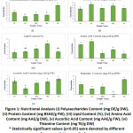 |
Figure 1: Nutritional Analysis. |
Mineral Analysis
Eight important macro as well as micro elements were quantified in five test samples using AAS, and the amounts were presented in Table 3. The macroelements like Ca, Mg, K and Na as well as microelements like Zn and B were found to be present in the stem of BO in highest amount with respect to other four samples. However, the peels of ST contain highest amount of Fe and Cu with respect to others.
Table 3: Result of Mineral Analysis (mg/g Dry Weight)
| Sample | Ca | Mg | K | Na | Fe | Cu | Zn | B |
| ST | 1.41 | 1.44 | 37.79 | 0.83 | 0.43 | 0.04 | 0.044 | 1.45 |
| CS | 7.04 | 2.60 | 37.59 | 2.03 | 0.08 | 0.01 | 0.047 | 3.35 |
| MA | 4.05 | 1.29 | 45.21 | 0.63 | 0.06 | 0.009 | 0.044 | 0.89 |
| BO | 17.51 | 2.73 | 100.52 | 2.30 | 0.07 | 0.006 | 0.044 | 8.75 |
| LS | 3.11 | 1.40 | 28.85 | 0.71 | 0.12 | 0.01 | 0.026 | 2.97 |
Phytochemical Analysis
Polyphenols
The total polyphenol content of these five samples was shown in Figure 2(i). It was clearly observed that the amount of polyphenol was ranged from 5.80±0.09 mg GAE/g DW (stem of BO) to 86.36±0.10 mg GAE/g DW (peel of LS) in the samples under study.
Flavonoids
From the Figure 2(ii), the maximum flavonoid amount (49.59±0.07 mg QE/g DW) was observed in the peel of LS, and the peel of CS showed the minimum amount (14.36±0.08 mg QE/g DW).
Alkaloids
Total alkaloids content of these five samples were estimated in Figure 2(iii). The highest amount was shown in the peels of LS (0.113±0.05 mg CE/g DW). On the other side, the peel of CS exhibited the minimum amount (0.086±0.02 mg CE/g DW).
Cardiac Glycosides
The cardiac glycoside content of these five samples was shown in Figure 2(iv). Among the samples, the higher amount (0.34±0.01 mg DE/g DW) was observed in the peel of LS. But a very negligible amount (0.07±0.02 mg DE/g DW) was presented in the stem of BO.
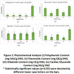 |
Figure 2: Phytochemical Analysis. |
The present study highlighted the dependence of the functional activity of the bioactive compounds to each other since they are highly correlated. The calculated correlations coefficient between the various bioactive compounds were represented in Table 4.
Table 4: Correlation between Bioactive Components.
| Correlation Parameters | Correlation Equation | The correlation coefficient (r) |
| Polyphenol & Flavonoid | y = 0.3121x + 23.568 | 0.5457 |
| Polyphenol & Alkaloid | y = 0.0003x + 0.0891 | 0.8862 |
| Polyphenol & Cardiac Glycoside | y = 0.0029x + 0.0928 | 0.9062 |
| Flavonoid & Alkaloid | y = 0.0006x + 0.0772 | 0.7241 |
| Flavonoid & Cardiac Glycoside | y = 0.0062x – 0.0309 | 0.7412 |
| Alkaloid & Cardiac Glycoside | y = 10.007x – 0.7957 | 0.9463 |
*Coefficient (r) values can range from +1 (positive relationship) to -1 (negative relationship)
Antioxidant Activity
DPPH Radical Scavenging Assay
From Figure 3(i), the percentage inhibition by quenching the DPPH radical ranged from 27.78±0.21% in peel sample of ST to 72.35±0.40% in peel sample of LS.
Simultaneously, the total antioxidant content of these five samples was also calculated. The peel of LS showed a significantly higher amount of antioxidant (37.57±0.22 mg AAE/g DW). The comparatively lower amount of antioxidant activity was detected in the stem of BO (13.91±0.13 mg AAE/g DW) as well as in the peel of ST (13.34±0.11 mg AAE/g DW).
H2O2 Radical Scavenging Assay
From Figure 3(ii), the percentage inhibition by H2O2 assay ranged from 60.87±0.22% in the stem of BO to 88.76±0.10% in peel sample of CS.
During the calculation of total antioxidant content, it was observed that the significantly higher amount of antioxidants were presented in the peel of CS (46.72±0.05mg GAE/g DW) and the peel of ST (46.20±0.09 mg GAE/g DW). The comparatively lower amount of antioxidant was exhibited in the stem of BO (30.80±0.12 mg GAE/g DW).
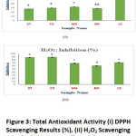 |
Figure 3: Total Antioxidant Activity. |
To establish a correlation between the phytonutrient and antioxidant activity, we have calculated a positive statistical relationship between bioactive compounds and antioxidant inhibitions, represented in Table 5.
Table 5: Correlation Coefficient (r) between the Bioactive Components and the Antioxidant Activities (DPPH and H2O2).
| Correlation Coefficient (r) | DPPH | H2O2 |
| Polyphenol | 0.9931 | 0.0307 |
| Flavonoid | 0.4952 | 0.0018 |
| Alkaloid | 0.8842 | 0.1296 |
| Cardiac Glycoside | 0.9015 | 0.0258 |
| Ascorbic Acid | 0.3159 | 0.2462 |
*Coefficient (r) values can range from +1 (positive relationship) to -1 (negative relationship)
Antimicrobial Activity
These five underutilized parts of the vegetables exhibited significant zone of inhibitions (mentioned in Figure 4) at a higher concentration against the tested S. aureus as well as E. coli, indicating these plant extracts have notable antibacterial properties compared with a standard antibiotic (streptomycin) and solvent (distilled water).
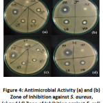 |
Figure 4: Antimicrobial Activity. |
The results (Table 6), highlighted the zone diameter, produced by the five samples against the two strains.
Table 6: Antimicrobial Activity: Zone of Inhibitions (mm)
| Organism Name | Staphylococcus aureus (J) | Escherichia coli (H) |
| Sample Name | Zone Diameter (mm) | Zone Diameter (mm) |
| Positive Control (PC.) | 18.3 | 20.3 |
| Negative Control (NC.) | Nil | Nil |
| ST | 6.3 | 6.5 |
| CS | 6.6 | 6.3 |
| MA | 6.8 | 7.7 |
| BO | 6.5 | 5.6 |
| LS | 8.3 | 8.2 |
Discussions
In this comparative study, the chosen five vegetables are widely used in India as well as in other countries. So the amounts of unused parts of these vegetables are huge, and hence discarded as organic waste from both households and food-processing industries. This study was done to decipher the possibility to reuse these agro-wastes by evaluating their bioactive compounds as well as antioxidant and antimicrobial activities. The results of proximate analysis, moisture content and relative water content of five samples were comparable to literature values of Gupta and Joshi.39,40 An investigation conducted by Khattak and Rahman, showed that the appreciable amount of polyphenol, flavonoids as well as antioxidant activity by DPPH and H2O2 were detected in the peel of S.T.41 The results obtained from the phytochemical analysis as well as antibacterial activity of the CS peel in the present assay was supported by a previous report by Foong et al.42
Agricultural wastes can be used in the production of single-cell protein as they are good sources of metabolites to support microbial growth.43 The peel of LS exhibited a significant amount of polysaccharide among all the five samples. Results were in line with the findings of Akter et al. 44 and showed the presence of high concentration of nutritional and phytochemical compounds in the powdered peel of LS. It was also found to be a rich reserve of minerals like calcium, potassium, magnesium, sodium and boron which was in congruence with the study of Ghosh & Chatterjee 40 and Rahman.45
Protein is crucial dietary components to supply nitrogen, potassium, sulfur and other substances for growth. As the peel of CS showed a high amount of protein and amino acids, it can be considered as a good source of nitrogen.46, 47According to Rodzali et al.48 the unripe banana peel possessed higher antioxidant and antimicrobial activities due to the presence of phenols, flavonoids and minerals like calcium and potassium which were aided with this current research data of MA. Surprisingly, the stem of BO showed a significant amount of lipid compared with other samples. It had also contained the highest amount of potassium and calcium which can correlate with Baloch et al.49 The study of Khan et al.50 and Ghosh et al.51 reported a positive correlation between the total antioxidant capacity with the plasma polyphenolic level, which was justified by the calculated records.
These bioactive compounds, as well as antioxidants, are used to enhance the nutritional and therapeutic potentiality of processed products as recorded in Siriwardhana et al.52 According to Hintz et al., in the food industry, plant antimicrobial compounds have potential use as bio-preservatives and bio-insecticides, with potential use against various foodborne pathogens.53 Therefore, these selected vegetable waste may be converted to wealth if used in proper way in food and pharmaceutical industries.
Conclusions
This study on underutilized parts of the vegetables highlighted that, they are not at all waste but may be converted to a wealth if they are used properly. Developing as well as under developed and economically weak Countries can implement the use of the agro-waste for mankind. In the present study, the peels of LS has acquired attention as a potential unused wealth and hence, it can be used for bioprospecting. More research in the domain of waste to wealth conversion related to present domain is still needed to fight against malnutrition and food scarcities around the world.
Acknowledgements
The authors are thankful to Dr Sukhendu Mondal, Assistant Professor, Department of Microbiology, and Calcutta University, for his support to do the antimicrobial activity study. The authors are highly obliged to Dr Madhusudan Mondal, Former Additional Director, Botanical Survey of India (BSI), Kolkata, and West Bengal for identifying the plant samples.
Funding Source
No external fund received for the study.
Conflict of Interest
The authors declare no conflict of interest.
References
- Sekeroglu N., Ozkutlu F., Deveci M., Dede O. and Yilmaz N. Evaluation of Some Wild Plants Aspect of Their Nutritional Values Used as Vegetable in Eastern Black Sea Region of Turkey. Asian Journal of Plant Sciences. 2006; 5(2):185-189.
CrossRef - Liu R. H. Health Benefits of Fruit and Vegetables are from Additive and Synergistic Combinations of Phytochemicals. American Journal of Clinical Nutrition. 2003; 78(3):517S-520S.
CrossRef - Kandari V. and Gupta S. Bioconversion of Vegetable and Fruit Peel Wastes in Viable Product. Journal of Microbiology and Biotechnology Research. 2012; 2(2):308-312.
- Sivakumar N. T. and Venkataraman R. Phytochemical and Pharmacological Studies on Plant Waste Materials. Der Pharmacia Sinica. 2010; 1(1):1-6.
- Parashar S., Sharma H. and Garg M. Antimicrobial and Antioxidant Activities of Fruits and Vegetable Peels: A Review. Journal of Pharmacognosy and Phytochemistry. 2014; 3(1):160-164.
- Rehman Z., Habib F. and Shah W. H. Utilization of Potato Peels Extract as a Natural Antioxidant in Soy Bean Oil. Food Chemistry. 2004; 85:215-220.
CrossRef - Mukherjee P. K., Nema N. K, Maity N. and Sarkar B. K. Phytochemical and Therapeutic Potential of Cucumber. Fitoterapia. 2012; 84:227-236.
CrossRef - Someya S., Yoshiki Y. and Okubo K. Antioxidant Compounds from Bananas (Musa Cavendish). Food Chemistry. 2002; 79(3):351-354.
CrossRef - Singh G., Asha K. and Sehgal S. Development and Nutritional Evaluation of Products Prepared from Dried Powder of Cauliflower Leaves. Journal of Food Science and Technology. 2005; 42(2):137-139.
- Modgil M., Modgil R. and Kumar R. Carbohydrate and Mineral Content of Chyote (Sechium edule) and Bottle Gourd (Lagenaria siceraria). Journal of Human Ecology. 2004; 15(2):157-159.
CrossRef - Oyeleke OA. Outlines of Food Analysis (2nd), London: Macmillian Publishers Ltd.; 1984: p. 27-30.
- Singh A. Practical Plant Physiology. New Delhi: Kalyani Publishers; 1977.
- Unani Pharmacopoeia. New Delhi: IMPCOPS Publication; 2004: p. 137.
- Aremu M. O., Olaofe O., Basu S. K., Abdulazeez G. and Acharya S. N. Processed Cranberry Bean (Phaseolus coccineus), Seed Flour for The African diet. Canadian Journal of Plant Sciences. 2010; 90:719-728.
CrossRef - Trease and Evans’ Pharmacognosy, 15th ed. London: Saunders Publishers; 1997: p. 42-44.
- Brain K. R. and Turner T. D. The Practical Evaluation of Phytopharmaceuticals. 2nd ed. Bristol: Wright Science Technica; 1975: p. 81- 82.
- Ayoola G. A., Coker H. A. B., Adesegun S. A., Adepoju-Bello A. A., Obaweya K., Ezennia E. C. and Atangbayila T. O. Phytochemical Screening and Antioxidant Activities of Some Selected Medicinal Plants Used for Malaria Therapy in Southwestern Nigeria. Tropical Journal of Pharmaceutical Research. 2008; 7(3):1019-24.
CrossRef - Yasuma A. and Ichikawa T. A New Histochemical Staining Method for Protein. Journal of Laboratory and Clinical Medicine. 1953; 41(2):296-9.
- Shanmugam B., Shanmugam K. R., Sahukari R., Subbaiah G. V., Korivi M. and Reddy K. S. Antibacterial Activity and Phytochemical Screening of Phyllanthus niruri in Ethanolic, Methanolic and Aqueous Extracts. International Journal of Pharmaceutical Sciences Review and Research. 2014; 27(2):85-89.
- Mace Gorbach S. L. Anaerobic Bacteriology for Clinical Laboratories. Pharmacognosy. 1963; 23:89-91.
- Shalini S. and Sampathkumar P. Phytochemical Screening and Antimicrobial Activity of Plant Extracts for Disease Management. International Journal of Current Science. 2012:209-18.
- Torres-Castillo J. A., Sinagawa-Garcia S. R., Martínez-Avila G. C. G., Lopez-Flores A. B., Sanchez-Gonzalez E. I., Aguirre-Arzola V. E., Torres-Acosta R. I., Olivares-Saenz E., Osorio-Hernandez E and Gutierrez-Diez A. Phytochemical Detection, Antioxidants, Enzymes and Antifungal Properties. FYTON. 2013; 82:193-202.
- Brinda P., Sasikala P. and Purushothaman K. K. Pharmacognostic Studies on Merugan Kizhangu. Med. Ethnobot. Res. 1981; 3:84-96.
- Ugochukwu S. C., Uche A. and Ifeanyi O. Preliminary Phytochemical Screening of Different Solvent Extracts of Stem Bark and Roots of Dennetia tripetala G. Baker. Asian J Plant Science and Res. 2013; 3(3):10-13.
- Harshal A. P. and Priscilla M. D’M. Spectrophotometric Estimation of Total Polysaccharides in Cassia tora Gum. Journal of Applied Pharmaceutical Science. 2011; 1(3):93-95.
- Walker J. M. The Protein Protocols Handbook (2nd). Totowa, New Jersey: Humana Press; 2002: p.7-9.
- Bligh E. G. and Dyer W. J. A Rapid Method for Total Lipid Extraction and Purification. J. Biochem. Physiol. 1959; 37:911-917.
CrossRef - Sircelj H., Tausz M., Grill D. and Batic F. Biochemical Responses in Leaves of Two Apple Tree Cultivars Subjected to Progressing Drought. J Plant Physiol. 2005; 162:1308-18.
CrossRef - Kashyap G. and Gautam DM Analysis of Vitamin C in Commercial and Naturals Substances by Iodometric Titration found in Nimar and Malwa region. Journal of Scientific Research in Pharmacy. 2012; 1(2):77-78.
- Dutta S., Dey P. and Chaudhuri K. T. Quantification and Correlation of the Bioactive Phytochemicals of Croton Bonplandianum Leaves of Sub-Himalayan Region of West Bengal. Asian Journal of Pharmaceutical and Clinical Research. 2013; 6(3):142-147.
- Thakur A., Vaidya D., Kaushal M. and Gupta A. Physicochemical Properties, Mineral Composition, FTIR Spectra and Scanning Electron Microscopy of Wild Apricot Kernel Press Cake. International Journal of Food Science and Nutrition. 2019; 4(2):140-143.
- Singleton V. L., Orthofer R. and Lamuela-Raventos R. M. Analysis of Total Phenols and Other Oxidation Substrates and Antioxidants by Means of Folin-Ciocalteau Reagent. Methods in Enzymology. 1999; 299:152-178.
CrossRef - Zhishen J., Mengcheng T. and Jianming W. The Determination of Flavonoid Contents in Mulberry and their Scavenging Effects on Superoxide Radicals. Food chem. 1999; 64:555-559.
CrossRef - John B., Sulaiman C. T., George S. and Reddy V. R. K. Spectrophotometric Estimation of Total Alkaloids in Selected Justicia Species. International Journal of Pharmacy and Pharmaceutical Sciences. 2014; 6(5):647-648.
- Solich P., Sedliakova V. and Karlicek R. Spectrophotometric Determination of Cardiac Glycosides by Flow-Injection Analysis. Analytica Chimica Acta. 1992; 269(2):199-203.
CrossRef - . Shen Q., Zhang B., Xu R., Wang Y., Ding X. and Li P. Antioxidant Activity In Vitro of Selenium-Contained Protein from The Se-enriched Bifidobacterium animalis Anaerobe 16. 2010:380-386.
CrossRef - Ruch R. J., Cheng S. J. and Klaunig J. E. Prevention of Cytotoxicity and Inhibition of Intercellular Communication by Antioxidant Catechins Isolated from Chinese Green Tea. Carcinogenesis. 1989; 10(6):1003-8.
CrossRef - Sen A. and Batra A. Evaluation of Antimicrobial Activity of Different Solvent Extracts of Medicinal Plant: Melia Azedarach International Journal of Current Pharmaceutical Research. 2012; 4(2):67-73.
- Gupta K. and Joshi V. K. Fermentative Utilization of Waste from Food Processing Industry. Verma L. R. and Joshi V. K. (Eds). Postharvest Technology of Fruits and Vegetables: Handling Processing Fermentation and Waste Management (Vol. 2). New Delhi, India: Indus Pub. Co.; 2000.
- Ghosh P and Chatterjee S. Evaluation of organoleptic, proximate parameters and analysis of the nutritional composition of five wild weeds: A search for low-cost nutraceuticals. International Journal of Pharmaceutical Sciences and Research. 2020; 11 (10):5170-5181.
- Khattak K. F. and Rahman T. U. Phytochemical Profiles, Nutrient Compositions, Extraction Yields and Antioxidant Activities of Seven Underground Vegetable’s Peels. World Journal of Pharmaceutical Sciences. 2015; 3(10):2146-2151.
- Foong F. H. N., Mohammad A. and Ichwan S. J. A. Biological Properties of Cucumber (Cucumis sativus ) Extracts. Malaysian Journal of Analytical Sciences. 2015; 19(6):1218-1222.
- Halliwell B. Dietary Polyphenols: Good, Bad, or Indifferent for Your Health? Cardiovascular Res. 2007; 73(2):341-7.
CrossRef - Akter F., Jahan E. A. and Mamun A. A. Physicochemical Properties and Phytochemical Screening of Lagenaria Siceraria Peel Extracts. Pharmacologyonline. 2019; 1:61-64.
- Rahman A. S. H. Bottle Gourd (Lagenaria siceraria): A Vegetable for Good Health. Nat Prod Rad. 2003; 2(5):249-56.
- Vora J. D., Rane L. and Kumar S. A. Biochemical, Anti-Microbial and Organoleptic Studies of Cucumber (Cucumis Sativus). International Journal of Science and Research. 2014; 3(3):662-664.
- Sarkar S., Mondal M., and Ghosh P et al. Quantification of total protein content from some traditionally used edible plant leave: A comparative study. Journal of Medicinal Plant Studies. 2020; 8 (4): 166-170.
CrossRef - Rodzali N. N. B., Hashim S. N. B. and Suib M. S. B. M. Phytochemical Screening and Antioxidant Activity of Unripe Canvedish and Dream Banana (Musa sp.) Fruits Peels. Journal of Academia UiTM Negeri Sembilan. 2018; 6(1):39-44.
- Baloch A. B., Xia X. and Sheikh A. Proximate and Mineral Compositions of Dried Cauliflower (Brassica Oleracea L.) Grown in Sindh, Pakistan. Journal of Food and Nutrition Research. 2015; 3(3):213-219.
CrossRef - Khan R. A., Khan M. R., Sahreen S., Shah N. A., Bokhari J., Rashid U. and Jan S. Phytotoxic Characterization of Various Fractions of Launaea nudicaulis. Journal of Medicinal Plants Research. 2012; 6(8):1403-1406.
CrossRef - Ghosh P, Das C, Biswas S, Nag SK, Dutta A, Biswas M, Sil S, Hazra L, Ghosh C, Das S, Saha M, Mondal N, Mandal S, Ghosh A, Karmakar S and Chatterjee S.. Phytochemical Composition Analysis and Evaluation of In Vitro Medicinal Properties and Cytotoxicity of Five Wild Weeds: A Comparative Study. F1000Research. 2020; 9: 493.
CrossRef - Siriwardhana N., Kalupahana N. S., Cekanova M., LeMieux M., Greer B. and Moustaid-Moussa N. Modulation of Adipose Tissue Inflammation by Bioactive Food Compounds. J Nutr Biochem. 2013; 24(4):613-23.
CrossRef - Hintz T., Matthews K. K. and Di R. The Use of Plant Antimicrobial Compounds for Food Preservation. Biomed Res Int. 2015:246264.
CrossRef
Accepted on: 28 Jan 2021
Second Review by: Vinotha Sanmugarajah Sri Lanka
Final Approval by: Prof. Daniel Cozzolino


Web of Science Coverage
Emerging Sources Citation Index (ESCI)
2024 Journal Impact Factor: 1.1
Scopus Journal Metrics
CiteScore 2025: 2.6
CiteScore Details
Sustainable Nutrition: Food Systems, Nutrient Retention, and Public Health Impact
![]()
This journal is a member of, and subscribes to the principles of, the Committee on Publication Ethics (COPE)


