Ethanolic Extract of Black Rice ‘Sembada Hitam’ Bran Protects the Cytotoxic Effect of H<sub>2</sub>O<sub>2</sub> on NIH3T3 Cells.
Galuh Oktavya1
, Hendry T. S. G. Saragih3, Ardaning Nuriliani4* Department of Tropical Biology, Faculty of Biology, Universitas Gadjah Mada, Yogyakarta, Indonesia.
Corresponding Author Email: ardaning@ugm.ac.id
DOI : http://dx.doi.org/10.12944/CRNFSJ.11.1.29
Download this article as:
![]()
Oxidative stress which is triggered by endogenous and exogenous stressors such as oxygen metabolism in mitochondria, radiation, drugs, and pollutants, negatively affect biological systems. Various pathophysiological conditions and the life span of organisms were affected by such condition. Secondary metabolites found in natural ingredients such as black rice have high antioxidant activity that can prevent oxidative stress. This study aimed to examine potency of the ethanolic extract of black rice (Oryza sativa L. 'Sembada Hitam') bran to protects H2O2-induced NIH3T3 cells. This research was focused to evaluate the potency of black rice bran’s (BRB’s) extract on cytotoxicity, apoptosis, and cell growth due to H2O2 induction. This study used a combination of H2O2 exposure at various concentrations (50, 100, 150, 200, and 300 μM) and BRB’s extract at various concentrations (7.81; 15.63; 31.25; 62.5; 125; 250; 500; and 1000 μg/mL). Our results showed that BRB’s extract at the concentration of 7.81 to 1000 μg/mL maintained NIH3T3 cells viability above 80% against 50 and 100 μM H2O2 exposure for 24 hours. These were in line with the apoptosis test results, which showed that the BRB’s extract suppressed apoptosis, especially the combination of BRB and H2O2 exposure at 62.5 μg/mL and 100 μM; 62.5 μg/mL and 200 μM, as well as 250 μg/mL and 100 μM, respectively. Moreover, the H2O2-induced NIH3T3 cells’ growth was maintained up to the fifth day under the BRB’s extract treatment. The result proved that pretreatment of BRB’s extract at the concentration of 62.5 μg/mL is highly effective as an anti-apoptotic and increases cell proliferation up to the fifth day on H2O2-induced NIH3T3 cells. Collecting all the results together, we suggested that BRB’s extract have a protective effect by maintaining NIH3T3 cell viability against H2O2 induction.
KEYWORDS:Apoptosis; Black Rice (Oryza Sativa L. ‘Sembada Hitam’) Bran; Cell Growth; Cell Viability; NIH3T3 Cells; Oxidative Stress
Introduction
Exposure of endogenous stressors through mitochondrial metabolic processes and exogenous stressors such as nutrients, lifestyle, chemical reagents, UV light, air pollution, molecular radiation, and microbiome can cause oxidative stress1,2,3. An imbalance of oxidants and antioxidants that triggers excessive ROS (reactive oxygen species) production induces oxidative stress which leads to signaling disturbances and various biomolecules damaged such as the disturbance of genome integrity to redox metabolism that affects biological processes1,3. Hydrogen peroxide (H2O2) are ROS that are produced mainly as byproducts of oxygen metabolism by mitochondria that act as cell signaling molecules to trigger adaptive responses that contribute to life extension4,5. ROS production increases significantly above the antioxidant defense capacity, causing antioxidant activity in biological systems decrease. It will result in oxidative damage leads to cell dysfunction and behavioral changes that consequences in premature senescence, abnormal proliferation, deregulation of inflammatory responses, cell tumorigenesis, and carcinogenic processes4,6. Excess of H2O2 exposure causes oxidative stress and decreases cell proliferation as well as cell viability leading to cell death7,8,9.
Various studies showed that secondary metabolites in plants such as black rice from many cultivars (Oryza sativa L. ‘Cempo Ireng’)10,11, Oryza sativa L. Indica12, Melik black rice, and Toraja local black rice 13 have high antioxidant activity14, which can eliminate oxidative stress. A high levels of nutrients in black rice such as protein, gluten, low sugar, fat, vitamin B, riboflavin, thiamine, vitamin E, iron, tocopherol, magnesium, niacin, phosphorus, zinc, and dietary fiber. Consumption of black rice can increase human life span, reduce inflammation and irritation, prevent anemia, treat various diseases (blood pressure, colds, urinary tract infections, heart attacks), and potentially as antiaging, anticancer, antidiabetic, as well as reduce the risk of obesity15.
‘Sembada Hitam’ is one of Indonesian cultivars of black rice planted in Yogyakarta. Previous study showed 2.5 μg/mL of the ethanolic extract of BRB (black rice bran) ‘Sembada Hitam’ contained high antioxidants, which significantly reduces MDA (malondialdehide) levels in HUVEC (human umbilical vein endothelial cells) induced by preeclampsia for 24 hours. The preeclampsia-induced MDA level of 8.7085 μM fell to 5.0895 μM, comparable with MDA control levels in normal pregnancy16. On the other hand, the ethanolic extract of ‘Sembada Hitam’ rice bran was reported to have a potential antiangiogenic effect in preeclampsia treatment17. However, the potency of ‘Sembada Hitam’ rice, especially in protecting cells from oxidative stress, has not been widely studied yet.
Materials and Methods
Extraction of Black Rice Bran
Black rice ‘Sembada Hitam’ purchased from the local farmer in Ngaglik, Sleman, Yogyakarta, Indonesia. Ten miligram of the bran was extracted by maceration with 85 mL of absolute ethanol (Sigma-Aldrich) and 15 mL of 1N HCl for 48 hours. The filtrate underwent remaceration twice overnight. Further, the filtrate was evaporated as described previously with slight modification17.
Cell Culture
NIH3T3 cells line obtained from the Laboratory of Parasitology, Faculty of Medicine, Public Health and Nursing, Universitas Gadjah Mada, Yogyakarta, Indonesia. This study was conducted under Ethical Number: KE-FK-1351-EC-2021. The cells were cultured using DMEM and purchased from Gibco (made in USA) with REF number 12800-058. DMEM for culture containing 10% fetal bovine serum (FBS) (Origin: Brazil, REF number 10270-106), 2% penicillin streptomycin (REF number 15140-122), and 0.5% amphotericin B (fungizone) (made in Israel, REF number 15290-018). Cells were harvested after 80% confluence using PBS for washing and 0.5% trypsin-EDTA (made in Canada, REF number 25200-056).
MTT Assay
NIH3T3 cells were cultured in 96-well plate with a density 3500 cells/well using DMEM and incubated for 24 hours at 37 °C with 5% CO2. After incubation, the cell was treated with various concentration (50; 100; 200; 300; 400; or 600 μM) of H2O2 for 24 hours with three replications. The cell also treated with the various concentration (7.81; 15.63; 31.25; 62.5; 125; 250; 500; or 1000 µg/mL) of ethanolic extract of black rice ‘Sembada Hitam’ bran for 24 hours with three replications. Cytoprotective study was performed with the combination of selected H2O2 concentration which caused reduction on the viability of NIH3T3 cells (50, 100, 150, 200, or 300 μM), and ethanolic extract of black rice ‘Sembada Hitam’ bran at the various concentration (7.81; 15.63; 31.25; 62.5; 125; 250; 500 or 1000 μg/mL), then each three replication incubated for the next 24 hours. Cytotoxicity and cytoprotective studies was performed by MTT assay. The percentage of cell viability was calculated based on the Cancer Chemoprevention Research Center formula18.
Apoptosis Assay
NIH3T3 cells were cultured in 24-well plate with a density 75000 cells/well and incubated for 24 hours at 37 °C with 5% CO2 Cells were treated with combination of selected H2O2 concentration (100 or 200 μM) and black rice bran’s extract selected concentration (62.5 or 250 μg/mL) for 24 hours with three replications. The cells were stained with 10 μL of acridin orange and propidium iodide AO/PI (1:1) on a cover slide for apoptotic detection. The green nuclei detected as a living cell and red nuclei as an apoptotic cell. A population of at least 200 cells per slide under a fluorescence microscope (Carl Zeiss/Axio Observe 21) were counted. The number of apoptotic cells were represented as percentage.
Cell Growth Assessment by Trypan Blue Staining
NIH3T3 cells were cultured in 6-well plate with a density 40000 cells/well and incubated for 24 hours at 37 °C with 5% CO2. After 24 hours incubation, the combination of selected H2O2 concentration (100 or 200 μM) and black rice bran’s extract selected concentration (62.5 or 250 μg/mL) were treated on NIH3T3 cells with two replications. The H2O2 induction was done for 24 hours whereas the black rice bran’s extract was treated continuously for another 5 days with a single passage after 3-days culture. The cells were counted at the 3rd and 5th day after trypsinization using trypan blue staining to analyze cell growth assay.
Statistical Analysis
Statistical data analysis was done by one-way analysis of variance (ANOVA) (p £ 0.05) followed by Tukey HSD test. The results were described as mean ± standard deviation (SD).
Results and Discussion
Cytotoxicity of Hydrogen Peroxide on NIH3T3 cells
Increasing levels of ROS from various endogenous and exogenous stressors such as hydrogen peroxide (H2O2) induces lipid peroxidation or DNA oxidation which modifies cellular biomolecules and causes oxidative stress19. We investigated the effect of H2O2 exposure at various concentrations on NIH3T3 cell viability. The results showed that induction of 25 to 600 μM H2O2 for 24 hours decreased the viability of NIH3T3 cells dose-dependent manner, especially 100 to 600 μM H2O2 showed significantly different results from the control. Three hundred μM H2O2 exposure significantly decreased cell viability up to 40%, whereas at 600 μM, increased cell death (Figure 1.). Our data indicated that H2O2 was toxic by decreasing the viability of NIH3T3 cells.
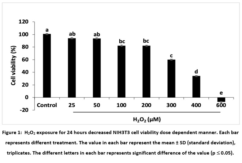 |
Figure 1: H2O2 exposure for 24 hours decreased NIH3T3 cell viability dose dependent manner. Each bar represents different treatment. |
The previous study also reported that H2O2 exposure to fibroblasts reduces the viability of MEF (mouse embryonic fibroblast) cells by around 30% at the concentration of 400 and 600 μM after 24 hours exposure7, around 60% in human dermal fibroblasts (BJ cells) (500 μM of H2O2 exposure)9, around 60% in H8F2p25LM cells (10-100 μM H2O2 exposure), and around 60% in MRC-5 cells (100-500 μM H2O2 exposure). 70, 80, 90, and 100 μM H2O2 exposured cause the lethal condition on H8F2p25LM cells20. NOX (NADPH oxidase) enzymes are activated at the ligand-receptor interactions (TNFα-TNFR or EGF-EGFR), and extracellular superoxide dismutase (SOD3) captures superoxide, providing H2O2 for imported by aquaporins (peroxiporins). This condition triggers proliferation, migration, and angiogenesis1. Endogenous and exogenous stress triggers excess H2O2 production and causes an antioxidant defense imbalance, leading to oxidative stress21. H2O2 triggers oxidative stress through downstream cellular signaling mechanisms22. Peroxiredoxin-2 acts as a primary H2O2 ultrasensitive receptor that specifically transmits oxidative equivalents to redox regulatory transcription. The STAT3 factor conveys redox signaling and H2O2 to accumulate locally at the target site. This causes tumor growth, metastases, inflammation, and fibrosis1. Excessive H2O2 accumulation causes mitochondrial membrane abnormalities and produces cellular oxidative stress, disrupts cell function, and integrity21.
Cytotoxicity of Black Rice ‘Sembada Hitam’ Bran’s Extract on NIH3T3 cells
In this study, exposures of H2O2 induced oxidative stress and decreased cell viability to cell death. Natural ingredients, such as black rice, contains high antioxidant activity and protect cells from oxidative stress. The results showed that 7.81 up to 1000 μg/mL of black rice bran extract’s had no cytotoxicity effect on NIH3T3 cells, especially at the concentration of 250 to 1000 μg/mL which showed highly cell viability. These results provided 7.81 to 1000 μg/mL of BRB ‘Sembada Hitam’ extract can be applied in further treatment (Figure 2.).
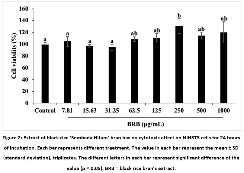 |
Figure 2: Extract of black rice ‘Sembada Hitam’ bran has no cytotoxic effect on NIH3T3 cells for 24 hours of incubation. Each bar represents different treatment. |
BRB’s extract treatment showed a slightly increasing cell viability, with no significant difference from the control group. ‘Sembada Hitam’ contains high anthocyanins (cyanidin-3-glucoside) 369.5 µg/100 g of rice seeds. Black rice also contains protein, fat, iron, tocopherol, zinc, and vitamin B such as thiamine and riboflavin, which were higher than brown rice and white rice23. Black rice extract slightly increases fibroblast cell viability (RDFs), with no significantly different from the control group22. The pericarp (outer layer) of black rice contains black pigments called anthocyanins, highly antioxidant activities that can prevent free radicals in the body 15.
Cytoprotective of Black Rice ‘Sembada Hitam’ Bran’s Extract on NIH3T3 cells Induced by H2O2
Based on the cytotoxicity result of BRB’s extract, it is known that the extract did not show a cytotoxic effect and maintained the viability of NIH3T3 cells. Further, we investigated whether the BRB’s extract has a cytoprotective effect against H2O2 exposure on NIH3T3 cells. The combination of H2O2 at various concentrations (50, 100, 150, 200, or 300 μM) and BRB’s extract at the concentration of 7.81 to 1000 μg/mL were exposed on NIH3T3 cells for 24 hours. Our results showed that 7.81 to 1000 μg/mL of BRB’s extract had a protective effect against 50 and 100 μM H2O2 exposure. Moreover, BRB’s extract significantly increased cell viability at 500 and 1000 μg/mL. However, higher concentrations of H2O2 exposure (150, 200, or 300 μM) combined with various concentrations of BRB’s extract were not effective to protect NIH3T3 cell viability. The combination of 200 μM H2O2 exposure and various concentrations of BRB’s extract decreased more than 60% of NIH3T3 cell viability. Whereas, the combination of 300 μM H2O2 exposure and various concentrations of BRB’s extract significantly caused a lethal condition on NIH3T3 cells (Figure 3.).
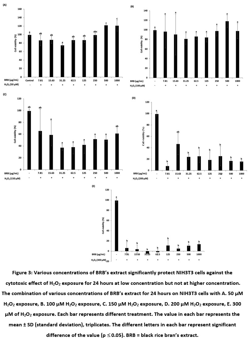 |
Figure 3: Various concentrations of BRB’s extract significantly protect NIH3T3 cells against the cytotoxic effect of H2O2 exposure for 24 hours at low concentration but not at higher concentration. |
The protective effect of anthocyanin extract in black rice on rat dermal fibroblasts (RDF) increases cell viability against H2O2-induced oxidative stress24. Boonyanuphong and U Tobgay19 also reported that the antioxidant activity of black glutinous rice was able to prevent H2O2 oxidative stress and reduce ROS levels on HT-29 cells. Anthocyanins regulate of PI3K/Akt and ERK1/2 expression as a cellular defense system against oxidative stress. Another study also reported the high antioxidant activity contained in the anthocyanin extract of black rice (Oryza sativa L. Indica) (25 ppm of an extract with an antioxidant capacity of 824 ± 17.24 μM) was able to inhibit oxidation reactions that could protect and prevent DPPH radicals11. In normal physiological conditions, the intracellular H2O2 produced by mitochondria is around 10 μM or below while the rate of production in organs is around 50 μM per minute per gram. However, the susceptibility of plasma membrane permeability to extracellular H2O2 can disrupt ROS homeostasis and modulate redox signaling pathways, so that H2O2 can diffuse into cells and be decomposed easily by intracellular antioxidants that triggers oxidative stress1,25. Induction of H2O2 at the concentrations of 150 and 200 μM on hADMSCs cells caused DNA damage26. Another study also reported cytoplasmic condensation and increased intracellular gap on human lens epithelial cells (HLE) shown at the concentration of 200 μmol/L H2O2 exposure represented apoptosis cells27.
Anti-apoptotic of Black Rice ‘Sembada Hitam’ Bran’s Extract on NIH3T3 Cells Induced by H2O2
The features of H2O2-induced apoptotic cells were detected using a double-staining method using AO-PI which detected green nuclei as a living cell and red nuclei as an apoptotic cell. Treatment of BRB’s extract at the concentration of 62.5 μg/mL was shown effective in suppressing apoptotic cells caused by H2O2 exposures at 100 and 200 μM. Whereas, 250 μg/mL of BRB’s extract was effective in suppressing cell apoptosis after H2O2 exposure for 24 hours at the concentration of 100 μM. In contrast, 250 μg/mL of BRB’s extract was not effective in suppressing cell apoptosis after H2O2 exposure for 24 hours at 200 μM. AO/PI double staining showed a change in the color and morphology of apoptotic cells. Control cells display a bright green color and intact nuclear structure, indicating healthy cells, whereas red stained cells indicate the presence of apoptotic cells. Our results showed that H2O2 induced cell death through an apoptotic pathway dose-dependent manner. Figure 4. shows a shift from viable cells to apoptotic cells represented red nuclei and live cells represented green nuclei. At the combination concentration of 200 μM H2O2 and 250 μg/mL of BRB’s extract showed that the red fluorescence in the cells significantly increased, indicates the number of cell death and shrinkage (Figure 4.). We predicted that the antioxidant activity of BRB’s extract has an anti-apoptotic role in H2O2-induced cell death.
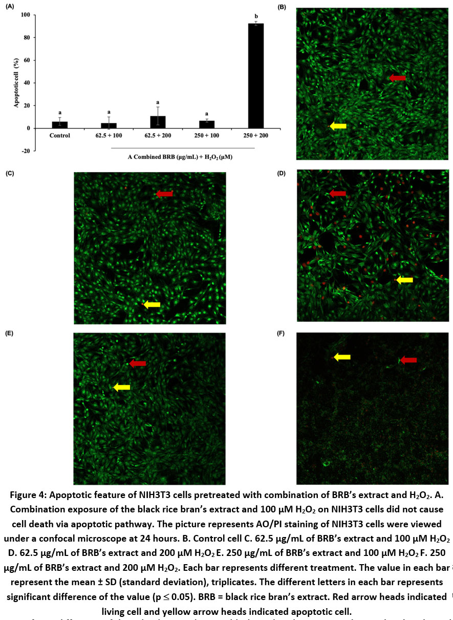 |
Figure 4: Apoptotic feature of NIH3T3 cells pretreated with combination of BRB’s extract and H2O2. A. Combination exposure of the black rice bran’s extract and 100 μM H2O2 on NIH3T3 cells did not cause cell death via apoptotic pathway. |
Anthocyanin form extract of black rice was effective in suppressing caspase-3 from H2O2 and induced apoptosis in RDF cells21,24. Cytochrome P450 enzyme (CYP) can convert toxic metabolites into ROS, such as H2O2 which can trigger oxidative stress to cause biological processes such as apoptosis. Cells that collectively reduce the oxidative state through an antioxidant system consisting of enzymatic antioxidants, can manipulate ROS levels by regulating gene expression and related signaling pathways to maintain redox balance and integrity of cellular components28. Antioxidant enzymes protect cell damage by decreasing the expression of pro-apoptotic proteins. In addition, Nrf2 regulation increases CAT expression which converts H2O2 produced by SOD into H2O and O2 to inhibit oxidation on HDF cells and causes anti-apoptotic conditions.
Our study showed that the BRB’s extract has antioxidant activity by protecting and maintaining cell viability from H2O2 stress at the concentrations of 100 μM or lower. Whereas, at the high concentrations of more than 100 μM, the BRB’s extract at the concentration of 250 μg/mL was not able to protect cell viability. It indicates that the higher concentration of the extract, it may has a prooxidant effect which causes a reduction in cell viability12. Various studies reported concentration-dependent antioxidant activity and prooxidants in natural antioxidants. The antioxidant activity of low concentrations of Trolox (2.5-15 μM) significantly decreases ROS production. However, the effect of prooxidants at the higher concentrations (20-160 μM) induces apoptosis29. The Sinlek rice bran extract had antioxidant activity at the concentration of 0.005-0.5 mg/mL and prooxidant activity at the higher concentrations (5-7.5 mg/mL) against 1 mM H2O2 induced oxidative stress on Caco-2 cells. Whereas, in Riceberry bran extract, at the concentrations of 15 and 17.5 mg/mL had antioxidant activity and at the concentration of 20 mg/mL prooxidant activity eliminated the protective effect30. Vitamin C, vitamin E, carotenoids, and polyphenols have an important role in the endogenous antioxidant defense system. Antioxidants in physiological dose ranges are able to protect against oxidative stress and excessive concentrations cause damage through prooxidant effects31. Moreover, prooxidant activity also catalyzed by Fe and Cu produces free radicals to mutagenesis in biological systems32.
Black Rice ‘Sembada Hitam’ Bran’s Extract Protects Cell Growth of NIH3T3 Cells Induced by H2O2
Protective effect of the BRB’s extract on cell survival against H2O2 exposure were assessed through cell growth assays on the 3rd and 5th day. The results showed that the cells were able to proliferate constantly after H2O2 exposure for 24 hours together with BRB’s extract treatment. The BRB’s extract at the concentration of 62.5 μg/mL significantly increased cell proliferation after 100 μM of H2O2 exposure for 24 hours. However, the cells which exposed with 200 μM of H2O2 started to stop their proliferation after 3 days culture. Interestingly, the treatment of BRB’s extract at the concentration of 250 μg/mL showed lower cells proliferation compared to that of 62.5 μg/mL against H2O2 exposure at the concentration of 100 and 200 μM (Figure 5.). Based on these results, we suggested that the BRB’s extract can prolong the life span of NIH3T3 cells up to 5 days after 24 hours exposure of H2O2 on the first day.
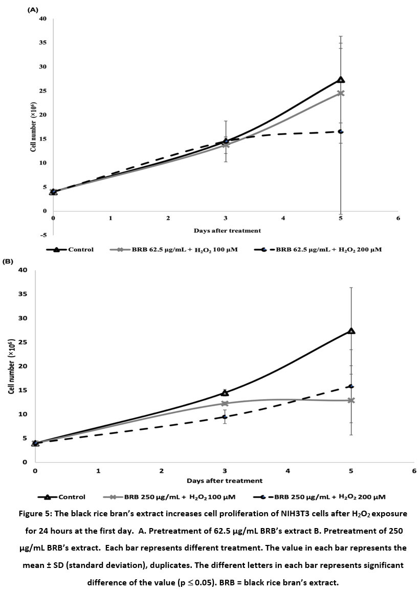 |
Figure 5: The black rice bran’s extract increases cell proliferation of NIH3T3 cells after H2O2 exposure for 24 hours at the first day. |
All kinds of oxidative damages reduce the replicative capacity and shorten the life span of the cells and organisms can be repaired by antioxidants. Natural and synthetic antioxidants can extend replicative life span through the activity of signaling pathways and proteasome activation33. The antioxidant effect of polyphenolic compounds in quercetin has been shown to protect IPEC-J2 cells from H2O2-induced apoptosis and 5 μg/mL of quercetin promote cell division34. Pretreatment of paeoniflorin (root extracted of Paeonia lactiflora Pall.) for 24 hours could attenuate apoptosis in PIG1 and PIG3V cells, but not effective for 48 hours treatment. Potency of paeoniflorin to counteract H2O2-induced oxidative damage on PIG1 and PIG3V cells were predicted by JNK and Nrf2/HO-1 pathway35. Moreover, H2O2 induced premature senescence and could be improved by over-expression of NAMPT on MEF cells7.
Conclusion
The BRB’s extract has potency to suppress apoptosis and maintain cell viability as well as cell growth against H2O2-induced oxidative stress. These results can be considered for application as a nutraceutical in chronic diseases mediated by oxidative stress and anti-aging cosmetic. The senescence regulation such as cell cycle arrest and DNA repair are required to further understand the cellular response.
Acknowledgement
None
Conflict of Interest
This article is the authors original work and is not under consideration for publication elsewhere. The corresponding author has full responsibility for the progress revisions until final approval of the manuscript. The authors declare no conflict of interest, or any personal business interest, affiliation, or activity with any entity.
Funding Sources
This research received funding from Hibah Rekognisi Tugas Akhir (RTA Grant), Universitas Gadjah Mada 2022 (Number: 5722/UN1.P.III/Dit-Lit/PT.01.05/2022) and Lecturer-Students Collaboration Research Grant, Faculty of Biology, UGM (Number: 1020/UN1/FBI/KSA/PT.01.03/2021 and 1185/UN1/FBI.1/KSA/PT.01.03/2022).
References
- Sies H., Berndt C., Jones D. P. Oxidative Stress. The Annual Review of Biochemistry. 2017;86(25):34.
CrossRef - Cosentino G., Plantamura I., Cataldo A., Iorio M. V. Micro RNA and Oxidative Stress Interplay in the Context of Breast Cancer Pathogenesis. IJMS. 2019;20(20):5143.
CrossRef - García-Sánchez A., Miranda-Díaz A. G., Cardona-Muñoz E. G. The Role of Oxidative Stress in Physiopathology and Pharmacological Treatment with Pro- and Antioxidant Properties in Chronic Diseases. Oxidative Medicine and Cellular Longevity. 2020;1-16.
CrossRef - Li Y. R,. Jia Z., Trush M. Defining ROS in Biology and Medicine. ROS. 2016;1(1).
CrossRef - Suryadinata R. V. Effect of Free Radicals on Inflammatory Processes in Chronic Obstructive Pulmonary Disease. AMNT. 2018;2(4):317.
- Krisdiantari N. Influence of Induction Phase of Chemotherapy on The Plasma Malondialdehyde as Oxidative Stress Biomarker in Acute Lymphoblastic Leukemia. UNHAS. 2018:1-65.
- Nuriliani A., Nakahata Y., Ahmed R., Khaidizar F. D., Matsui T., Bessho Y. Over‐Expression of Nicotinamide Phosphoribosyltransferase In Mouse Cells Confers Protective Effect Against Oxidative And ER Stress‐Induced Premature Senescence. Genes Cells. 2020;25(8):593-602.
CrossRef - Chidawanyika T., Supattapone S. Hydrogen Peroxide-induced Cell Death in Mammalian Cells. Journal of Cellular Signaling. 2021;2(3):6.
CrossRef - Buranasudja V., Muangnoi C., Sanookpan K., Halim H., Sritularak B., Rojsitthisak P. Eriodictyol Attenuates H2O2-Induced Oxidative Damage in Human Dermal Fibroblasts through Enhanced Capacity of Antioxidant Machinery. Nutrients. 2022;14(12):2553.
CrossRef - Suryanti V., Riyatun., Suharyana S., Sutarno., Saputra O. A. Antioxidant Activity And Compound Constituents of Gamma-Irradiated Black Rice (Oryza sativa L.) var. Cempo Ireng Indigenous of Indonesia. Biodiversitas. 2020;21(9).
CrossRef - Hetharia G. E., Briliannita A., Astuti M., Marsono Y. Antioxidant Extraction Based on Black Rice (Oryza Sativa L. Indica) to Prevent Free Radical. Mater Sci Eng. 2020;823(1):012002.
CrossRef - Han M., Bae J., Ban J., Shin H., Lee D., Chung J. Black Rice (Oryza sativa L.) Extract Modulates Ultraviolet-Induced Expression of Matrix Metalloproteinases and Procollagen In A Skin Cell Model. Int J Mol Med. 2018. http://doi.org/10.3892/ijmm.2018.3508
CrossRef - Fatchiyah F., Sari D. R. T., Safitri A., Cairns J. R. Phytochemical Compound and Nutritional Value in Black Rice from Java Island, Indonesia. Systematic Reviews in Pharmacy. 2020;11(7):8.
- Arifin A. S., Yuliana N. D., Rafi M. Antioxidant Activity of Pigmented Rice and Impact on Health. j pangan. 2019;28(1):11-22.
CrossRef - Kumar N., Murali R. D. Black Rice: A Novel Ingredient in Food Processing. Journal of Nutrition & Food Sciences. 2020;10(771):7.
- Christanto D. R., Mose J. C., Yuniarti T., Bestari M. B., Purwestri Y. A., Fauziah P. N. The Role of Black Rice Bran (Oryza Sativa L. “Sembada Hitam”) on Levels of Malondialdehyde in Induction Human Umbilical Vein Endothelial Cell Serum Preeclampsia. OJOG. 2020;10(12):1686-1692.
CrossRef - Christanto D. R., Mose J. C., Yuniarti T., Bestari M. B., Fauziah P. N., Purwestri Y. A., Munthe J. N. Anti-angiogenic Effect of Black Rice Bran (Oryza Sativa L. ‘Sembada Hitam’) on Soluble Fms-Like Tyrosine Kinase and Placental Growth Factor in Preeclampsia. Systematic Reviews in Pharmacy. 2021;12(1):4.
- Fitriasari A., Dewi D., Ikawati M., Meiyanto E. Protokol In Vitro. UGM. Cancer Chemoprevention Research Center (CCRC) Faculty of Pharmacy; 2010.
- Boonyanuphong P, Tobgay U. Protective Effect of Two Thai Pigmented Rice Cultivars Against H2O2-Inducedoxidative Damage In HT-29 Cell Culture. Food Res. 2022;6(1):27-33. http://doi.org/10.26656/fr.2017.6(1).206
CrossRef - Pieńkowska N., Bartosz G., Pichla M., Grzesik-Pietrasiewicz M., Gruchala M., Sadowska-Bartosz I. Effect of Antioxidants on The H2O2-Induced Premature Senescence of Human Fibroblasts. Aging. 2020;12(2):1910-1927. http://doi.org/10.18632/aging.102730
CrossRef - Zhao X., Fang J., Li S., Gaur U., Xing X., Wang H., Zheng W. Artemisinin Attenuated Hydrogen Peroxide (H2O2)-Induced Oxidative Injury in SH-SY5Y and Hippocampal Neurons via the Activation of AMPK Pathway. IJMS. 2019;20(11):2680. http://doi.org/10.3390/ijms20112680
CrossRef - Palungwachira P., Tancharoen S., Phruksaniyom C., Klungsaeng, S., Sichan R., Kikuchi K. Antioxidant and Anti-Inflammatory Properties of Anthocyanins Extracted from Oryza sativa L. in Primary Dermal Fibroblasts. Oxidative Medicine and Cellular Longevity. 2019;2019:1-18. http://doi.org/10.1155/2019/2089817
CrossRef - Kristamtini., Indrasari S. D., Widyayanti S., Andriyanto R., Sumarno. Molecular, Morphological, and Biochemical Identification of Sembada Merah and Sembada Hitam Rice (Oryza sativa L.). J Phys: Conf Ser. 2021;1918(5):052017. http://doi.org/10.1088/1742-6596/1918/5/052017
CrossRef - Palungwachira P., Tancharoen S., Dararat P., Nararatwanchai T. Anthocyanins Isolated From Oryza Sativa L. Protect Dermal Fibroblasts From Hydrogen Peroxide-Induced Cell Death. J Nat Sc Biol Med. 2020;11(1):45. http://doi.org/10.4103/jnsbm.JNSBM_171_19
CrossRef - Shaw P., Kumar N., Sahun M., Smits E., Bogaerts A., Privat-Maldonado A. Modulating the Antioxidant Response for Better Oxidative Stress-Inducing Therapies: How to Take Advantage of Two Sides of the Same Medal?. Biomedicines. 2022;10(4):823. http://doi.org/10.3390/biomedicines10040823
CrossRef - Valverde M., Lozano-Salgado J., Fortini P., Rodriguez-Sastre M. A., Rojas E., Dogliotti E. Hydrogen Peroxide-Induced DNA Damage and Repair through the Differentiation of Human Adipose-Derived Mesenchymal Stem Cells. Stem Cells International. 2018:1-10. http://doi.org/10.1155/2018/1615497
CrossRef - Shentu X. C., Ping X. Y., Cheng Y. L., Zhang X., Tang Y. L., Tang X. J. Hydrogen Peroxide-Induced Apoptosis of Human Lens Epithelial Cells is Inhibited by Parthenolide. Int J Ophthalmol. 2018. http://doi.org/10.18240/ijo.2018.01.03
CrossRef - He L., He T., Farrar S., Ji L., Liu T., Ma X. Antioxidants Maintain Cellular Redox Homeostasis by Elimination of Reactive Oxygen Species. Cell Physiol Biochem. 2017;44(2):532-553.
CrossRef - Giordano M. E., Caricato R., Lionetto M. G. Concentration Dependence of the Antioxidant and Prooxidant Activity of Trolox in HeLa Cells: Involvement in the Induction of Apoptotic Volume Decrease. Antioxidants. 2020;9(11):1058.
CrossRef - Jittorntrum B., Chunhabundit R., Kongkachuichai R., Srisala S., Visetpanit Y. Cytoprotective and Cytotoxic Effects of Rice Bran Extracts on H2O2– Induced Oxidative Damage in Human Intestinal Caco-2 Cells. Thai J Toxicology. 2009;24(2):92-100.
CrossRef - Kocyigit A., Selek S. Exogenous Antioxidants are Double-edged Swords. Bezmialem Science. 2016;4(2):70-75.
CrossRef - Eghbaliferiz S., Iranshahi M. Prooxidant Activity of Polyphenols, Flavonoids, Anthocyanins and Carotenoids: Updated Review of Mechanisms and Catalyzing Metals: Prooxidant Activity of Polyphenols and Carotenoids. Phytother Res. 2016;30(9):1379-1391.
CrossRef - Sadowska-Bartosz I., Bartosz G. Effect of Antioxidants on the Fibroblast Replicative Lifespan In Vitro. Oxidative Medicine and Cellular Longevity. 2020;2020:1-15.
CrossRef - Chen Z., Yuan Q., Xu G., Chen H., Lei H., Su J. Effects of Quercetin on Proliferation and H2O2-Induced Apoptosis of Intestinal Porcine Enterocyte Cells. Molecules. 2018;23(8).
CrossRef - Yuan J., Lu Y., Wang H, Feng X., Jiang S., Gao X., Qi R., Wu Y., Chen H. Paeoniflorin Resists H2O2-Induced Oxidative Stress in Melanocytes by JNK/Nrf2/HO-1 Pathway. Front Pharmacol. 2020;11:536.
CrossRef
Accepted on: 07 Dec 2022
Second Review by: Md. Mohibbullah Bangladesh
Final Approval by: Dr Aly El.Sheikha


Web of Science Coverage
Emerging Sources Citation Index (ESCI)
2024 Journal Impact Factor: 1.1
Scopus Journal Metrics
CiteScore 2025: 2.6
CiteScore Details
Sustainable Nutrition: Food Systems, Nutrient Retention, and Public Health Impact
![]()
This journal is a member of, and subscribes to the principles of, the Committee on Publication Ethics (COPE)


