Omega-3 Fatty Acids and COVID-19: Prevention or Adjuvant Therapy?
Maria Alessandra Gammone* and Nicolantonio D’Orazio
Human and Clinical Nutrition Unit, Department of Medical, Oral and Biotechnological Sciences, University G. D’Annunzio.
Corresponding Author E-mail: m.alessandra.gammone@gmail.com
DOI : http://dx.doi.org/10.12944/CRNFSJ.11.2.07
Download this article as:
![]()
The mechanisms of COVID-19 complications are multifactorial, including long-term tissue dam-ages from direct viral attack, dysregulation of both immunity and renin-angiotensin-aldosterone system and coagulation system, unresolved systemic inflammation and oxidative stress. Omega-3 polyunsaturated fatty acids (omega-3 or n-3 PUFAs) might have favorable effects on immunity, inflammation, oxidative stress at different stages of SARS-CoV-2 infection. Omega-3 and their metabolites including specialized proresolvin mediators, have shown effects in reducing pro-inflammatory cytokines, accelerating the resolution of chronic inflammation and restoring tissue homeostasis, and therefore offer a promising strategy against COVID-19. This article will discuss the inflammatory condition during COVID-19 pandemic, focus on the mechanisms that may contribute to the likely benefits of omega-3 and provide potential recommendations to pro-mote strategies for wellness.
KEYWORDS:COVID-19; Inflammation; omega-3; n-3 polyunsaturated fatty acids; SARS-CoV-2
Introduction
Comorbidities may predispose individuals to high-risk infective diseases such as SARs-CoV-2, with more severe complications, making interventions more complicated 1. An important comorbidity prone to infectious diseases, particularly to SARs-CoV-2 infection and to COVID-19, is obesity, an alarming pandemic 3 that predisposes them to comorbidities, such as diabetes mellitus, hypertension or metabolic syndrome and that adds a higher risk for poor outcomes and mortality in case of impact with novel viruses 1,4. Recent studies suggest that omega-3 PUFAs may interact at different stages of SARS-CoV-2 infection, particularly in contrasting the viral entry and replica-tion phases, where persistent viral infection may be responsible for the sustained in-flammatory state of long COVID.
There is growing evidence for the beneficial effects of omega-3 PUFAs and their metabolites, namely specialized pro-resolving mediators (SPMs), including ameliora-tion of uncontrolled inflammatory responses, reduction of oxidative stress and mitiga-tion of coagulopathy. Therefore, the nutritional status of omega-3 is crucial for the overall immune response, tissue inflammation and repair, which may be beneficial for the condition of long COVID.
Protective and supportive therapies may be helpful to improve COVID-19 pa-tients’ prognosis. In this respect, the beneficial effect of n-3 polyunsaturated fatty ac-ids, (n-3 PUFA or omega-3) includes the reduction of uncontrolled phlogistic reactions, oxidative stress and coagulopathies as well. In the light of their favorable safety pro-file, it is rational to consider n-3 PUFAs as a potential preventive strategy or adjuvant therapy in order to improve COVID-19 patients’ outcomes.
The role of inflammation and obesity as a comorbid condition
A persistent pathological inflammatory process is generated during some comor-bidities, such as diabetes mellitus, hypertension, metabolic syndrome and obesity: in particular, adipose tissue constitutes an autonomous endocrine organ, releasing large amounts of “adipokines” 5, bioactive peptides with a central role in vascular homeo-stasis, regulation of appetite, glucose and lipid metabolism, and immunity. Adipokines can target different organs and influence phlogosis responses and can exert pro-inflammatory or anti-inflammatory actions 6. Leptin is the leading adipokine; it promotes the displacement of local macrophages in the white adipose tissue (WAT) determining a shift toward a pro-inflammatory profile and decreases regulatory T-cells, also inducing Th17 polarization 7,8. Hyperleptinemia is a typical obesity marker 9, with leptin resistance upsetting the endothelial signals, contributing to a pro-inflammatory microenvironment, and predisposing to cardiac and vascular com-plications 10. Adiponectin, which is the antagonist adipokine with an-ti-inflammatory activity, is inversely linked to the amount of adipose tissue in obese subjects 11: a low adiponectin level is correlated to higher inflammatory mediators (particularly CRP and IL-6) levels and to several obesity-related metabolic diseases 12-15. Adipocyte hypertrophy is correlated to unbalanced intracellular signaling: the c-Jun NH2-terminal kinase (JNK) and the nuclear factor-kB (NF-kB) pathways are ac-tivated; enlarged omental adipocytes are hyper-responsive to TNF-α, determining ad-ipokines over-excretion 16,17. Also, oxidative stress and hypoxia (due to hypoperfu-sion of the expanding adipose tissue) contribute to the obese pro-inflammatory micro-environment 18,19 by decreasing the mRNA levels of adiponectin, while increasing the levels of both pro-inflammatory genes (TGF, TNF-α, PAI-1, IL-1, IL-6, MCP-1) and hypoxia response genes (glucose transporter 1, HIF-1, VEGF): this exacerbates the in-flammation in adipose tissue, contributing to obesity-related implications. This chronic inflammatory state creates a natural background predisposing obese subjects to nega-tive outcomes if an additional inflammatory stimulus (such as a virus) is introduced. In H1N1 a link between obesity and higher mortality was evidenced for body mass in-dex (BMI)>45 kg/m2 (OR 4.2; CI 1.9-9.4). Most recently, these findings were supported during the COVID-19 pandemic 2: obesity tripled the risk of hospitalization of those infected with SARs-CoV-2. More than 30% adult COVID-19 hospitalizations had obe-sity as a comorbid condition 20. In a healthcare cost model, 20.3% of patients with BMI>40 kg/m2 needed intensive care treatment, including invasive mechanical venti-lation compared with 6.6% of those with BMI <25 kg/m2 21. In infected patients it was reported not only the presence of dysregulated inflammation but also a pro-thrombotic state 22, with evidence of venous thrombocytope-nia/thromboembolism, renal failure, and disseminated intravascular coagulation in many ARDS patients. The patients with microthrombi showed more comorbidities such as overweight/obesity (64%), hypertension (62%), and cardiovascular disease (53%) 23. Endothelial hyper-activation enhances signaling pathways and leads to the generation of vascular adhesion molecules and proinflammatory cytokines, addressing inflammatory cells to both endothelium and underlying tissues 24,25. Further, both endothelium and adipose tissue produce plasminogen activator inhibitor-1(PAI-1): a higher levels of PAI-1 is typical of obesity and can determine hypofibrinolysis, thus contributing to poor outcomes in these patients 26,27.
Nutrients in COVID-19 prevention and Treatment
COVID-19 patients often suffer from harmful consequences (Table 1), because of a prominent systemic inflammation, with the outcome of COVID-19 patients being closely related to their nutritional status. In this respect, an adequate intake of nutri-ents can be helpful in order to prevent the infection and support the immune system during COVID-19 acute phase, but also in the post-acute phase, thus contrasting the various long-lasting symptoms typical of the so-called “long COVID”. For example, vitamin C contrasts inflammation and stimulates the immune response by regulating both cytokine secretion and histamine release, decreases oxidative stress, and regulates of T and B lymphocytes differentiation/proliferation 28. Numerous observational studies demonstrated that insufficient levels of vitamin D are related to COVID-19 se-verity 29-31; in addition, patients treated with a high-dose cholecalciferol supple-mentation displayed faster negativization 32, decreased access to intensive care 33 and increased survival among COVID-19 hospitalized patients than those without supplementation 34. Similarly, the glycoprotein lactoferrin can modulate the in-flammatory process by inhibiting the production of proinflammatory cytokines and by regulating the expression of iron homeostasis proteins (such as ferritin, ceruloplasmin and transferrin receptor 1) 35. Among the most helpful nutrients in the prevention and treatment of COVID-19, omega-3 fatty acids can play a pivotal role, as well as in the therapy of many inflammation-related diseases. Among the physiological process-es of phlogosis resolution is the enzymatic conversion of omega-3 eicosapentaenoic acid (EPA) and docosahexaenoic acid (DHA) into specialized pro-resolution mediators (including resolvins and protectins) participating in the resolution of phlogistic status and helping solve the cytokine storm and COVID-19 associated complications 36.
Overview and potential role of omega-3 in COVID-19 4.1
Immunomodulating Effect
A weak and ineffectual immune system often provides the chance for pathogens to bring severe illness. Omega-3 fatty acids, present in walnuts, flaxseeds and seafood (such as sardines, halibut, tuna, salmon, mackerel, but also in marine sponges, algae, crustaceans and krill), are responsible for numerous cellular functions such as signal-ing and cell-to-cell interaction 37. An adequate dietary intake of these polyunsatu-rated fatty acids can modulate the immune answer, by improving omega-6 and ome-ga-3 ratio. Their anti-phlogistic action is mediated by the inhibition of both 5-lipoxygenase/ leukotriene B4-B5 pathway and NF-κB pathway with a reduced ex-pression of cell surface adhesion molecules and a reduced production of interleukins (IL-1 and IL-6) from neutrophils 38. Omega-3 were displayed to decrease the genera-tion of pro-inflammatory cytokines from macrophages infected with Pseudomonas aeruginosa 39 and to increase phagocytic capacity of macrophages: in fact, microor-ganisms-mediated activation of the macrophage TLR4 signaling cascade depends on membrane lipid composition whose structures change after the incorporation of EPA and DHA 40. A 45-days double blind randomized study divided the healthy adult volunteers in two groups: placebo versus 3g DHA daily supplementation. The supple-mented group showed a minor post-exercise stress-induced IL-2 release from periph-eral mononuclear cells41; this is certainly helpful in the resolution of upper respira-tory tract infections 42. Similarly, a 45-days 1.6g-1.8g daily supplementation dis-played to enhance NK cell activity 43, reduce prostaglandins E2 levels and stimulate interferon-gamma secretion 44 with a substantial immune reinforcement, potentially able to prevent and to mitigate COVID-19 infection. In the same way, an increased in-take of omega-3 determines their increased incorporation into cell membranes, thus replacing arachidonic acid: this mechanism may enhance inflammation resolution in athlete post-exercise45 as well as in COVID-19 patients. Additionally, some potential antiviral activities of DHA-derived mediators have been reported: protectin D1 (a member of the class of specialized proresolving mediators generated by the oxygena-tion of DHA) and 17-HDHA (an autoxidation product of DHA) demonstrated to in-hibit respectively influenza A and H1N1 viral replication in mice46. Similar results were displayed against Zika virus 47, coxsakievirus and enterovirus with a signifi-cant viral load attenuation in human cells48.
Antinflammatory effect
A plethora of anti-inflammatory mechanisms have been attributed to omega-3 (Table 1). Firstly, they modulate the expression of adhesion molecules and inflamma-tory cytokines by activating anti-inflammatory transcription factors (PPARα/γ) and stopping TLR4-mediated activation of NF-κB. Secondly, omega-3 are metabolized into leukotrienes (with anti-inflammatory activities) by cyclooxygenases and lipoxygenas-es. Additionally, their metabolism produces proresolving mediators (Figure 1) with powerful antiphlogistic activities, expecially resolvins, protectins and maresins: they inhibit the migration of polymorphonuclear cells and the generation of both reactive oxygen species and chemokines, stimulating tissue regeneration and restoration of tis-sue homeostasis 49, which may be really helpful in limiting cytokine storm during COVID-19. Further, an intersection between innate immune inflammatory and mito-chondria has also been reported: the mitochondrial dysfunction can trigger uncon-trolled inflammatory answers50 determining the secondary injury aggravation in COVID-1951. At the same time the hypersecretion of inflammatory mediators trig-gers further intracellular cascades, altering mitochondrial functions: IL-6 and IFN-γ stimulate mitochondrial ROS production and determine mitochondrial membrane permeabilization until cell death; IL-1β and TNF-α inhibit mitochondrial oxidative phosphorylation and ATP production with exacerbation of cell injury52. In this re-spect, omega-3 displayed overabundance of beneficial effects against inflammation in many trials. Rats on n-3 PUFA enriched diet presented a reduction not only in pulmo-nary microvascular permeability and lung neutrophil accumulation but also decreased concentrations of arachidonic acid-derived metabolites (such as prostaglandin E2 and thromboxane B2) in alveolar macrophages, compared to n-6 PUFA enriched diet 53. In another study, pre-incubation with DHA of rhinovirus-infected epithelial cells de-creased the release of IL-6 and IFN-γ-inducible protein, and suppressed the vi-rus-induced inflammation54. In intensive care unit patients (with severe sepsis or septic shock requiring mechanical ventilation), a DHA enriched diet significantly ameliorated clinical outcomes with a lower mortality rate, in comparison to the control groups55. Similarly, a high-dose EPA diet (9 daily grams for 7 days) was evaluated in early-stage sepsis. This meta-analysis evidenced a noticeable improvement in oxy-genation of ventilated patients with acute respiratory distress syndrome. The lower rates of organ failures and severe sepsis development were associated to reduced levels of CRP, IL-6 and procalcitonin 56 with a reduction of intensive care stay by about two days57. Further studies defined the benefit of EPA and DHA supplementation (from 4 to 6 grams per day) in severe COVID-19, inhibiting cytokine secretion and mitigating the inflammatory state58. This antinflammatory activity could be partic-ularly precious in high-risk populations with underlying health conditions, such as di-abetes, obesity, hypertension, oncologic diseases and old age59, which could trigger the detrimental outcome often associated with severe COVID-19.
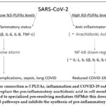 |
Figure 1: The connection n-3 PUFAs, inflammation and COVID-19 outcomes: |
N-3 PUFAs can replace the pro-inflammatory arachidonic acid in cell membranes or be metabo-lized to specialized pro-resolving mediators (SPMs): this down-regulates the NF-kB pathways and inhibits the synthesis of pro-inflammatory cytokines.
Anti-Arrhythmic, Vasodilator and Anti-thrombotic Effect
While systemic inflammation, respiratory complications and multi-organ dys-function determine a noticeable morbidity and mortality, cardiovascular complica-tions (such as myocarditis, acute myocardial infarction, dysrhythmias and thrombo-embolic accidents) during COVID-19 can also occur 60 typically in older subjects with comorbidities. However numerous cases of large vessels occlusion were reported also in young patients 61 because of significant coagulation anomalies, such as in-creased d-dimer, prolonged prothrombin time, and abnormal platelet levels 62. Omega-3 are known to contrast cardiovascular risk factors, such as hypertension, hy-perlipidemia and abnormal heart rhythm 63 reducing the risk of cardiac death for both hemodialysis or atrial fibrillation patients and healthy subjects without anamnes-tic cardiovascular diseases 64. In fact, they penetrate into cell membranes altering the lipid raft structure and function: this leads to improved intracellular organelle and cellular functions, higher arrhythmic thresholds, modified autonomic tone and atten-uated hypertension. Their anti-arrhythmic action can be explained by different mech-anisms: the modulation of L-type calcium, sodium and potassium channels 65, the inhibition of thromboxane generation 66, the capability to lower the plasmatic con-centration of non-esterified fatty-acids, which had previously displayed proarrhythmic activities 67. Additionally, omega-3 inhibit chemotactic answer of immune cells and adhesion molecules interaction/expression on endothelial cells, thus contrasting the development of blood clots in vessels: this mechanism, together with the an-ti-inflammatory activity, can explain the anti-thrombotic properties of omega-3 (Table 1). These mechanisms, responsible for omega-3 anti-atherogenic effects 68, may con-trast the development of blood clots in arteries during COVID-19, considering its pro-coagulant status and high risk of thromboembolic complications 69. Another re-search in a Japanese population evidenced that higher fish intakes were inversely as-sociated with intracerebral hemorrhaging 70. Omega-3 may influence membrane fluidity, interacting with Peroxisome Proliferator-Activated Receptors (PPARs) an-drepresent a substrate for lipoxygenase, cicloxygenase and cytochrome P450 71. As a result, n-3 PUFAs can induce hemodynamic modifications, improve endothelial func-tion and arterial compliance, decrease arrhythmias risk, and inhibit inflammatory pathways. Strong evidence suggests that DHA is more efficient in decreasing blood pressure, heart rate, platelet aggregation, and improving both the endothelial health and HDL/LDL ratio 71 thus decreasing global cardiovascular risk, so that the daily supplementation of omega-3 can be recommended for cardiovascular prevention.
Impact on respiratory system
The role of omega-3 in respiratory affections has already been evidenced in asthma and exercise-induced bronchoconstriction where the presence of epithelial in-jury and phlogosis in the airways was found 72. A decreased bronchial phlogosis due to an omega-3 dietary supplementation was clearly reported 73,74. For example, the low incidence of asthma and other chronic respiratory diseases in Eskimos may be due to the great intake of fat fish among this population 75. More specifically, a daily ad-ministration of 3.2 grams of EPA and 2.0 grams of DHA for 3 weeks decreased the concentration of pro-inflammatory cytokines (IL-1β and TNF-α) in the sputum also displaying that both fish oil and anti-leukotrienes medication were independently ef-fective in mitigating hyperpnea-induced and exercise-induced bronchoconstriction as well as airway inflammation 76. A randomized clinical trial evidenced that a high daily omega-3 dietary intake mitigated lung inflammation with a meliorated oxygena-tion in critical acute lung injury: the meta-analysis of outcome data displayed that the use of an inflammation-modulating diet in patients with acute respiratory distress syndrome increased ventilator-free days and significantly decreased mortality at 28-day interval 77. Similarly, an open-label trial showed the efficacy of parenteral nutrition with fish oil in modulating inflammatory response and cytokine production in patients with respiratory distress during sepsis: after 3 days the omega-3/omega-6 ratio was reversed with EPA and DHA prevalent over arachidonic acid, and omega-3 incorporation into mononuclear leukocyte membranes. Additionally, critical patients with acute lung injury and acute respiratory distress syndrome are prone not only to a major risk of sepsis but also to cardiac arrest: omega-3 can promote resolution of in-flammation and precondition the heart against septic cardiomyopathy because of their antioxidant and immuno-modulating activity 78.
Effect on the Renin Angiotensin Aldosterone system
Renin Angiotensin Aldosterone system (RAAS) has been focused on for several years because of its pivotal role in the physiology and pathophysiology of cardiovas-cular disease: it is involved in blood pressure regulation, fluid, and electrolyte balance through action on kidney and blood vessels. Angiotensin converting enzyme (ACE), is a critical regulator of RAAS by converting Angiotensin I (Ang-I) to Angiotensin II (Ang-II), which is the most powerful biologically active product of RAAS. Ang-II in-creases blood pressure stimulates aldosterone secretion, which results in sodium reab-sorption and potassium excretion. However, a second ACE (ACE2) is a negative regu-lator of RAAS: it opposes the effect of ACE in the heart, kidneys, and lungs and con-verts Ang-II to Angiotensin, which is a vasodilator, antihypertrophic, antithrombotic peptide 79. In fact, in almost all the cardiovascular pathological conditions there is a disturbance in ACE/ACE2 ratio, usually due to a down-regulation in ACE2 levels.
Additionally, ACE2 serves as a receptor for SARS-CoV-2 entry into target cells by binding of the spike protein to ACE2 and a specific transmembrane serine protease 2 (TMPRSS2) required for the spike protein priming, which also leads to down-regulation of ACE280. Interestingly, poor outcomes of COVID-19 have been observed in patients with pre-existing cardiovascular diseases, who have already defi-ciency in ACE2 and increased ACE/ACE2 ratio81.
Omega-3 supplementation was associated to a significant decrease in serum levels of both ACE/ACE2 ratio and ACE (Table 1), displaying that they may reduce suscepti-bility to COVID-19 and reduce disease severity and its complication 82.
Table 1: Potential molecular mechanisms of omega-3 PUFAs on COVID-19 complications
|
Target |
Mechanisms |
Effects |
References |
|
Inflammation |
Specialized proresolving mediators Reduction of cytokine storm Reprogramming pheripheral blood cell transcriptome |
Phlogosis resolution |
49-54 |
|
Immune dysregulation |
|
Innate immunity enhancement |
38,40,41 |
|
Viral invasion |
Tissue damage |
Reduced exacerbation of cell injury |
52 |
|
Coagulopathy |
Inhibition of platelet aggregation |
Thrombosis prevention |
63-69 |
|
ACE2/Ang1-7 imbalance |
Inhibition of angiotensin-converting enzyme Reduced angiotensin II formation Activation of endothelial nitric oxide synthase generation Suppression of TGF-beta expression |
ACE2/Ang1-7 rebalance Ameliorated vasodilation |
79-81 |
|
Oxidative stress |
Increase of antioxidant enzyme |
Antioxidant effects |
78 |
|
Psico-social impact |
Release of neurotransmitters with hypotalamus-pituitary-adrenal axis effects |
Reduction of chronic fatigue, depression and post-traumatic stress disorder |
83 |
Conclusion
This review highlights the molecular mechanisms of omega-3 PUFAs mediated resistance against COVID-19 based on available evidence. In addition to preserving or repairing the brain structure and function by interacting with phospholipid metabo-lism and the known shift in the pattern of lipid metabolites to a more an-ti-inflammatory metabolite profile, omega-3 PUFAs and/or their biologically active metabolites have the potential to improve oxidative stress, and immune dysregulation; maladaptation of the RAAS and coagulation system; and psychosocial stress from changes in health, financial status, or social life. Despite these promising effects of omega-3 PUFAs, additional epidemiological, experimental, and RCTs are needed to test, validate, and translate these proposed effects in the context of long COVID.
The administration of omega-3 fatty acids in COVID-19 patients aims to target both the improvement of health status and the prevention of potential complications: their anti-inflammatory action can stimulate macrophage phagocytic effort, disable enveloped virus, modulate cell signaling, attenuate coagulopathy, shift the lipid me-tabolites towards an anti-inflammatory pattern and globally mitigate an uncontrolled immune response secondary to the infection.
Acknowledgment
The authors acknowledge the Department of Medical, Oral and Biotech-nological Sciences, of University G. D’Annunzio for the support.
Conflicts of Interest
The authors declare no conflict of interest.
Funding Sources
The author(s) received no financial support for the research, authorship, and/or publication of this article.
References
- Marincu L., Bratosin F., Vidican I., Bostanaru A.C., Frent S., Cerbu B., Turaiche M., Tirnea L., Timircan M. Predictive value of comorbid conditions for COVID-19 mortality. J Clin Med. 2021; 10(12): 2652.
CrossRef - Louie J.K., Acosta M., Sauel M.C., Rchechter R., Vugia D.J., Harriman K., Matyas B.T. California Pandemic (H1N1) Working Group. A novel risk factor for a novel virus: Obesity and 2009 pandemic influenza A (H1N1). Clin Infect Dis. 2011; 52(3): 301-312.
CrossRef - Poitou C., Mosbah H., Clément K. Mechanisms in endocrinology: Update on treatments for patients with genetic obesity. Eur J Endocrinol. 2020; 183(5): R149–66.
CrossRef - World Health Organization. 2021. Obesity. https://www.who.int/ health-topics/obesity.
- Francisco V., Ruiz-Fernández C., Pino J., Mera A., González-Gay M.A., Gómez R. Adipokines: linking metabolic syndrome, the immune system, and arthritic diseases. Biochem Pharmacol. 2019; 165: 196–206.
CrossRef - Lelis D., Freitas D.F., deMachado A.S., Crespo T.S., Santos S.H.S. Angiotensin (1-7), adipokines and inflammation. Metab Clin Exp. 2019; 95: 36–45.
CrossRef - Gammone M.A., D’Orazio N. Anti-obesity activity of the marine carotenoid fucoxanthin. Mar Drugs. 2015; 13(4): 2196-214.
CrossRef - Abella V., Scotece M., Conde J., Pino J., Gonzalez-Gay M.A., Gómez-Reino J.J. Leptin in the interplay of inflammation, metabolism and immune system disorders. Nat Rev Rheumatol. 2017; 13: 100–9.
CrossRef - deGit K.C.G., Peterse C., Beerens S., Luijendijk M.C.M., vanDerPlasse G., laFleur S.E. Is leptin resistance the cause or the consequence of diet-induced obesity? Int J Obes. 2018; 42(8): 1445–1457.
CrossRef - Cui H., López M., Rahmouni K. The cellular and molecular bases of leptin and ghrelin resistance in obesity. Nat Rev Endocrinol. 2017; 13(6): 338–351.
CrossRef - Fang H., Judd R.L. Adiponectin regulation and function. Compr Physiol. 2018; 8: 1031–63.
CrossRef - Kumada M., Kihara S., Sumitsuji S., Kawamoto T., Matsumoto S., Ouchi N. Association of hypoadiponectinemia with coronary artery disease in men. Arterioscler Thromb Vasc Biol. 2003; 1: 1.
CrossRef - Riccioni G., Gammone M.A., Currenti W., D’Orazio N. Effectiveness and safety of dietetic supplementation of a new nutraceutical on lipid profile and serum inflammation biomarkers in hypercholesterolemic patients. Molecul. 2018; 23(5): 1168.
CrossRef - Gammone M.A., Pluchinotta F.R., Bergante S., Tettamanti G., D’Orazio N. Prevention of cardiovascular diseases with carotenoids. Front Biosci (Schol Ed). 2017; 9: 165-171.
CrossRef - Gammone M.A., Riccioni G., Massari F., D’Orazio N. Beneficial effect of ivabradine against cardiovascular diseases. Front Biosci (Schol Ed). 2020; 12: 161-172.
CrossRef - Maury E., Brichard S.M. Adipokine dysregulation, adipose tissue inflammation and metabolic syndrome. Molec Cell Endocr 2010; 314(1): 1-16.
CrossRef - Maury E., Noel L., Detry R., Brichard S.M. In vitro hyper-responsiveness to TNF-alpha contributes to adipokine dysregulation in omental adipocytes of obese subjects. J Clin Endocrinol Metab. 2009; 2: 1123-1135.
- Gammone M.A., Riccioni G., D’Orazio N. Marine Carotenoids against Oxidative Stress: Effects on Human Health. Mar Drugs. 2015; 13(10): 6226-46.
CrossRef - Pasarica M., Sereda O.R., Redman L.M., Albarado D.C., Hymel D.T., Roan L.E., Rood J.C., Burk D.H., Smith S.R. Reduced adipose tissue oxygenation in human obesity: evidence for rarefaction, macrophage chemotaxis and inflammation without an angiogenic response. Diabet. 2009; 58: 718–725.
CrossRef - Center of Disease Control and Prevention. 2021. Overweight & obesity: Obesity, race/ethnicity, COVID-19. https://www.cdc.gov/obesity/ data/obesity-and-covid-19.
- Czernichow S., Bain S.C., Capehorn M., Bøgelund M., Madsen M.E., Yssing C., McMillan A.C., Cancino A.P., Panton U.H. Costs of the COVID19 pandemic associated with obesity in Europe: A health-care cost model. Clin Obes. 2021; 11(2):e12442.
CrossRef - Campbell C.M., Kahwash R. Will complement inhibition be the new target in treating COVID-19 related systemic thrombosis? Circulat. 2020; 41(22):1739-1741.
CrossRef - Gammone M.A., D’Orazio N. COVID-19 and obesity: overlapping of two pandemics. Obes Facts. 2021; 14(6):579-585.
CrossRef - Iba T., Levy J.H., Levi M., Thachil J. Coagulopathy in COVID-19. J Thromb Haemost. 2020; 18(9):2103-2109.
CrossRef - Sengenes C., Miranville A., Lolmede K., Curat C.A., Bouloumie A. The role of endothelial cells in inflamed adipose tissue. J Intern Med. 2007; 262:415–421.
CrossRef - Bikdeli B., Madhavan M.V., Gupta A., Jimenez D., Burton J.R., DerNigoghossian C. Global COVID-19, Thrombosis Collaborative Group: pharmacological agents targeting thromboinflammation in COVID-19. Review and implications for future research. Thromb Haemost. 2020; 120(7):1004-1024.
CrossRef - Gammone M.A., D’Orazio N. Obesity and COVID-19: A detrimental intersection. Front Endocrinol. 2021; 12:6526-39.
CrossRef - Chen Y., Luo G., Yuan J., Wang Y., Yang X., Wang X., Li G., Liu Z., Zhong N. Vitamin C mitigates oxidative stress and tumor necrosis factor-alpha in severe community-acquired pneumonia and LPS-induced macrophages. Mediat Inflamm. 2014;426740.
CrossRef - D’Avolio A., Avataneo V., Manca A., Cusato J., DeNicolò A., Lucchini R., Keller F., Cantù M. 25-Hydroxyvitamin D concentrations are lower in patients with positive PCR for SARS-CoV-2. Nutr. 2020; 12:1359.
CrossRef - Meltzer D.O., Best T.J., Zhang H., Vokes T., Arora V., Solway J. Association of vitamin D status and other clinical characteristics with COVID-19 test results. JAMA Netw Open. 2020; 3:e2019722.
CrossRef - Vassiliou A.G., Jahaj E., Pratikaki M., Orfanos S.E., Dimopoulou I., Kotanidou A. Low 25-hydroxyvitamin D levels on admission to the intensive care unit may predispose COVID-19 pneumonia patients to a higher 28- day mortality risk: A pilot study on a Greek ICU cohort. Nutr. 2020; 12:3773.
CrossRef - Rastogi A., Bhansali A., Khare N., Suri V., Yaddanapudi N., Sachdeva N., Puri G.D., Malhotra P. Short term high-dose vitamin D supplementation for COVID-19 disease: a randomised, placebo-controlled study (SHADE study). Postgrad Med J. 2020; 98:87–90.
CrossRef - Nogues X., Ovejero D., Pineda-Moncusí M., Bouillon R., Arenas D., Pascual J., Ribes A., Guerri-Fernandez R., Villar-Garcia J., Rial A. Calcifediol treatment and COVID-19-related outcomes. J Clin Endocrinol Metab. 2021; 106:e4017–e4027.
CrossRef - Loucera C., Peña-Chilet M., Esteban-Medina M., Muñoyerro-Muñiz D., Villegas R., Lopez-Miranda J., Rodriguez-Baño J., Túnez I., Bouillon R., Dopazo J. Real world evidence of calcifediol or vitamin D prescription and mortality rate of COVID-19 in a retrospective cohort of hospitalized Andalusian patients. Sci Rep. 2021; 11:23380.
CrossRef - Cutone A., Lepanto M.S., Rosa L., Scotti M.G., Rossi A., Ranucci S., DeFino I., Bragonzi A., Valenti P., Musci G. Aerosolized bovine lactoferrin counteracts infection, inflammation and iron dysbalance in a cystic fibrosis mouse model of pseudomonas aeruginosa chronic lung infection. Int J Mol Sci. 2019; 20:21-28.
CrossRef - D’Angelo S., Motti M.L., Meccariello R. Omega-3 and Omega-6 Polyunsaturated Fatty Acids, Obesity and Cancer. Nutr. 2020; 12:2751.
CrossRef - Gammone M.A., Riccioni G., Parrinello G., D’Orazio N. Omega-3 polyunsaturated fatty acids: benefits and endpoints in sport. Nutr. 2018; 11(1):46.
CrossRef - D’Orazio N., Gemello E., Gammone M.A., DeGirolamo M., Ficoneri C., Riccioni G. Fucoxantin: A treasure from the sea. Mar Drugs. 2012; 10:604–616.
CrossRef - Schoeniger A., Fuhrmann H., Schumann J. LPS- or Pseudomonas aeruginosa-mediated activation of the macrophage TLR4 signaling cascade depends on membrane lipid composition. Peer J. 2016; 4:e1663.
CrossRef - Hellwing C., Tigistu-Sahle F., Fuhrmann H., Kakela R., Schumann J. Lipid composition of membrane microdomains isolated detergent-free from PUFA supplemented RAW264.7 macrophages. J Cell Physiol. 2018; 233, 2.
CrossRef - Gray P., Gabriel B., Thies F., Gray S.R. Fish oil supplementation augments post-exercise immune function in young males. Brain Behav Immun. 2012; 26:1265–1272.
CrossRef - Gleeson M., Bishop N.C. The T cell and NK cell immune response to exercise. Ann Transplant. 2005; 10:43–48.
- Kawabata F., Neya M., Hamazaki K., Watanabe Y., Kobayashi S., Tsuji T. Supplementation with eicosapentaenoic acid-rich fish oil improves exercise economy and reduces perceived exertion during submaximal steady-state exercise in normal healthy untrained men. Biosci. Biotechnol. Biochem. 2014; 78:2081–2088.
CrossRef - Andrade P.M.M., Ribeiro B.G., Bozza M.T., Costa-Rosa L.F.B., DoCarmo M.G.T. Effects of the fish-oil supplementation on the immune and inflammatory responses in elite swimmers. Prost Leuk Essent Fatty Acids. 2007; 77:139–145.
CrossRef - Nieman, D.C.; Mitmesser, S.H. Potential impact of nutrition on immune system recovery from heavy exertion: a metabolomics perspective. Nutr.2017; 9: 513.
CrossRef - Ramon S., Baker S.F., Sahler J.M., Kim N., Feldsott E.A., Serhan C.N., Phipps R.P. The specialized proresolving mediator 17-HDHA enhances the antibody mediated immune response against influenza virus: a new class of adjuvant? J Immunol. 2014; 193:6031–6040.
CrossRef - Braz-De-Melo H.A., Pasquarelli-do-Nascimento G., Correa R., dasNevesAlmeida R., deOliveira-Santos I., Prado P.S., Magalhaes K.G. Potential neuroprotective and anti-inflammatory effects provided by omega-3 (DHA) against Zika virus infection in human SH-SY5Y cells. Sci Rep. 2019; 9:20119.
CrossRef - Yan B., Zou Z., Chu H., Chan G., Tsang J.O., Lai P.M., Yuen K.Y. Lipidomic profiling reveals significant perturbations of intracellular lipid homeostasis in enterovirus-infected cells. Int J Mol Sci. 2019; 20(23):5952.
CrossRef - Zhang H.W., Wang Q., Mei H.X., Zheng S.X., Ali A.M., Wu Q.X., Jin S.W. RvD1 ameliorates LPS-induced acute lung injury via the suppression of neutrophil infiltration by reducing CXCL2 expression and release from resident alveolar macrophages. Int Immunopharm 2019; 76:105877.
CrossRef - Mohanty A., Tiwari-Pandey R., Pandey N.R. Mitochondria: the indispensable players in innate immunity and guardians of the inflammatory response. J Cell Communic Signal. 2019; 13:303–318
CrossRef - Keshavarz-Bahaghighat, H.; Darwesh, A.M.; Sosnowski, D.K., Seubert, J.M. Mitochondrial dysfunction and inflammaging in heart failure: novel roles of CYP-derived epoxylipids. Cells.2020; 9(7):1565.
CrossRef - Jo E.K., Kim J.K., Shin D.M., Sasakawa C. Molecular mechanisms regulating NLRP3 inflammasome activation. Cell Molec Immunol. 2016; 13:148–159.
CrossRef - Darwesh M., Bassiouni W., Sosnowski D.K. Can N-3 polyunsaturated fatty acids be considered a potential adjuvant therapy for COVID-19-associated cardiovascular complications? Pharmacol Therap. 2021; 219:107703.
CrossRef - Saedisomeolia A., Wood L.G., Garg M.L., Gibson P.G., Wark P.A. Antinflammatory effects of long-chain n-3 PUFA in rhinovirus-infected cultured airway epithelial cells. Br J Nutr. 2009; 101:533–540.
CrossRef - Pontes-Arruda A., Aragao A.M., Albuquerque J.D. Effects of enteral feeding with eicosapentaenoic acid, gamma-linolenic acid, and antioxidants in mechanically ventilated patients with severe sepsis and septic shock. Crit Care Med. 2006; 34:2325–2333.
CrossRef - Hosny M., Nahas R., Ali S., Elshafei S.A., Khaled H. Impact of oral omega-3 fatty acids supplementation in early sepsis on clinical outcome and immunomodulation. Ind J Crit Care Med. 2013; 1:119–126.
CrossRef - Pradelli L., Mayer K., Klek S., Omar-Alsaleh A.J., Clark R.A.C., Rosenthal M.D., Muscaritoli M. Omega-3 fatty-acid enriched parenteral nutrition in hospitalized patients: systematic review with meta-analysis and trial sequential analysis. J Parenter Enter Nutr. 2020; 44:44–57.
CrossRef - Bistrian B.R. Parenteral fish-oil emulsions in critically Ill COVID-19 emulsions. J Parenter Enter Nutr. 2020; 44(7):1168.
CrossRef - Torrinhas R.S., Calder P.C., Waitzberg D.L. Response to bistrian BR. Parenteral fish-oil emulsions in critically Ill COVID-19 emulsions. J Parenter Enteral Nutr. 2020; 44(7):1169–1170.
CrossRef - Long B., Brady W.J., Gottlieb M. Cardiovascular complication in COVID-19. Am J Emerg Med. 2020; 38(7):1504-1507.
CrossRef - Oxley T.J, Mocco J., Majidi S., Kellner C.P., Shoirah H., Singh I.P., DeLeacy R.A., Shigematsu T., Ladner T.R., Yaeger K.A., Skliut M., Weinberger J., Dangayach N.S., Bederson J.B., Tuhrim S., Fifi J.T. Large-vessel stroke as a presenting feature of Covid-19 in the young. N Engl J Med 2020; 382(20):e60.
CrossRef - Tang N., Li D., Wang X. Sun Z. Abnormal coagulation parameters are associated with poor prognosis in patients with novel coronavirus pneumonia. J Thromb Haemost. 2020; 18 (4):844-847.
CrossRef - Yokoyama M., Origasa H., Matsuzaki M., Japan M.Y. EPA lipid intervention study (JELIS) investigators. Effects of eicosapentaenoic acid on major coronary events in hypercholesterolaemic patients: a randomized open label, blinded endpoint analysis. Lancet. 2007; 369:1090–1098.
CrossRef - Friedman A.N., Yu Z., Denski C., Tamez H., Wenger J., Thadhani R., Li Y., Watkins B. Fatty acids and other risk factors for sudden cardiac death inpatients starting hemodialysis. Am J Nephrol 2013; 38:12–18.
CrossRef - Kang J.X., Leaf A. Antiarrhythmic effects of polyunsaturated fatty acids: recent studies. Circulat. 1996; 94:1774–1780.
CrossRef - Nair S.S.D., Leitch J.W., Falconer J., Garg M.L. Prevention of cardiac arrhythmia by dietary (n-3) polyunsaturated fatty acids and their mechanism of action. J Nutr 1997; 127:383–393.
CrossRef - Gammone M.A., D’Orazio N. Cocoa overconsumption and cardiac rhythm: potential arrhythmogenic trigger or beneficial pleasure? Curr Res Nutr Food Sci. 2021; 9(1):40-51.
CrossRef - Calder P.C. The role of marine omega-3 (n-3) fatty acids in inflammatory processes, atherosclerosis and plaque stability. Mol Nutr Food Res. 2012; 56:1073–1080.
CrossRef - Synowiec A., Szczepanski A., Barreto-Duran E.; Lie L.K., Pyrc K. Severe acute respiratory syndrome coronavirus 2 (SARS-CoV-2): a systemic infection. Clin Microbiol. Rev 2021; 34(2):e00133-20.
CrossRef - Raatz S.K., Silverstein J.T., Jahns L., Picklo M.J. Issues of fish consumption for cardiovascular disease risk reduction. Nutr. 2013; 5:1081–1097.
CrossRef - Cottin S., Sanders T., Hall W. The differential effects of EPA and DHA on cardiovascular risk factors. Proc Nutr Soc. 2011; 70:215–231.
CrossRef - Gammone M.A., Riccioni G., Parrinello G., D’Orazio, N. Omega-3 Polyunsaturated Fatty Acids: Benefits and Endpoints in Sport. Nutr. 2018; 11(1):46.
CrossRef - Schubert R., Kitz R., Beermann C. Effect of n-3 polyunsaturated fatty acids in asthma after low-dose allergen challenge. Int Arch Allergy Immunol. 2009; 148:321–329.
CrossRef - Biltagi M.A., Baset A.A., Bassiouny M., Kasrawi M.A., Attia M. Omega-3 fatty acids, vitamin C and Zn supplementation in asthmatic children: A randomized self- controlled study. Acta Paediatr. 2009; 98:737–742.
CrossRef - Horrobin D.F. Low prevalences of coronary heart disease (CHD), psoriasis, asthma and rheumatoid arthritis in Eskimos: Are they caused by high dietary intake of eicosapentaenoic acid (EPA), a genetic variation of essential fatty acid (EFA) metabolism or a combination of both? Med Hypothes. 1987; 22:421–428.
CrossRef - Tecklenburg-Lund S., Mickleborough T.D., Turner L.A., Fly A.D., Stager J.M., Montgomery G.S. Randomized controlled trial of fish oil and montelukast and their combination on airway inflammation and hyperpnea-induced bronchoconstriction. PLoS ONE. 2010; 5:e13487.
CrossRef - Pontes-Arruda A., Demichele S., Seth A., Singer P. The use of an inflammationmodulating diet in patients with acute lung injury or acute respiratory distress syndrome: a meta-analysis of outcome data. J Parent Enteral Nutr. 2008; 32:596–605.
CrossRef - Leger T., Azarnoush K., Traore A., Cassagnes L., Rigaudiere J. P., Jouve C., Demaison L. Antioxidant and cardioprotective effects of EPA on early low-severity sepsis through UCP3 and SIRT3 upholding of the mitochondrial redox potential. Oxid Med Cell Longev. 2019; e 9710352.
CrossRef - Bosso M., Thanaraj T.A., Abu-Farha M. The Two Faces of ACE2:The Role of ACE2 Receptor and Its Polymorphisms in Hypertension and COVID-19. Molec Ther Methods &Clin Devel. 2020;18(1):321-327.
CrossRef - Cheng H., Wang Y., Wang G.Q. Organ-protective effect of angiotensin-converting enzyme 2 and its effect on the prognosis of COVID-19. J Med Virol. 2020;92(9):726-730.
CrossRef - Viana S.D., Nunes S., Reis F. ACE2 imbalance as a key player for the poor outcomes in COVID-19 patients with age-related comorbidities – Role of gut microbiota dysbiosis. Ageing Res Rev. 2020;62:101123.
CrossRef - Daboul S.M., Abusamak M., Mohammad B.A., Alsayed A.R., Habash M., Mosleh I., Al-Shakhshir S., Issa R., Abu-Samak M. The effect of omega-3 supplements on the serum levels of ACE/ACE2 ratio as a potential key in cardiovascular disease: A randomized clinical trial in participants with vitamin D deficiency. Pharm Pract (Granada). 2023;21(1):2761.
CrossRef - Chang J.P.C., Chang S.S., Chen H.T., Chien Y.C., Yang H.T. et al. Omega-3 polyunsaturated fatty acids (n-3 PUFAs), somatic and fatigue symptoms in cardiovascular diseases comorbid major depressive disorder (MDD): A randomized controlled trial. Brain, Behavior, and Immun. 2023; 112:125-131.
CrossRef
Accepted on: 30 Jul 2023
Second Review by: Sepideh Hesami
Final Approval by: Dr Ardiansyah


Web of Science Coverage
Emerging Sources Citation Index (ESCI)
2024 Journal Impact Factor: 1.1
Scopus Journal Metrics
CiteScore 2025: 2.6
CiteScore Details
Sustainable Nutrition: Food Systems, Nutrient Retention, and Public Health Impact
![]()
This journal is a member of, and subscribes to the principles of, the Committee on Publication Ethics (COPE)


